. 2017 Jan 18;91(3):e01398-16.
doi: 10.1128/JVI.01398-16. Print 2017 Feb 1.

Pre-mRNA Processing Factor Prp18 Is a Stimulatory Factor of Influenza Virus RNA Synthesis and Possesses Nucleoprotein Chaperone Activity

Affiliations

Pre-mRNA Processing Factor Prp18 Is a Stimulatory Factor of Influenza Virus RNA Synthesis and Possesses Nucleoprotein Chaperone Activity

M Minakuchi et al. J Virol. .

Abstract

The genome of influenza virus (viral RNA [vRNA]) is associated with the nucleoprotein (NP) and viral RNA-dependent RNA polymerases and forms helical viral ribonucleoprotein (vRNP) complexes. The NP-vRNA complex is the biologically active template for RNA synthesis by the viral polymerase. Previously, we identified human pre-mRNA processing factor 18 (Prp18) as a stimulatory factor for viral RNA synthesis using a Saccharomyces cerevisiae replicon system and a single-gene deletion library of Saccharomyces cerevisiae (T. Naito, Y. Kiyasu, K. Sugiyama, A. Kimura, R. Nakano, A. Matsukage, and K. Nagata, Proc Natl Acad Sci USA, 104:18235-18240, 2007, https://doi.org/10.1073/pnas.0705856104). In infected Prp18 knockdown (KD) cells, the synthesis of vRNA, cRNA, and viral mRNAs was reduced. Prp18 was found to stimulate in vitro viral RNA synthesis through its interaction with NP. Analyses using in vitro RNA synthesis reactions revealed that Prp18 dissociates newly synthesized RNA from the template after the early elongation step to stimulate the elongation reaction. We found that Prp18 functions as a chaperone for NP to facilitate the formation of NP-RNA complexes. Based on these results, it is suggested that Prp18 accelerates influenza virus RNA synthesis as an NP chaperone for the processive elongation reaction.

Importance: Templates for viral RNA synthesis of negative-stranded RNA viruses are not naked RNA but rather RNA encapsidated by viral nucleocapsid proteins forming vRNP complexes. However, viral basic proteins tend to aggregate under physiological ionic strength without chaperones. We identified the pre-mRNA processing factor Prp18 as a stimulatory factor for influenza virus RNA synthesis. We found that one of the targets of Prp18 is NP. Prp18 facilitates the elongation reaction of viral polymerases by preventing the deleterious annealing of newly synthesized RNA to the template. Prp18 functions as a chaperone for NP to stimulate the formation of NP-RNA complexes. Based on these results, we propose that Prp18 may be required to maintain the structural integrity of vRNP for processive template reading.

Keywords: host factor; protein chaperones; ribonucleoprotein; viral RNA synthesis.


Figures

FIG 1
FIG 1
Stimulation of viral RNA synthesis by Prp18 in vitro. (A) Purified GST and GST-Prp18 were separated by SDS-PAGE and visualized by Coomassie brilliant blue staining. (B) Effect of Prp18 on cell-free RNA synthesis. Cell-free RNA synthesis was carried out by using vRNP (3 ng of NP equivalents) as an enzyme source and v53 in the presence of increasing amounts of GST (lane 2, 30 ng; lane 3, 100 ng; lane 4, 300 ng) and GST-Prp18 (lane 5, 30 ng; lane 6, 100 ng; lane 7, 300 ng). RNA products from endogenous viral RNA present in vRNP are shown at the top of a 10% PAGE gel in the presence of 8 M urea, and RNA products from the 53-mer model vRNA are indicated by an arrowhead.
FIG 2
FIG 2
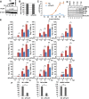
Effect of Prp18 knockdown on viral RNA synthesis. (A) HeLa cells were transfected with control siRNA or siPrp18. At 72 h posttransfection, cell lysates were subjected to Western blotting using anti-Prp18 and anti-β-actin antibodies. (B) Seventy-two hours after transfection of siRNA, the viability of control and Prp18 KD cells was determined by trypan blue staining. The averages and standard deviations determined from three independent experiments are shown. (C) Seventy-two hours after transfection of siRNA, control and Prp18 KD cells were infected with influenza virus at an MOI of 0.01. The culture supernatants collected at 12, 24, 36, 48, 60, and 72 hpi were subjected to plaque assays to examine the production of infectious virions. The average titers and standard deviations determined from three independent experiments are shown. (D) Seventy-two hours after transfection of siRNA, control (lanes 1, 3, 5, and 7) and Prp18 KD (lanes 2, 4, 6, and 8) cells were infected with influenza virus at an MOI of 3. At 3, 5, and 7 hpi, the cell lysates were subjected to Western blotting with anti-PB1, anti-PB2, anti-PA, anti-NP, and anti-β-actin antibodies. (E) Control and Prp18 KD cells were infected with influenza virus at an MOI of 3 and incubated for 3, 5, and 7 h. Total RNA was prepared from cells, and quantitative RT-PCR was carried out with primer sets specific for segment 3 vRNA, segment 3 cRNA, segment 3 mRNA, segment 5 vRNA, segment 5 cRNA, segment 5 mRNA, segment 7 vRNA, segment 7 cRNA, and segment 7 mRNA. The results were normalized to the level of 18S rRNA. The averages and standard deviations determined in three independent experiments are shown. The level of significance was determined by Student's t test (*, P < 0.01; **, P < 0.05; ***, P < 0.5). (F) HeLa cells were transfected with control siRNA or siPrp18. At 72 h posttransfection, cells were infected with influenza virus at an MOI of 3 and incubated for 5 h. Total RNA was prepared from cells, and quantitative RT-PCR was carried out with primer sets specific for M1 mRNA and M2 mRNA. The results were normalized to the level of 18S rRNA. The ratio of the amount of M2 mRNA to that of M1 mRNA was also determined in three independent experiments, with standard deviations. The level of significance was determined by Student's t test (*, P < 0.01; **, P < 0.05).
FIG 3
FIG 3
Effect of Prp18 on primary transcription. (A) HeLa cells were transfected with control siRNA or siPrp18. At 48 h posttransfection, cells were transfected with the pCAGGS-rPrp18-Myc (containing a silent mutation within the siRNA target sequence) and pCAGGS-empty plasmids. After 24 h posttransfection, cells were superinfected with influenza virus at an MOI of 3 and incubated for 4 h in the presence of cycloheximide (CHX). Total RNAs were subjected to quantitative RT-PCR with a primer set specific for segment 5 mRNA. The results are normalized to the level of 18S rRNA. The averages and standard deviations determined from three independent experiments are shown. The level of significance was determined by Student's t test (*, P < 0.01). (B) Western blotting was conducted by using infected cells prepared as described above for panel A. Cell lysates were loaded onto a 10% SDS-PAGE gel and subjected to Western blotting with anti-Prp18 antibody.
FIG 4
FIG 4
Prp18 interacts with NP and viral polymerase in vivo and in vitro. (A) HEK293T cells were transfected with either pCAGGS or pCAGGS-Prp18-Myc. At 48 h posttransfection, these cells were infected with influenza virus at an MOI of 3 and incubated for 6 h. Cell lysates were prepared and incubated with either control IgG-conjugated (lanes 3 and 5) or anti-Myc antibody-conjugated agarose (lanes 4 and 6). Pulldown products were visualized by immunoblotting using anti-PB1, anti-PB2, anti-PA, anti-NP, and anti-Myc antibodies. (B) GST pulldown assays were carried out by using purified GST-Prp18 (lanes 5 to 7) and either vRNP (lanes 1, 3, and 6) or mnRNP (lanes 2, 4, and 7). Western blot analyses were carried out with anti-GST, anti-PB1, anti-PB2, anti-PA, and anti-NP antibodies. (C) GST pulldown assays were conducted by using purified GST (lane 1, 0.6 pmol), GST-Prp18 (lane 2, 0.6 pmol; lane 3, 2 pmol; lane 4, 6 pmol), and His-NP (lanes 1 to 4, 1.25 pmol). Western blotting was carried out with anti-His and anti-GST antibodies.
FIG 5
FIG 5
Prp18 stimulates viral RNA synthesis through interaction with NP. (A) Schematic diagram of Prp18 deletion mutants. a.a, amino acids. (B) Intracellular localization of GST-tagged Prp18 deletion mutants. HeLa cells were transfected with plasmids expressing GST-Prp18, GST-Prp18C1, GST-Prp18C2, and GST-Prp18Δ71. At 24 h posttransfection, cells were subjected to indirect-immunofluorescence assays with anti-GST antibody. DAPI, 4′,6-diamidino-2-phenylindole. (C) GST pulldown assays of GST-Prp18 deletion mutants. HEK293T cells were transfected with plasmids expressing GST-Prp18, GST-Prp18C1, GST-Prp18C2, and GST-Prp18Δ71. At 24 h posttransfection, cells were infected with influenza virus at an MOI of 3. At 6 hpi, cell lysates were prepared and subjected to GST pulldown assays. Coprecipitated proteins were analyzed by Western blotting with anti-PB1, anti-PB2, anti-PA, anti-NP, and anti-GST antibodies. nls, nuclear localization signal. (D) HEK293T cells were transfected with plasmids expressing NP and either GST-Prp18, GST-Prp18C1, GST-Prp18C2, or GST-Prp18Δ71. At 24 h posttransfection, cell lysates were prepared and subjected to a GST pulldown assay. Coprecipitated proteins were analyzed by Western blotting with anti-NP and anti-GST antibodies. (E) Purified recombinant GST, GST-Prp18, GST-Prp18C2, and GST-Prp18Δ71 were separated by 10% SDS-PAGE and visualized by staining with Coomassie brilliant blue. (F) Effect of Prp18 on cell-free RNA synthesis. Cell-free RNA synthesis was carried out with 0.3 pmol (lanes 1, 4, 7, and 10), 1 pmol (lanes 2, 5, 8, and 11), and 3 pmol (lanes 3, 6, 9, and 12) of GST (lanes 1 to 3), GST-Prp18C2 (lanes 4 to 6), GST-Prp18WT (lanes 7 to 9), and GST-Prp18Δ71 (lanes 10 to 12), as described in the legend of Fig. 1.
FIG 6
FIG 6
Prp18 stimulates viral RNA synthesis after the early elongation process. (A) Time course of cell-free viral RNA synthesis. Cell-free viral RNA synthesis was carried out in the presence of either GST (lanes 2 to 5) or GST-Prp18 (lanes 6 to 9), as described in the legend of Fig. 1. After 0-, 1-, 2-, and 4-min incubations, samples were collected and analyzed by using 8 M urea–10% PAGE gels. (B) Limited elongation assays were carried out in the presence of either GST (lanes 1 to 3) or GST-Prp18 (lanes 4 to 6) in the absence of UTP. Samples were analyzed by using 8 M urea–15% PAGE gels. (C) RNase T2 digestion assay. Cell-free RNA synthesis was carried out in the presence of either GST (lanes 1 to 3) or GST-Prp18 (lanes 4 to 6). The newly synthesized RNAs were digested with 6.6 U (lanes 2 and 5) and 20 U (lanes 3 and 6) of RNase T2. The digested RNAs were analyzed by using 8 M urea–4% PAGE gels. (D) The signal intensities of each lane were measured by using ImageJ software. Averages and standard deviations determined from three independent experiments are shown.
FIG 7
FIG 7
Prp18 facilitates NP-RNA complex formation in vitro. (A) Purification of the GST-Prp18 and His-NP proteins. Details are described in Materials and Methods. Purified proteins were loaded onto a 10% SDS-PAGE gel and visualized by Coomassie brilliant blue staining. (B) Gel shift assays were carried out with 5′-32P-labeled v53 and His-NP. NP-RNA complexes were separated on a 0.6% agarose gel and detected by autoradiography. (C and D) His-NP (lanes 1 to 7, 12.5 fmol) and increasing amounts of GST-Prp18 (lane 2, 0.013 pmol; lane 3, 0.04 pmol; lane 4, 0.13 pmol; lane 5, 0.4 pmol; lane 6, 1.3 pmol; lanes 7 and 8, 4 pmol) were incubated at 30°C for 30 min. After further incubation with 5′-32P-labeled v53, samples were separated on a nondenaturing gel and subjected to autoradiography. In panel D, the band intensities were quantitatively measured by using ImageJ software, and the averages and standard deviations determined from three independent experiments are shown. The level of significance was determined by Student's t test (*, P < 0.01; **, P < 0.05). AU, arbitrary units.

Similar articles

Cited by

References

    1. Yamanaka K, Ishihama A, Nagata K. 1990. Reconstitution of influenza virus RNA-nucleoprotein complexes structurally resembling native viral ribonucleoprotein cores. J Biol Chem 265:11151–11155. - PubMed
    1. Honda A, Ueda K, Nagata K, Ishihama A. 1988. RNA polymerase of influenza virus: role of NP in RNA chain elongation. J Biochem 104:1021–1026. - PubMed
    1. Compans RW, Content J, Duesberg PH. 1972. Structure of the ribonucleoprotein of influenza virus. J Virol 10:795–800. - PMC - PubMed
    1. Arranz R, Coloma R, Chichon FJ, Conesa JJ, Carrascosa JL, Valpuesta JM, Ortin J, Martin-Benito J. 2012. The structure of native influenza virion ribonucleoproteins. Science 338:1634–1637. doi:10.1126/science.1228172. - DOI - PubMed
    1. Moeller A, Kirchdoerfer RN, Potter CS, Carragher B, Wilson IA. 2012. Organization of the influenza virus replication machinery. Science 338:1631–1634. doi:10.1126/science.1227270. - DOI - PMC - PubMed

LinkOut - more resources