. 2017 Jan 1;28(1):41-53.
doi: 10.1091/mbc.E16-07-0557. Epub 2016 Nov 16.

Reconstituting regulation of the canonical Wnt pathway by engineering a minimal β-catenin destruction machine

Affiliations

Reconstituting regulation of the canonical Wnt pathway by engineering a minimal β-catenin destruction machine

Mira I Pronobis et al. Mol Biol Cell. .

Abstract

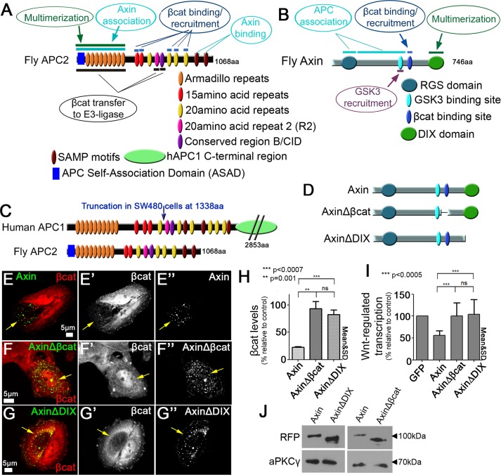
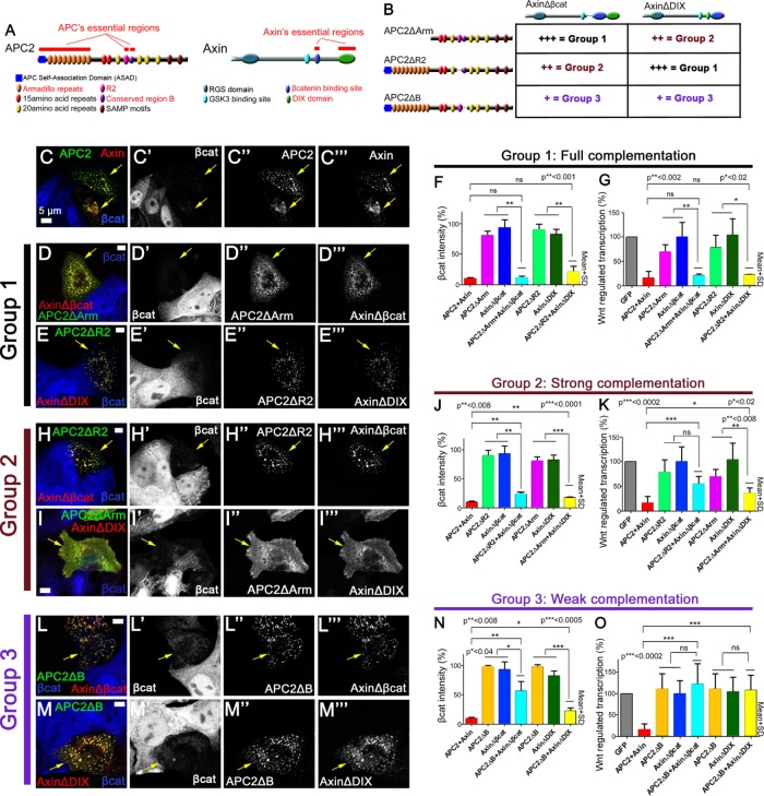
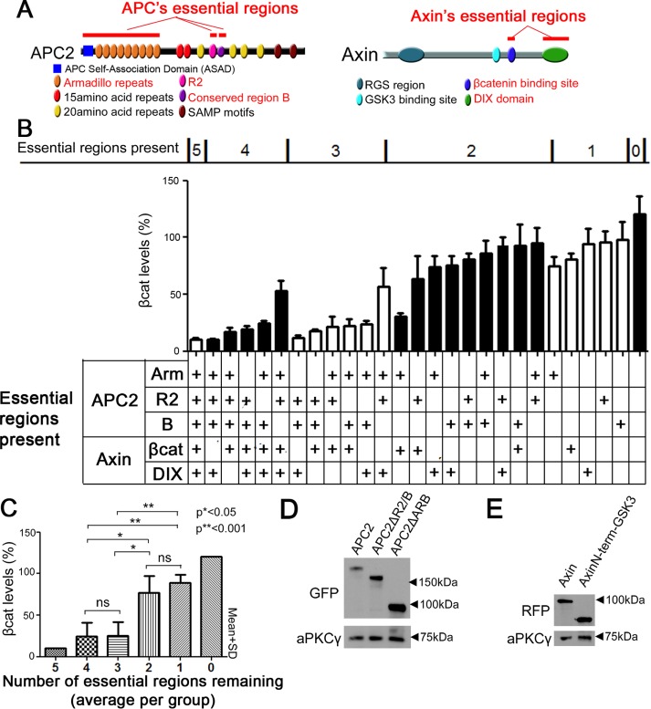
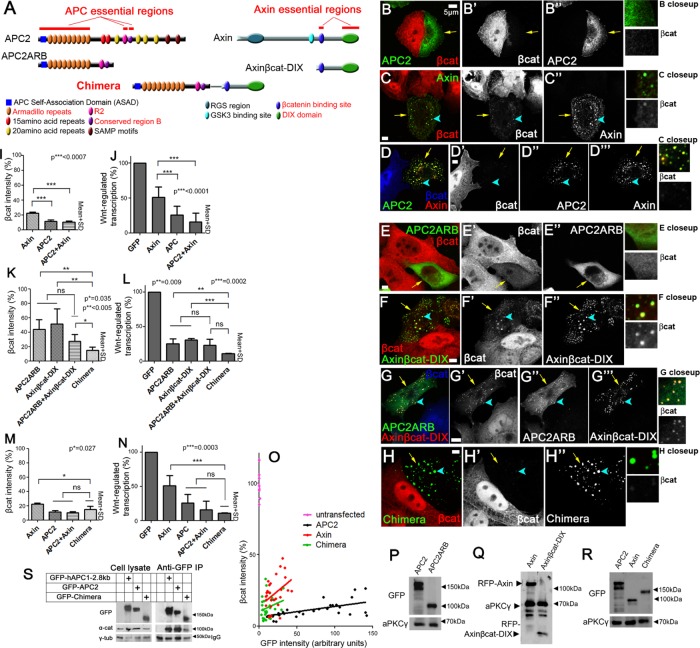
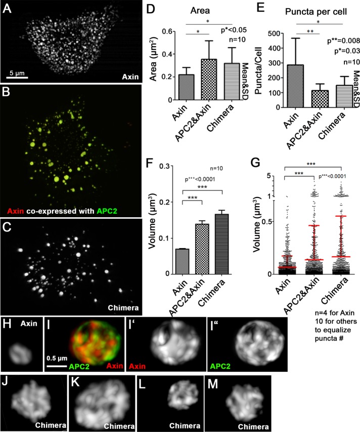
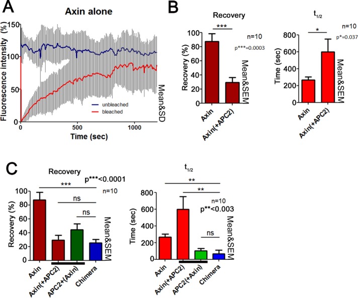
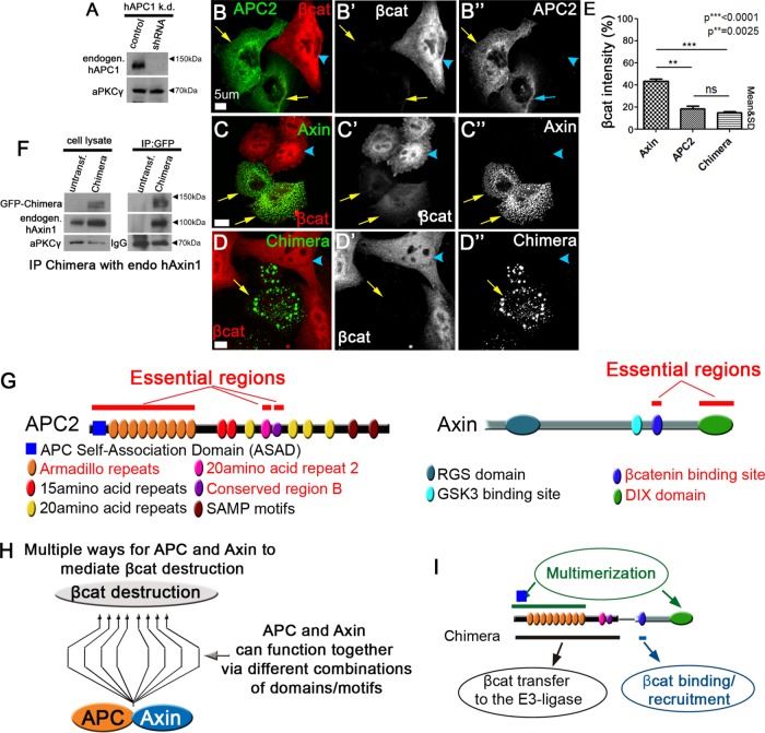
Negatively regulating key signaling pathways is critical to development and altered in cancer. Wnt signaling is kept off by the destruction complex, which is assembled around the tumor suppressors APC and Axin and targets β-catenin for destruction. Axin and APC are large proteins with many domains and motifs that bind other partners. We hypothesized that if we identified the essential regions required for APC:Axin cooperative function and used these data to design a minimal β-catenin-destruction machine, we would gain new insights into the core mechanisms of destruction complex function. We identified five key domains/motifs in APC or Axin that are essential for their function in reconstituting Wnt regulation. Strikingly, however, certain APC and Axin mutants that are nonfunctional on their own can complement one another in reducing β-catenin, revealing that the APC:Axin complex is a highly robust machine. We used these insights to design a minimal β-catenin-destruction machine, revealing that a minimized chimeric protein covalently linking the five essential regions of APC and Axin reconstitutes destruction complex internal structure, size, and dynamics, restoring efficient β-catenin destruction in colorectal tumor cells. On the basis of our data, we propose a new model of the mechanistic function of the destruction complex as an integrated machine.


Figures

FIGURE 1:
FIGURE 1:
APC and Axin have regions with potentially redundant functions, and Axin has two domains/motifs essential for βcat destruction. (A) Drosophila APC2. Top, regions that may have redundant functions. Bottom, unique functions of APC2. (B) Drosophila Axin. Top, regions that may have redundant functions. Bottom, regions with unique functions. (C) Comparison of human APC1 and fly APC2, indicating truncated protein present in SW480 cells. (D) Schematic representation of Axin mutants. (E) Wild-type Axin can reduce βcat levels. Axin-RFP expressed in SW480 cells and stained for βcat via antibody. Arrows, transfected cells. (F, G) AxinΔβcat-RFP (F) and AxinΔDIX-RFP (G) cannot facilitate βcat destruction. (H) Deleting either the βcat-binding site or DIX domain of Axin impairs its ability to reduce βcat levels. Quantification of βcat relative to untransfected cells in 10 cells each measured in three independent experiments. (I) Wnt-regulated transcription remains high in cells expressing Axin mutants that delete the βcat-binding or DIX domain. Quantification of TOP/FOPflash reporter gene assay of indicated constructs in SW480 cells. Triplicates were measured in three independent experiments. Student’s t test was used. (J) Immunoblot of indicated constructs to compare expression levels. aPKCγ is a loading control.
FIGURE 2:
FIGURE 2:
Certain APC and Axin mutants that are nonfunctional on their own can complement one another to facilitate βcat destruction. (A) Schematic representation of APC2 and Axin. Essential regions are highlighted in red. (B) Combinations of APC2 and Axin mutants tested. Groups 1–3 exhibit different levels of complementation. (C–E, H–I, L–M) Immunofluorescence, wild-type or mutant versions of GFP-APC2 and Axin-RFP expressed in SW480 cells and stained for βcat via antibody (blue). Arrows = transfected cells. (C) Wild-type APC2 and Axin form cytoplasmic complexes and effectively reduce βcat. (D, E) The pairs GFP-APC2ΔArm + AxinΔβcat-RFP (D) and GFP-APC2ΔR2 + AxinΔDIX-RFP (E) colocalize in puncta and show full complementation in βcat reduction, and they are thus categorized into group 1. (F) Quantification, βcat fluorescence intensity relative to untransfected control SW480 cells, group 1. Constructs are indicated. Ten cells each in three independent experiments. (G) Quantification, Wnt regulated transcription in SW480 cells of group 1. Group 1 APC and Axin mutants are indistinguishable from wild-type APC2 plus Axin in inhibiting Wnt-regulated transcription. Three triplicates measured in three independent experiments. (H, I) The pairs GFP-APC2ΔR2 + AxinΔβcat-RFP (H) and GFP-APC2ΔArm + AxinΔDIX-RFP (I) show strong complementation in βcat reduction and are thus categorized into group 2. Of interest, whereas GFP-APC2ΔR2 and AxinΔβcat-RFP colocalize in puncta (H), GFP-APC2ΔArm and AxinΔDIX-RFP (I) do not form large cytoplasmic puncta. (J) Quantification of βcat levels, group 2 mutants, as in F. (K) Quantification of Wnt-regulated transcription in group 2, as in G. (L, M) The pairs GFP-APC2ΔB + AxinΔβcat-RFP (L) and GFP-APC2ΔB + AxinΔDIX-RFP (M) show weak complementation in βcat reduction and are categorized into group 3. (N) Quantification of βcat levels of group 3 mutants. (O) Quantification of Wnt-regulated transcription in group 3. Student’s t test was used.
FIGURE 3:
FIGURE 3:
The essential regions in APC and Axin each make important but partially overlapping contributions to a fully functional destruction complex. (A) Schematic representation of APC2 and Axin. Essential regions are indicated in red. (B) The essential regions in APC2 and Axin work cooperatively to ensure efficient reduction of βcat levels. Coexpression of indicated APC2 and Axin mutants in SW480 cells, followed by quantification of βcat fluorescence intensity. Top, mutants are sorted into groups that are defined by how many essential regions remain present. Middle, βcat average intensity. Bottom, table describing which essential regions remain present. (C) For βcat reduction, up to two essential regions are largely dispensable. Columns represent the averages of the groups that were defined in B. Student’s t test was used. (D) Immunoblot analysis of expression levels of a subset of the APC2 mutants used; others are given in other figures or in Roberts et al. (2011, 2012) or Pronobis et al. (2015). (E) Expression levels of indicated Axin mutant.
FIGURE 4:
FIGURE 4:
The APC:Axin Chimera reduces βcat as efficiently as wild-type APC plus Axin. (A) Schematic, APC and Axin constructs plus Chimera. Top, wild-type APC and Axin with essential regions indicated. Middle, mutants incorporating essential regions alone. Bottom, essential regions were combined into a single polypeptide to create the Chimera. (B–H) Immunofluorescence, GFP-tagged or RFP-tagged wild-type or mutant constructs misexpressed in SW480 cells and stained for βcat via antibody. Arrows point to transfected cells. Close-ups of a transfected cell are shown to the right to reveal which constructs retain βcat in puncta. (B) Wild-type GFP-APC2 reduces βcat levels. (C) Wild-type GFP-Axin also reduces βcat levels, but some βcat remains in puncta (arrowheads, close-up). (D) Coexpressing GFP-APC and Axin-RFP effectively reduces βcat levels. (E) GFP-APC2ARB, which consists of APC’s three essential regions, can moderately reduce βcat levels, but βcat remains higher than is seen after wild-type APC2 transfection. (F) Axinβcat-DIX-RFP also moderately reduces βcat levels, and detectable βcat remains in the puncta (arrowhead, close-up). (G) Coexpressing APC2ARB and Axinβcat-DIX does not further decrease βcat levels, and βcat remains in puncta (arrowhead, close-up). (H) Expressing the GFP-Chimera leads to strong reduction of βcat, and no βcat is seen in puncta (arrowhead, close-up). (I, K, M) Quantification, βcat fluorescence intensity in SW480 cells transfected with indicated constructs, normalized to untransfected cells. Ten cells each in three independent experiments were measured. (J, L, N) Quantification of Wnt-regulated transcription in SW480 cells (TOPflash activity, normalized to untransfected cells). Three triplicates were measured in three independent experiments. (I) Axin cannot reduce βcat levels as effectively as APC2 or APC2 + Axin. (J) Axin cannot inhibit Wnt-regulated transcription as effectively as APC2 or APC2 + Axin. (K) Mutants carrying only the essential regions of APC2 or Axin only moderately reduce βcat levels, whereas covalently linking the essential regions of APC and Axin into the Chimera increases βcat reduction. (L) The Chimera strongly inhibits Wnt-regulated transcription. (M) The Chimera reduces βcat levels better than wild-type Axin. (N) Wnt-regulated transcription is as effectively inhibited by the Chimera as it is by wild-type APC2 + Axin or APC2. Student’s t test was used. (O) APC2, Axin, and the Chimera are not dosage dependent in βcat degradation. GFP levels (reflecting expression level of APC or Axin construct) vs. βcat signal in individual cells expressing each construct. βcat signal is normalized to nearby untransfected cells. A set of individual values of untransfected cells shows the degree of variability among cells in the same population. Thirty cells total for each condition. Both the Chimera and APC2 are more effective at reducing βcat than is Axin over a wide range of expression levels. (P–R) Immunoblot, expression levels of indicated constructs. aPKCγ is a loading control. Relative expression levels vary somewhat from experiment to due to transfection efficiency. (S) α-Catenin coimmunoprecipitates with the Chimera. Left, cell lysates from cells expressing the indicated constructs. γ-Tubulin serves as a loading control. Right, anti-GFP immunoprecipitates from cells expressing the indicated constructs. Bottom, effectiveness of antibody pull down.
FIGURE 5:
FIGURE 5:
The Chimera mimics the APC2:Axin complex in internal structure and size. (A–C) SIM images. SW480 cells transfected with Axin-RFP (A), GFP-APC2 plus Axin-RFP (B), or GFP-Chimera (C). (D) The cross-sectional area of the chimeric complexes is similar to those produced by APC2 plus Axin and larger than those produced by Axin alone. (E) The number of puncta in Chimera-transfected cells is similar to that in APC2 + Axin–transfected cells and reduced relative to cells expressing Axin alone. Area and number obtained using LSM 710 images and the ImageJ Particle Analyzer. Ten cells quantified per construct. (F, G) Chimera puncta are similar in volume to APC2:Axin complexes and larger than those produced by Axin alone. Puncta volume assessed from SIM images of indicated constructs using Imaris Software (Bitplane). (F) Average puncta volume of indicated constructs. Ten cells used for each condition. (G) Volume comparison of equal numbers of puncta from Axin-transfected (n = 4 cells quantified), APC2 + Axin–transfected (n = 10 cells), or Chimera-transfected (n = 10 cells) cells. Fewer Axin cells were analyzed to equalize puncta number. Student’s t test was used. (H–M) SIM close-up three-dimensional projections of puncta from cells like those in A–C. (H, I) APC2 coexpression (I) leads to a more internal complex structure of Axin puncta than that of puncta assembled from Axin alone (H). (J–M) The Chimera has a complex internal structure resembling that of APC:Axin complexes. Representative images of GFP-Chimera expressed in two different SW480 cells.
FIGURE 6:
FIGURE 6:
Dynamics of the Chimera are similar to that of APC in APC2 + Axin complexes. (A–C) FRAP analysis of complexes formed by Axin-RFP alone, Axin-RFP coexpressed with GFP-APC2, or GFP-Chimera in SW480 cells. Ten complexes from three independent experiments. (A) Axin complexes are dynamic. Example of FRAP traces of complexes formed by Axin-RFP in SW480 cells. Unbleached (blue) and bleached cells (red). (B) Axin’s dynamics slows when APC is coexpressed. GFP-APC2 and Axin-RFP were expressed in SW480 cells. Left, recovery plateau; right, t1/2. (C) APC2 and the Chimera have similar dynamics. The recovery plateau of the Chimera is similar to that of both Axin and APC2. However, the time needed for the Chimera to recover is more similar to that of APC2.
FIGURE 7:
FIGURE 7:
High levels of the truncated endogenous APC1 are not essential for function of the Chimera and new models of the destruction complex and its key functions. (A) Immunoblot assessing level of knockdown in SW480 cells stably transduced with shRNA targeting the endogenous truncated human APC1. aPKC serves as a loading control; representative of three experiments. (B–D) SW480 cells stably transduced with shRNA targeting the endogenous truncated human APC1 and then transfected with the indicated GFP-tagged constructs. Arrows, transfected cells; arrowheads, untransfected cells. (E) Quantification, βcat fluorescence intensity in cells like those in B–D. Constructs are indicated. Ten cells each in three independent experiments. Student’s t test was used. (F) The Chimera coimmunoprecipitates endogenous human Axin. Left, cell lysates from cells expressing the indicated constructs. aPKC serves as a loading control. Right, anti-GFP immunoprecipitates, bringing down the GFP-tagged-Chimera. Bottom, effectiveness of antibody pull down. (G) Essential regions of APC2 and Axin. (H) APC and Axin can use different combinations of domains/motifs to secure low levels of βcat. The nature of the combination defines the efficiency of the complex. (I) The Chimera helps define all the essential functions of the APC:Axin destruction complex.

Similar articles

Cited by

References

    1. Ahmed Y, Nouri A, Wieschaus E. Drosophila Apc1 and Apc2 regulate Wingless transduction throughout development. Development. 2002;129:1751–1762. - PubMed
    1. Akong K, Grevengoed E, Price M, McCartney B, Hayden M, DeNofrio J, Peifer M. Drosophila APC2 and APC1 play overlapping roles in wingless signaling in the embryo and imaginal discs. Dev Biol. 2002;250:91–100. - PubMed
    1. Brangwynne CP, Eckmann CR, Courson DS, Rybarska A, Hoege C, Gharakhani J, Julicher F, Hyman AA. Germline P granules are liquid droplets that localize by controlled dissolution/condensation. Science. 2009;324:1729–1732. - PubMed
    1. Cadigan KM, Peifer M. Wnt signaling from development to disease: insights from model systems. Cold Spring Harb Perspect Biol. 2009;1:a002881. - PMC - PubMed
    1. Choi SH, Estarás C, Moresco JJ, Yates JR, Jones KA. α-Catenin interacts with APC to regulate β-catenin proteolysis and transcriptional repression of Wnt target genes. Genes Dev. 2013;27:2473–2488. - PMC - PubMed

Publication types

LinkOut - more resources