Review
. 2016 Dec;22(12):1795-1807.
doi: 10.1261/rna.057950.116.

Unmasking the U2AF homology motif family: a bona fide protein-protein interaction motif in disguise

Affiliations
Review

Unmasking the U2AF homology motif family: a bona fide protein-protein interaction motif in disguise

Sarah Loerch et al. RNA. 2016 Dec.

Abstract

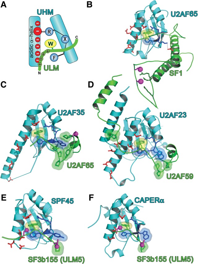
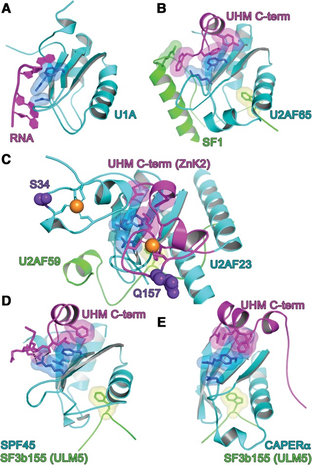
U2AF homology motifs (UHM) that recognize U2AF ligand motifs (ULM) are an emerging family of protein-protein interaction modules. UHM-ULM interactions recur in pre-mRNA splicing factors including U2AF1 and SF3b1, which are frequently mutated in myelodysplastic syndromes. The core topology of the UHM resembles an RNA recognition motif and is often mistakenly classified within this large family. Here, we unmask the charade and review recent discoveries of UHM-ULM modules for protein-protein interactions. Diverse polypeptide extensions and selective phosphorylation of UHM and ULM family members offer new molecular mechanisms for the assembly of specific partners in the early-stage spliceosome.

Keywords: RRM; SF3B1; U2AF1; UHM; protein–protein interaction.


Figures

FIGURE 1.
FIGURE 1.
Multiple UHM–ULM interactions guide the early stages of spliceosome assembly and other pathways. Experimentally confirmed phosphorylation sites of the ULM-containing regions are marked by pink circles. “W” represents ULM tryptophans. Thick gray arrow represents the transition during spliceosome assembly. Thin black arrows represent known UHM–ULM interactions. Arrows toward SF3b155 indicate the general ULM-containing region rather than specific binding sites.
FIGURE 2.
FIGURE 2.
UHM–ULM recognition. (A) Schematic representation of consensus UHM (cyan)–ULM (green) interactions. (B–F) Structures of UHM–ULM complexes, including (B) U2AF65 UHM–SF1 ULM (PDB ID: 4FXW); (C) U2AF35 UHM–U2AF65 ULM (PDB ID: 1JMT); (D) U2AF23 UHM–U2AF59 ULM (PDB ID: 4YH8); (E) SPF45 UHM–SF3b155 ULM5 (PDB ID 2PEH); and (F) CAPERα UHM–SF3b155 ULM5 (PDB ID: 4OZ1). Magenta spheres mark phosphorylation sites: the SF1 and SF3b155 sites in B,E,F have been confirmed experimentally, whereas the human U2AF65 site in C is the predicted phosphorylation of a TP motif. The surface of the central ULM tryptophan is colored yellow, UHM RXF-motifs are blue, and acidic UHM α-helical residues are red; orange spheres are zinc ions on the back surface of the U2AF23 UHM in D.
FIGURE 3.
FIGURE 3.
RNP interactions. (A) N-terminal RRM of U1A prototype bound to RNA ligand. The RNP surfaces of UHM (cyan)–ULM (green) complexes, including (B) U2AF65 UHM–SF1 ULM (PDB ID: 4FXW); (C) U2AF23 UHM–U2AF59 ULM (PDB ID: 4YH8); (D) SPF45 UHM–SF3b155 ULM5 (PDB ID 2PEH); and (E) CAPERα UHM–SF3b155 ULM5 (PDB ID: 4OZ1). The conserved aromatic residue at RNP1 position 5 typically is masked by a C-terminal extension (magenta) of the core UHM fold. The key residues at positions 2 of RNP2 and positions 3/5 of RNP1 are colored blue. Orange spheres represent zinc ions. Indigo, space-filling residues in C correspond to MDS-relevant mutations of the human homolog. Views are following an ∼120° rotation about the y-axis relative to Figure 2.

Similar articles

Cited by

References

    1. Adam SA, Nakagawa T, Swanson MS, Woodruff TK, Dreyfuss G. 1986. mRNA polyadenylate-binding protein: gene isolation and sequencing and identification of a ribonucleoprotein consensus sequence. Mol Cell Biol 6: 2932–2943. - PMC - PubMed
    1. Afroz T, Skrisovska L, Belloc E, Guillen-Boixet J, Mendez R, Allain FH. 2014. A fly trap mechanism provides sequence-specific RNA recognition by CPEB proteins. Genes Dev 28: 1498–1514. - PMC - PubMed
    1. Avis JM, Allain FH, Howe PW, Varani G, Nagai K, Neuhaus D. 1996. Solution structure of the N-terminal RNP domain of U1A protein: the role of C-terminal residues in structure stability and RNA binding. J Mol Biol 257: 398–411. - PubMed
    1. Bae E, Reiter NJ, Bingman CA, Kwan SS, Lee D, Phillips GN Jr, Butcher SE, Brow DA. 2007. Structure and interactions of the first three RNA recognition motifs of splicing factor prp24. J Mol Biol 367: 1447–1458. - PMC - PubMed
    1. Bauer WJ, Heath J, Jenkins JL, Kielkopf CL. 2012. Three RNA recognition motifs participate in RNA recognition and structural organization by the pro-apoptotic factor TIA-1. J Mol Biol 415: 727–740. - PMC - PubMed

Publication types