. 2016 Dec;22(12):1844-1858.
doi: 10.1261/rna.056523.116. Epub 2016 Oct 20.

Solution structure of the 5'-terminal hairpin of the 7SK small nuclear RNA

Affiliations

Solution structure of the 5'-terminal hairpin of the 7SK small nuclear RNA

Sarah Bourbigot et al. RNA. 2016 Dec.

Abstract

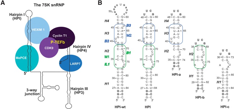
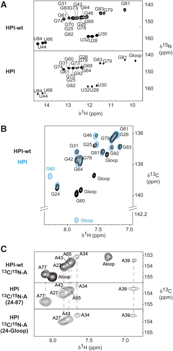
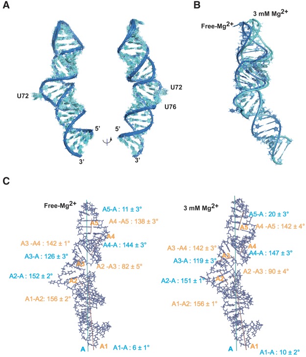
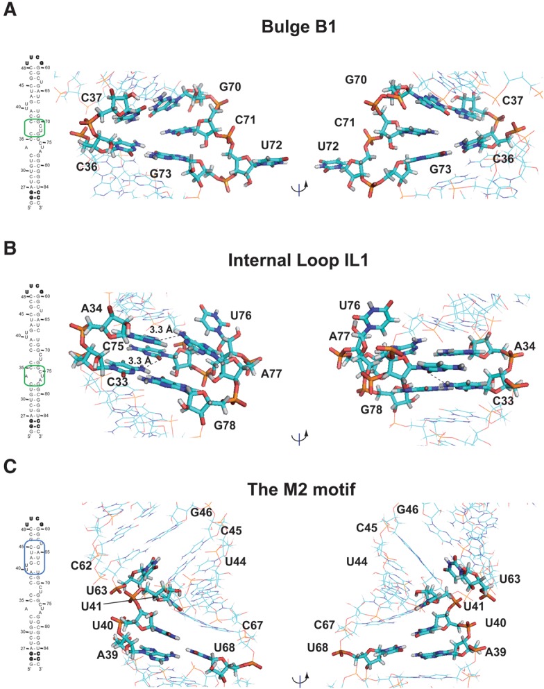
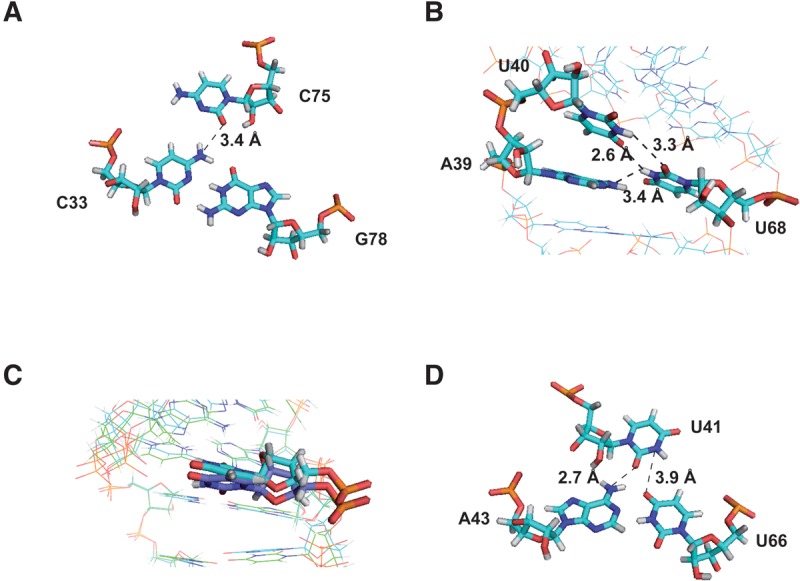
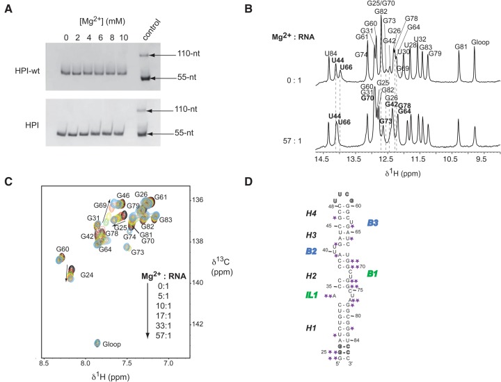
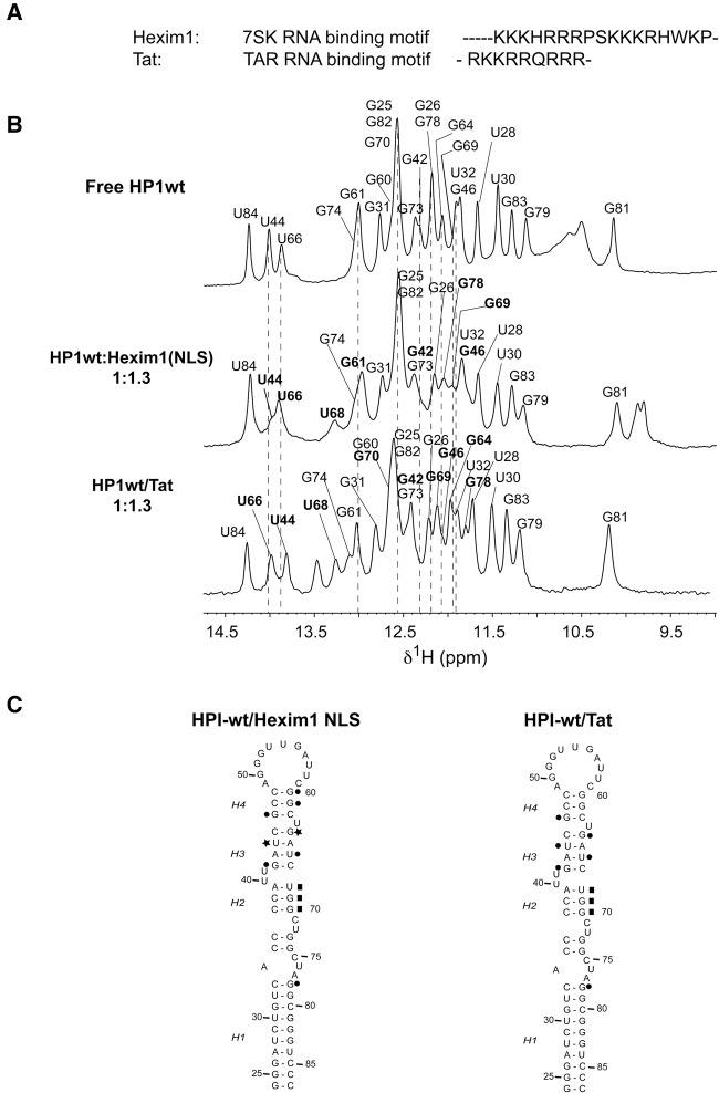
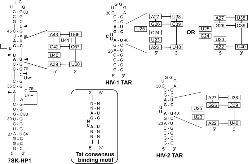
The small nuclear 7SK RNA regulates RNA polymerase II (RNA Pol II) transcription, by sequestering and inhibiting the positive transcription elongation factor b (P-TEFb). P-TEFb is stored in the 7SK ribonucleoprotein (RNP) that contains the three nuclear proteins Hexim1, LaRP7, and MePCE. P-TEFb interacts with the protein Hexim1 and the 7SK RNA. Once P-TEFb is released from the 7SK RNP, it activates transcription by phosphorylating the C-terminal domain of RNA Pol II. P-TEFb also plays a crucial role in the replication of the human immunodeficiency virus HIV-1, through its recruitment by the viral transactivator Tat. Previous work demonstrated that the protein Tat promotes the release of P-TEFb from the 7SK RNP through direct binding to the 7SK RNA. Hexim1 and Tat proteins both comprise conserved and similar arginine-rich motifs that were identified to bind the 7SK RNA at a repeated GAUC site located at the top of the 5'-terminal hairpin (HPI). Here, we report the solution structure of this region as determined by nuclear magnetic resonance, to identify HPI structural features recognized by Hexim1 and Tat. The HPI solution structure displays an elongated shape featuring four helical segments interrupted by one internal loop and three bulges with distinct folds. In particular, the repeated GAUC motif adopts a pre-organized geometry. Our results suggest that the binding of Hexim1 and Tat to the 7SK RNA could originate from a conformational selection of this motif, highlighting how RNA local structure could lead to an adaptive recognition of their partners.

Keywords: 7SK; Hexim; NMR; RNA; TAR; Tat.


Figures

FIGURE 1.
FIGURE 1.
The 7SK RNA and its 5′-terminal hairpin HPI. (A) Schematic representation of the 7SK RNP complex. P-TEFb comprises the cyclin T1 and the cyclin-dependent kinase CDK9. (B) Sequences and secondary structures of (i) the 7SK 5′-terminal hairpin (HPI-wt), (ii) HPI, and (iii) the three subdomain constructs (HPI-a, HPI-b, and HPI-c) used for nuclear magnetic resonance (NMR) studies. Outlined nucleotides at 5′-end were changed to improve transcription efficiency. The 11-nt apical loop of HPI-wt was substituted by a thermo-stable UUCG tetraloop (HPI). The RNA oligonucleotides HPI-a, HPI-b, and HPI-c comprise nucleotides 35–74, 29–38/69–82, and 24–36/73–87, respectively. Both HPI-a and HPI-b contain two additional G-C base pairs. The internal loop IL1 comprises (34/75–77) nucleotides. The bulges B1, B2, and B3 comprise (71/72), (40/41), and (63) residues, respectively. The M1 motif (green) contains the helix H2, the internal loop IL1, and the bulge B1. The M2 motif (blue) comprises the helix H3 and bulges B2 and B3.
FIGURE 2.
FIGURE 2.
HPI-wt and HPI adopt the same conformation. (A) 1H–15N HSQC spectra showing imino protons region of HPI-wt (top) and HPI (bottom). All imino protons were assigned via sequential nuclear Overhauser effects (NOEs) observed in 2D-NOESY experiments. In the HPI construct, imino protons of the UUCG apical loop resonate at the expected chemical shifts typically observed, which ensures a proper folding of the RNA (Varani et al. 1991; Molinaro and Tinoco 1995). (B) 1H–13C HSQC spectra showing the aromatic H8-C8 of selectively labeled [13C/15N-G] HPI-wt (black) and HPI (blue), recorded at 30°C. (C) 1H–13C HSQC spectra showing the aromatic H2–C2 correlations of the selectively labeled [13C/15N-A] HPI-wt (top), the selectively labeled [13C/15N-A] HPI (middle), and the segmentally and selectively labeled [13C/15N-A(G24-G60)] HPI (bottom). HPI was selectively labeled at all A positions (middle), and segmentally and selectively labeled at A positions in the G24-G60 segment (bottom). Assignment of aromatic H2–C2 of HPI-wt had been previously reported (Lebars et al. 2010). Assignment of HPI was based on analysis of NOESY spectra recorded at 4°C, 10°C, 15°C, 20°C, 30°C in 90/10 H2O/D2O and at 20°C and 30°C in 99.9% D2O. Spectra were recorded in 50 mM sodium phosphate buffer at pH 6.2.
FIGURE 3.
FIGURE 3.
Three-dimensional structure of HPI. (A) Superimposition of 10 converged NMR structures. Bases and sugars are shown in light blue and the backbone in dark blue. (B) Structures of HPI calculated using RDCs measured in the absence of Mg2+ (dark blue) and with 3 mM Mg2+ (light blue). The regions H1 are superimposed. (C) Inter-angle axis measurement for the free-Mg2+ structure (left) and 3 mM Mg2+ model (right). Overall axes (A) of RNAs are shown in blue. Five stems were defined using the 3DNA software (Colasanti et al. 2013). Helical axes for the five stems were generated using MOLMOL software and are indicated in pink (Koradi et al. 1996). Angles between each stem and the overall axis are indicated in blue. Angles between helices are indicated in orange.
FIGURE 4.
FIGURE 4.
Bulges and internal loop. (A) View of the bulge B1 in the M1 Motif. (B) View of the internal loop IL1 in the M1 motif. (C) View of the M2 motif. Broken lines indicate possible hydrogen bonds.
FIGURE 5.
FIGURE 5.
Hydrogen bond networks. (A) Base triple involving C33:G78:C75 in the free-Mg2+ structure. (B) Zoom on A39, U40, and U68 residues in the free-Mg2+ structure. (C) Comparison of the U41 residue from Mg2+-free (dark blue) and Mg2+-bound structures (light blue). (D) Base triple involving A43:U66:U41 in the 3 mM Mg2+ model. Distances consistent with the formation of hydrogen bonds are displayed. Broken lines indicate possible hydrogen bonds.
FIGURE 6.
FIGURE 6.
Effect of magnesium on HPI. (A) Native gel electrophoresis on 10% polyacrylamide. HPI-wt (top) and HPI (bottom) were loaded in the buffer used for NMR studies with increasing magnesium concentration. The last lane (control) contains a thio-modified RNA at its 5′-end, which allows the visualization of a monomer (55-nt: S-RNA) and a dimer (110-nt: RNA-S-S-RNA). (B) Titration of HPI by magnesium by monitoring exchangeable protons. The assignment of imino protons based on the analysis of NOESY experiments is indicated for the Mg2+-free RNA (top) and the 10 mM Mg2+-RNA corresponding to a ratio of 57:1 (bottom). Broken lines and bold assignments indicate imino protons that undergo chemical shifts changes. (C) Magnesium titration of HPI using nonexchangeable protons. Overlays of regions of 1H–13C HSQC spectra showing the aromatic H8-C8 of selectively labeled [13C/15N-G] HPI at 30°C, Mg2+-free (black) and upon successive additions of magnesium. Arrows indicate the cross-peaks that shifted upon addition of magnesium. (D) Summary of the effects observed in 1H and 13C experiments on HPI upon Mg2+ binding. Small effects are indicated by one star, while stronger effects are highlighted using two stars.
FIGURE 7.
FIGURE 7.
Interaction with Tat peptide and comparison with TAR RNA. (A) Comparison of Hexim1 and Tat RNA binding domains. (B) Titration of HPI RNA by Hexim1 NLS domain and Tat peptide. The imino protons region of 1D spectra recorded at 15°C in 50 mM sodium phosphate buffer in 90/10 H2O/D2O at pH 6.4. The assignment of imino proton based on analysis of NOESY experiments is indicated for the free HPI-wt (top), the HPI-wt:Hexim1(NLS) complex (middle), and the HPI-wt:Tat complex (bottom; Supplemental Fig. S10B). The assignment of HPI-wt:Hexim1(NLS) complex has already been reported in our previous work (Lebars et al. 2010). Broken lines and bold assignments indicate imino protons that undergo chemical shift changes. (C) Summary of the effects observed on HPI upon interaction. The results obtained with Hexim1 NLS have been previously reported (Lebars et al. 2010). The opening of base pairs is indicated with stars. The stabilization of base pairs is indicated with squares. Residues for which imino protons undergo chemical shifts changes upon addition of peptide are highlighted with circles.
FIGURE 8.
FIGURE 8.
Comparison with TAR RNA from HIV-1 and HIV-2. Sequences and secondary structures of HPI, HIV-1 TAR, and HIV-2 TAR. The Tat consensus binding motif is encircled. Bold nucleotides indicate the Tat binding motif. For HPI: Black arrows indicate that the nucleotide is involved in a stacking interaction; white arrows indicate that the residue is looped outside the helix and lines indicate that U41 and C75 are positioned into the major groove. Schematic representations of the Tat and Hexim binding site are shown. Solid and dotted horizontal lines indicate stable and weak base-pairings, respectively. Representations of HIV-1 TAR and HIV-2 TAR in the free form are based on structures from previous works (Aboul-ela et al. 1996; Long and Crothers 1999; Franck et al. 2009).

Similar articles

Cited by

References

    1. Aboul-ela F, Karn J, Varani G. 1996. Structure of HIV-1 TAR RNA in the absence of ligands reveals a novel conformation of the trinucleotide bulge. Nucleic Acids Res 24: 3974–3981. - PMC - PubMed
    1. Abu A, Petrov A, Stombaugh J, Zirbel CL, Leontis NB. 2012. Comprehensive survey and geometric classification of base triples in RNA structures. Nucleic Acids Res 40: 1407–1423. - PMC - PubMed
    1. Barboric M, Lenasi T. 2010. Kick-sTARting HIV-1 transcription elongation by 7SK snRNP deporTATion. Nat Struct Mol Biol 17: 928–930. - PubMed
    1. Barboric M, Yik JH, Czudnochowski N, Yang Z, Chen R, Contreras X, Geyer M, Matija Peterlin B, Zhou Q. 2007. Tat competes with HEXIM1 to increase the active pool of P-TEFb for HIV-1 transcription. Nucleic Acids Res 35: 2003–2012. - PMC - PubMed
    1. Barrandon C, Bonnet F, Nguyen VT, Labas V, Bensaude O. 2007. The transcription-dependent dissociation of P-TEFb-HEXIM1–7SK RNA relies upon formation of hnRNP-7SK RNA complexes. Mol Cell Biol 27: 6996–7006. - PMC - PubMed

Publication types

MeSH terms

LinkOut - more resources