. 2016 Nov 15;8(11):2754-2776.
doi: 10.18632/aging.101082.

Aging is associated with an expansion of CD49fhi mammary stem cells that show a decline in function and increased transformation potential

Affiliations

Aging is associated with an expansion of CD49fhi mammary stem cells that show a decline in function and increased transformation potential

Qiaoxiang Dong et al. Aging (Albany NY). .

Abstract

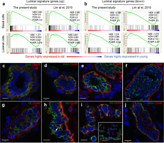
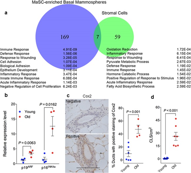
Breast cancer incidence increases during aging, yet the mechanism of age-associated mammary tumorigenesis is unclear. Mammary stem cells are believed to play an important role in breast tumorigenesis, but how their function changes with age is unknown. We compared mammary epithelial cells isolated from young and old mammary glands of different cohorts of C57BL6/J and BALB/c mice, and our findings revealed that old mammary glands were characterized by increased basal cell pool comprised of mostly CD49fhi cells, altered luminal-to-basal cell ratio, and irregular ductal morphology. More interestingly, basal stem cells in old mice were increased in frequency, but showed a functional decline of differentiation and increased neoplastic transformation potential. Gene signature enrichment analysis revealed a significant enrichment of a luminal cell gene expression signature in the basal stem cell-enriched population from old mice, suggesting some luminal cells were expressing basal markers. Immunofluorescence staining confirmed the presence of luminal cells with high CD49f expression in hyperplastic lesions implicating these cells as undergoing luminal to basal phenotypic changes during aging. Whole transcriptome analysis showed elevated immune and inflammatory responses in old basal stem cells and stromal cells, which may be the underlying cause for increased CD49fhi basal-like cells in aged glands.

Keywords: aging; breast cancer; mammary stem cell; neoplasia.


Conflict of interest statement

The authors declare no conflict of interests.

Figures

Figure 1
Figure 1. Mammary epithelial cell population and stem/progenitor cell frequency change during aging
(a) Cells with high levels of CD49f are MaSC-enriched basal cells, and cells with high levels of CD24 are luminal progenitor-enriched luminal cells. Representative flow cytometry analysis of mammary epithelial cells from young (3 mo) and old (26 mo) virgin C57BL6/J mice. (b-d) Mammary epithelial cell population changes during aging. Percent basal cells (CD24loCD49fhi) (b), percent luminal cells (CD24hiCD49flo) (c), and luminal-to-basal cell ratio (d) of Lin- mammary epithelial cells isolated from young and old virgin C57BL/6 (age, 2-4 mo vs. 25-32 mo; n = 15 vs. 9) and BALB/c mice (age, 2-4 mo vs. 17 mo; n = 8 vs. 5). (e-f) Stem/progenitor cell frequency changes during aging. Average frequencies of MaSC (e) expressed as % sphere formation and differentiation-initiating cells from basal cells (SFD-ICb), luminal progenitor cells (f) expressed as % sphere formation and differentiation-initiating cells from luminal cells (SFD-ICl), and luminal progenitor cells (g) expressed as % colony forming cell (CFC) from luminal cells in young and old virgin C57BL/6 (age, 2-3 mo vs. 25-26 mo; n = 6) and BALB/c mice (age, 2-4 mo vs. 17 mo; n = 8 vs. 5). (h-k) Representative examples of whole mount carmine alum staining of mammary glands collected from young (h) and old (i-k) virgin C57BL6/J mice. Scale bars, 1 mm
Figure 2
Figure 2. Stem cell function and transformation potential in young and old mammary glands
(a) Immunohistochemistry staining of β-casein showing more aveoli with light staining of β-casein in regenerated outgrowths derived from old MaSCs (26-29 mon.) than in those derived from young MaSCs (7-9 mon.) isolated from DsRed-C57BL6/J donor mice (n = 3 paired regenerated outgrowths from young and old MaSCs injected in the right or left side of the same recipient mice; paired t-test). (b) Frequency of preneoplastic lesions (expressed as % hyperplasia) in primary and MaSC-regenerated glands from young (7-9 mon.) and old (26-29 mon.) DsRed-C57BL6/J mice. (c) Frequency of preneoplastic lesions (expressed as % hyperplasia) and neoplastic lesions (% atypical ductal hyperplasia/ductal carcinoma in situ [ADH/DCIS]) in primary and MaSC-regenerated glands from young (2-4 mon.) and old (17-27 mon.) BALB/c mice.
Figure 3
Figure 3. Aged basal cells were enriched with cells expressing luminal gene signature and contains K8+ luminal cells
(a and b) Gene set enrichment analysis. Genes that are expressed in at least one sample (normalized number of reads > 1) were rank ordered according to their fold changes between young (4-6 mon.) and old (30-32 mon.), with genes highly expressed in old cells on the left. Two sets (the present study and that of Lim et al., 2010) of luminal signature genes were analyzed and indicated as black bars in the plots. The luminal cell signature genes were significantly enriched in the old basal cells, and no significant enrichment was seen in young or old luminal cells. NES, normalized enrichment score. NOM, nominal p-value. FDR, false discovery rate. FEWR, familywise error rate p-value. (c-j) Immunostaining of representative mammary ducts from old (26-31 mon., n = 6) C57BL6/J mice showing that basal cells were CD49fhi, SMA+ and K8 in the majority normal (c, e) and hyperplastic ducts (d, f), but the presence of CD49fhi luminal cells (K8+, SMA) in a few hyperplastic lesions (g-j). The inset in Panel i shows dislodged cells from the ductal wall at a lower magnification than the main panel. Scale bars, 10 μm.
Figure 4
Figure 4. Elevated senescent phenotype and inflammatory response in old mammary glands
(a) Venn diagram of differentially expressed genes during aging (by comparing samples from old and young mice) in MaSC-enriched basal spheres and stromal cells annotated with the top 10 enriched biological processes from Database for Annotation, Visualization and Integrated Discovery (DAVID) platform. The p value of each biological process is shown on the right. (b) There was greater expression of p19ARF and p16INK4a RNA in old stromal cells (26 mon.) than their younger (4 mon.) counterparts measured with real-time RT-PCR (n = 4). (c) Quantification of cyclooxygenase 2 staining in primary mammary glands from young (4 mon.; n = 6) and old (26 mon.; n = 4) C57BL/6 mice showing higher expression in old glands. The data are presented as percent ductal structures that had positive staining for cyclooxygenase 2 (defined as more than 25% cells stained positive within a particular ductal structure) in one tissue section of each mammary gland. Scale bar, 100 μm. (d) Quantification of macrophage crown-like structure (CLS) in primary mammary glands from young and old C57BL/6 mice showing higher numbers of CLS/cm2 in old (26 mon.; n = 7) than young glands (2-4 mon.; n = 8).

References

    1. Blagosklonny MV. Aging, stem cells, and mammalian target of rapamycin: a prospect of pharmacologic rejuvenation of aging stem cells. Rejuvenation Res. 2008;11:801–08. doi: 10.1089/rej.2008.0722. - DOI - PubMed
    1. de Magalhães JP. How ageing processes influence cancer. Nat Rev Cancer. 2013;13:357–65. doi: 10.1038/nrc3497. - DOI - PubMed
    1. Benz CC. Impact of aging on the biology of breast cancer. Crit Rev Oncol Hematol. 2008;66:65–74. doi: 10.1016/j.critrevonc.2007.09.001. - DOI - PMC - PubMed
    1. Antoniou A, Pharoah PD, Narod S, Risch HA, Eyfjord JE, Hopper JL, Loman N, Olsson H, Johannsson O, Borg A, Pasini B, Radice P, Manoukian S, et al. Average risks of breast and ovarian cancer associated with BRCA1 or BRCA2 mutations detected in case Series unselected for family history: a combined analysis of 22 studies. Am J Hum Genet. 2003;72:1117–30. doi: 10.1086/375033. - DOI - PMC - PubMed
    1. Garbe JC, Pepin F, Pelissier FA, Sputova K, Fridriksdottir AJ, Guo DE, Villadsen R, Park M, Petersen OW, Borowsky AD, Stampfer MR, Labarge MA. Accumulation of multipotent progenitors with a basal differentiation bias during aging of human mammary epithelia. Cancer Res. 2012;72:3687–701. doi: 10.1158/0008-5472.CAN-12-0157. - DOI - PMC - PubMed

Publication types

MeSH terms

Substances