. 2016 Nov 17:7:13415.
doi: 10.1038/ncomms13415.

A natural light-driven inward proton pump

Affiliations

A natural light-driven inward proton pump

Keiichi Inoue et al. Nat Commun. .

Abstract

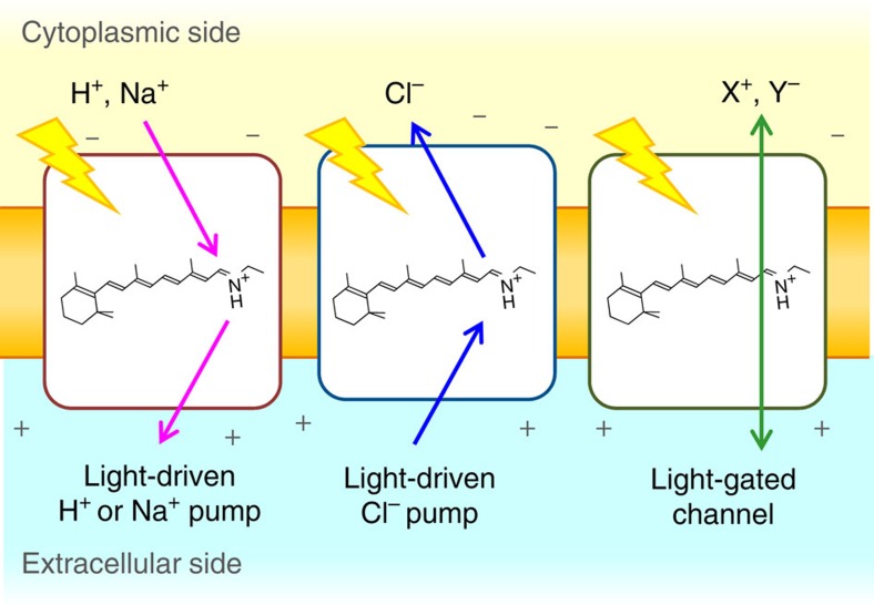
Light-driven outward H+ pumps are widely distributed in nature, converting sunlight energy into proton motive force. Here we report the characterization of an oppositely directed H+ pump with a similar architecture to outward pumps. A deep-ocean marine bacterium, Parvularcula oceani, contains three rhodopsins, one of which functions as a light-driven inward H+ pump when expressed in Escherichia coli and mouse neural cells. Detailed mechanistic analyses of the purified proteins reveal that small differences in the interactions established at the active centre determine the direction of primary H+ transfer. Outward H+ pumps establish strong electrostatic interactions between the primary H+ donor and the extracellular acceptor. In the inward H+ pump these electrostatic interactions are weaker, inducing a more relaxed chromophore structure that leads to the long-distance transfer of H+ to the cytoplasmic side. These results demonstrate an elaborate molecular design to control the direction of H+ transfers in proteins.


Figures

Figure 1
Figure 1. Ion-transporting microbial rhodopsins.
Light-driven outward cation pumps (left) and inward anion pumps (middle) function as active transporters, while light-gated channels conduct cations or anions in a passive manner (right).
Figure 2
Figure 2. Amino-acid sequence alignment of important residues and phylogenetic tree of microbial rhodopsins.
(a) Important residues for the function of microbial rhodopsins. BR, KR2, FR and ASR are outward H+ pump, outward Na+ pump, inward Cl pump, and photochromic sensor, respectively. Dashed rectangles indicate the positions of NDQ and NTQ motifs and residues unique for XeR. (b) Phylogenetic tree of selected microbial rhodopsins. The rhodopsins included in the phylogenetic tree: bacteriorhodopsin from Halobacterium salinarum (BR), archaerhodopsin-1, -2 and -3 from Halorubrum sodomense (AR1, AR2 and AR3), bacteriorhodopsin and middle rhodopsin from Haloquadratum walsbyi (HwBR, MR), halorhodopsin from H. salinarum, Salinibacter ruber and Natronomonas pharaonis (HsHR, SrHR, NpHR), PoXeR, xenorhodopsin from Haloplasma contractile (HcXeR), ASR, sensory rhodopsin II from N. pharaonis and H. salinarum (NpSRII and HsSRII), sensory rhodopsin I from S. ruber, Haloarcula vallismortis and H. salinarum (SrSRI, HvSRI and HsSRI), proteorhodopsin from Krokinobacter eikastus, Gillisia limnaea, Nonlabens dokdonensis and Vibrio sp. AND4 (KR1, GlPR, NdPR, VsPR), blue-absorbing proteorhodopsin from uncultured bacterium (BPR), green-absorbing proteorhodopsin from uncultured marine gamma proteobacterium (GPR), rhodopsin from Exiguobacterium sibiricum (ESR), thermophilic rhodopsin from Thermus thermophilus (TR), xanthorhodopsin from S. ruber (XR), rhodopsin from Gloeobacter violaceus PCC 7421 (GR), putative Cl pumping rhodopsin from Citromicrobium sp. JLT1363, Citromicrobium bathyomarinum and Sphingopyxis baekryungensis (CsClR, CbClR, SpClR), Cl- pump from Fulvimarina pelagi and Nonlabens marinus S1-08 (FR and NmClR), PoClR, PoNaR, two putative NaRs from Truepera radiovictrix (TrNaR1 and TrNaR2), putative NaR from Indibacter alkaliphilus (IaNaR), NaR from N. dokdonensis, G. limnaea and K. eikastus (NdNaR, GlNaR and KR2).
Figure 3
Figure 3. Light-driven H+ transport activity of PoXeR.
(a) E. coli C43(DE3) strain cells in which the expression of PoNaR, PoClR and PoXeR was induced (upper), and ion-pumping activity was assayed by observing pH changes (lower). Light is on between 0 and 150 s. (b) Electrophysiological measurements of PoXeR-driven photocurrent in ND7/23 cells (left) and IV plot of the current at pHo 7.2 and 9.0.
Figure 4
Figure 4. Molecular properties of PoXeR.
(a) Absorption spectra of dark- and light-adapted PoXeR (purple and orange solid lines, respectively). Dashed lines represent the calculated spectra of 100% all-trans and 100% 13-cis forms. (b) High-speed AFM image of PoXeR reconstituted in a nanodisc. (c) HPLC pattern of retinal extracted from PoXeR.
Figure 5
Figure 5. Photoreaction dynamics of PoXeR.
(a) Transient difference absorption spectra of dark-adapted PoXeR (left) and absorption difference time-evolution at specific wavelengths (centre). Decay-associated spectra obtained by global fitting of the changes in transient absorption (right). The inset shows the difference spectrum between light-adapted and dark-adapted state. (b) The process of dark-adaptation estimated by the recovery of absorption at 585 nm. (c) The photocycle of PoXeR. Values of the time-constants of each process are the mean±s.d.
Figure 6
Figure 6. Inward H+ pumping activity and light-induced FTIR spectra of PoXeR and its mutants.
(a) The crystal structure of ASR (PDB ID: 1XIO) is shown with the side chains of the acidic residues used in PoXeR mutagenesis studies. The residue numbering of PoXeR is shown. (b) H+ transport activity of PoXeR mutants in E. coli cells after normalization for the amount of protein. Light is on between 0 and 150 s. (c) The initial slopes of light-induced pH changes shown in b. (d) Light-induced difference FTIR spectra of WT PoXeR and the mutants at T=230 K and pH 8.0. Spectra are measured in H2O (black) and D2O (red).
Figure 7
Figure 7. Analysis of H+ uptake in PoXeR.
(a) Time-evolution of the accumulation of M intermediate at various pH values. (b) Light-induced difference FTIR spectra between PoXeR13C and PoXeRAT states at 277 K and pH 8.0.
Figure 8
Figure 8. Model for H+ transfer pathway in PoXeR.
The H+ transfer pathway in PoXeR suggested by the results of the present study. The primary H+ transfer occurs from the Schiff base to D216, and E35 affects this reaction. The Schiff base is not directly reprotonated from the aqueous phase, but there is an internal H+ donor, yet to be identified.
Figure 9
Figure 9. Mechanism of inward H+ transport in PoXeR and outward H+ transport in BR.
Schematic description of H+ transport in the photocycle of PoXeR compared with the photocycle of BR. In BR, only one double bond at C13=C14 isomerizes during the outward H+ pumping photocycle (trans to cis photoisomerization, and cis to trans thermal isomerization). In contrast, retinal isomerization is more complex to facilitate the inward H+ pump in PoXeR: (i) C13=C14 trans to cis photoisomerization, (ii) C15=N anti to syn thermal isomerization, and (iii) C13=C14 cis to trans and C15=N syn to anti thermal isomerization by a bicycle-pedal motion.

References

    1. Ernst O. P. et al.. Microbial and animal rhodopsins: structures, functions, and molecular mechanisms. Chem. Rev. 114, 126–163 (2014). - PMC - PubMed
    1. Deisseroth K. Optogenetics: 10 years of microbial opsins in neuroscience. Nat. Neurosci. 18, 1213–1225 (2015). - PMC - PubMed
    1. Oesterhelt D. & Stoeckenius W. Rhodopsin-like protein from the purple membrane of Halobacterium halobium. Nat. New Biol. 233, 149–152 (1971). - PubMed
    1. Matsuno-Yagi A. & Mukohata Y. Two possible roles of bacteriorhodopsin; a comparative study of strains of Halobacterium halobium differing in pigmentation. Biochem. Biophys. Res. Commun. 78, 237–243 (1977). - PubMed
    1. Schobert B. & Lanyi J. K. Halorhodopsin is a light-driven chloride pump. J. Biol. Chem. 257, 10306–10313 (1982). - PubMed

Publication types