. 2016 Nov 17:6:37255.
doi: 10.1038/srep37255.

Modulation of light-driven arousal by LIM-homeodomain transcription factor Apterous in large PDF-positive lateral neurons of the Drosophila brain

Affiliations

Modulation of light-driven arousal by LIM-homeodomain transcription factor Apterous in large PDF-positive lateral neurons of the Drosophila brain

Naoto Shimada et al. Sci Rep. .

Abstract

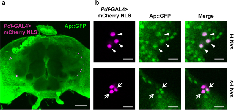
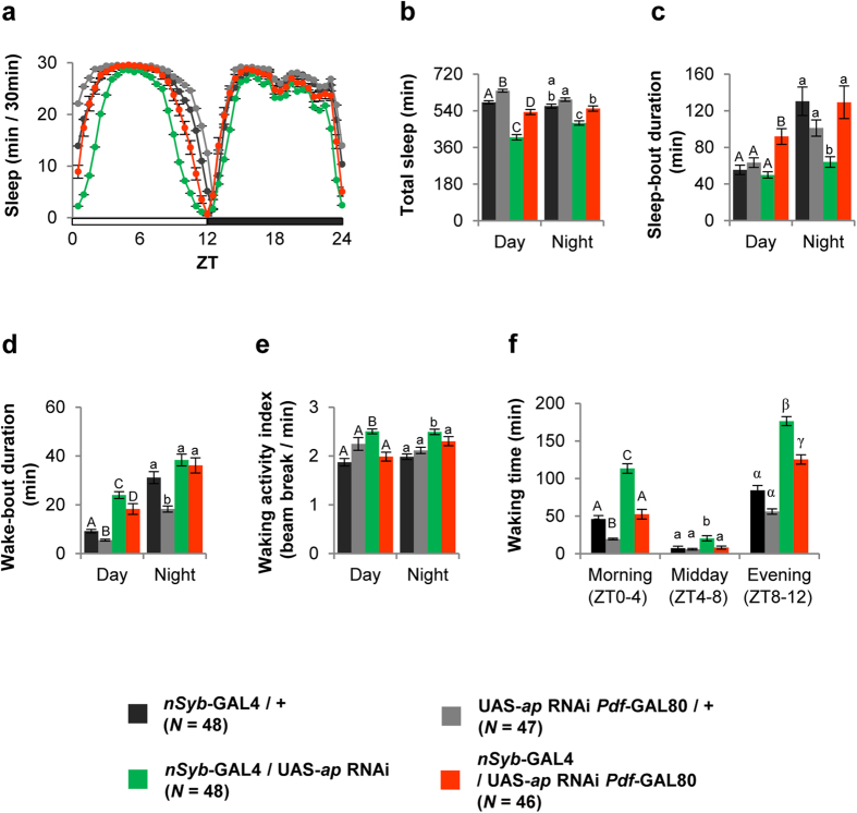
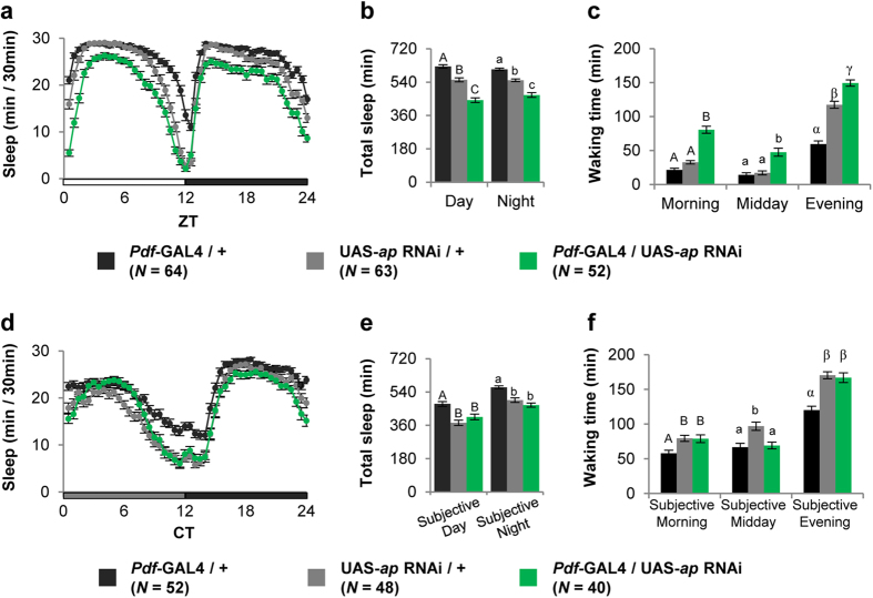
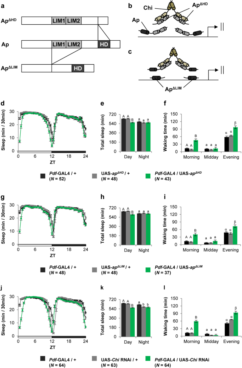
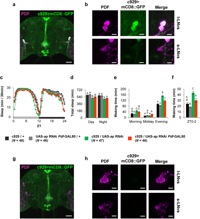
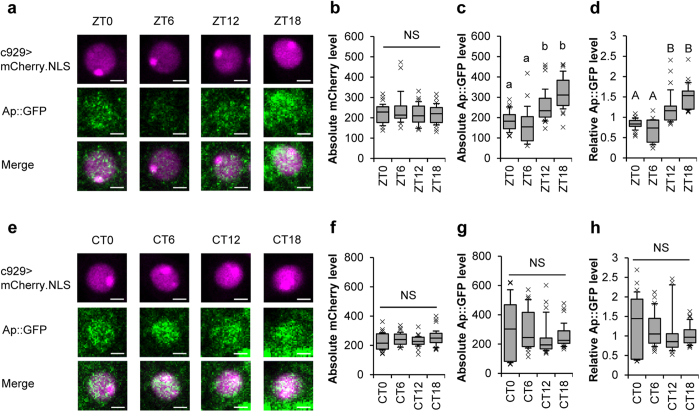
Apterous (Ap), the best studied LIM-homeodomain transcription factor in Drosophila, cooperates with the cofactor Chip (Chi) to regulate transcription of specific target genes. Although Ap regulates various developmental processes, its function in the adult brain remains unclear. Here, we report that Ap and Chi in the neurons expressing PDF, a neuropeptide, play important roles in proper sleep/wake regulation in adult flies. PDF-expressing neurons consist of two neuronal clusters: small ventral-lateral neurons (s-LNvs) acting as the circadian pacemaker and large ventral-lateral neurons (l-LNvs) regulating light-driven arousal. We identified that Ap localizes to the nuclei of s-LNvs and l-LNvs. In light-dark (LD) cycles, RNAi knockdown or the targeted expression of dominant-negative forms of Ap or Chi in PDF-expressing neurons or l-LNvs promoted arousal. In contrast, in constant darkness, knockdown of Ap in PDF-expressing neurons did not promote arousal, indicating that a reduced Ap function in PDF-expressing neurons promotes light-driven arousal. Furthermore, Ap expression in l-LNvs showed daily rhythms (peaking at midnight), which are generated by a direct light-dependent mechanism rather than by the endogenous clock. These results raise the possibility that the daily oscillation of Ap expression in l-LNvs may contribute to the buffering of light-driven arousal in wild-type flies.


Figures

Figure 1
Figure 1. Ap expression in adult brain.
Nuclei of PDF neurons were visualized using mCherry.NLS. Pdf-GAL4/ap::GFP; UAS-mCherry.NLS/+ flies were used. mCherry.NLS is shown in magenta, Ap::GFP is shown in green, and the overlap is shown in white. Adult brains were dissected at ZT18 after the flies were entrained to more than 3 LD cycles. (a) Stacked confocal image showing a front view of the adult brain. A scale bar represents 50 μm. (b) Confocal section image at the level of PDF neurons of the adult brain. Scale bars represent 10 μm. Triangles, l-LNvs; arrows, s-LNvs.
Figure 2
Figure 2. Pan-neural knockdown of ap promotes arousal under LD cycles.
We generated transgenic flies (UAS-ap RNAi Pdf-GAL80) with both the UAS-ap RNAi and Pdf-GAL80 constructs in the second chromosome. Black circles and bars, nSyb-GAL4/+; gray circles and bars, UAS-ap RNAi Pdf-GAL80/+; green circles and bars, nSyb-GAL4/UAS-ap RNAi; orange circles and bars, nSyb-GAL4/UAS-ap RNAi Pdf-GAL80. All sleep/wake parameters (daily sleep pattern, total sleep amount, sleep-bout duration, wake-bout duration, waking time, and waking activity index) were analyzed using the data averaged over 3 days of LD. Error bars show S.E.M. in each figure. Bars with the same letter indicate values that are not significantly different (P > 0.05). (a) Daily sleep patterns of control and experimental flies. (b) Total sleep amount during day and night. (c) Sleep-bout durations during day and night. (d) Wake-bout durations during day and night. (e) Waking activity indices during day and night. (f) Waking times in the morning (ZT0–4), midday (ZT4–8), and evening (ZT8–12).
Figure 3
Figure 3. PDF neuron-specific knockdown of ap promotes arousal in LD cycles.
Sleep/wake parameters were analyzed using the data averaged over 3 days of LD or DD. Error bars show S.E.M. in each figure. Bars with the same letter indicate values that are not significantly different (P > 0.05). (a and d) Daily sleep patterns of control and experimental flies. (b and e) Total sleep amount during day and night. (c and f) Waking times in the morning (ZT0–4 or CT0–4), midday (ZT4–8 or CT4–8), and evening (ZT8–12 or CT8–12). (a–c) Sleep was measured for 3 days in LD cycles after the flies were entrained to 3 LD cycles. (d–f) Sleep was measured in DD after the flies were entrained to 3 LD cycles.
Figure 4
Figure 4. Targeted expression of truncated forms of Ap and Chi RNAi in PDF neurons promotes arousal.
Sleep/wake parameters were analyzed using the data averaged over 3 days of LD. Error bars show S.E.M. in each figure. Bars with the same letter indicate values that are not significantly different (P > 0.05). (a) Schematic representation of wild-type and truncated forms of AP. (b and c) Possible model of dysfunction of Ap/Chi through expression of ApΔLIM and ApΔHD. Sleep/wake parameters obtained using (df) Pdf-GAL4/UAS-apΔHD flies, (gi) Pdf-GAL4/UAS-apΔLIM flies, and (jl) Pdf-GAL4/UAS-Chi RNAi flies. (d,g and j) Daily sleep patterns of control and experimental flies. (e,h and k) Total sleep amount during day and night. (f, i and l) Waking times in the morning (ZT0–4), midday (ZT4–8), and evening (ZT8–12).
Figure 5
Figure 5. Knockdown of ap in l-LNvs promotes arousal.
(a and b) c929-driven GFP (green) and PDF immunolabeling (magenta). (a) Stacked confocal image showing a front view of the adult brain. A scale bar represents 50 μm. (b) Confocal section image at the level of PDF neurons of the adult brain. Scale bars represent 10 μm. (cf) c929/UAS-ap RNAi (green) and c929/UAS-ap RNAi Pdf-GAL80 (orange) flies were used. c929/+ (black) and UAS-ap RNAi Pdf-GAL80/+ (gray) flies were used as the control. (c) Daily sleep patterns of control and experimental flies. (d) Total sleep amount during day and night. (e) Waking times in the morning (ZT0–4), midday (ZT4–8), and evening (ZT8–12). (f) Waking time during the period between ZT0 and ZT2. (g and h) PDF immunolabeling (magenta) in c929/Pdf-GAL80; UAS-mCD8::GFP/+ flies. (g) Stacked confocal image showing a front view of the adult brain. A scale bar represents 50 μm. (h) Confocal section image at the level of PDF neurons of the adult brain. Scale bars represent 10 μm.
Figure 6
Figure 6. Knockdown of ap in l-LNvs does not affect PDF expression and number of PDF-releasing sites.
(a) Real-time qRT-PCR analysis of Pdf mRNA expression level using Pdf-GAL4/+, UAS-ap RNAi/+, and Pdf-GAL4/UAS-ap RNAi males. Mean ± SEM values were calculated from five replicates. NS, not significant; **P < 0.01. (b) Intensity ratio of PDF signals in l-LNvs and s-LNvs (l-LNvs/s-LNvs). c929/UAS-ap RNAi (green bar) and c929/UAS-GFP RNAi (gray bar) flies were used. N = 7–8 in each bar. (c and d) PDF immunolabeling in the whole brain. Stacked confocal image showing a front view of the adult brain. A scale bar represents 50 μm. (e) Cell number in l-LNvs. (f and g) PDF immunolabeling in the optic lobe. Stacked confocal image of an optic lobe. Scale bars represent 20 μm. (c and f) Pdf-GAL4/UAS-ap RNAi flies were used. (d and g) Pdf-GAL4/UAS-GFP RNAi flies were used as the control. (h) Number of varicosities in l-LNvs. The number of spots of diameters (0.5, 1, and 2 μm) was counted for each diameter. Pdf-GAL4/UAS-ap RNAi (green bar) and Pdf-GAL4/UAS-GFP RNAi (gray bar) flies were used. 11–12 brains were used for each genotype.
Figure 7
Figure 7. Ap is rhythmically expressed in l-LNvs under LD cycle conditions.
(a) Ap::GFP expression in one of the l-LNvs at ZT0, 6, 12, and 18. c929/ap::GFP; UAS-mCherry.NLS/+ flies (6 days old) were used. Adult male brains were collected at 4 time points (ZT0, 6, 12, and 18) after the flies were entrained to 6 LD cycles. A scale bar represents 2 μm. (b) Absolute mCherry.NLS levels at ZT0, 6, 12, and 18. In l-LNvs, the c929-driven mCherry.NLS expression level remained nearly constant throughout the day. Thus, we used the mCherry.NLS as an internal standard. (c) Absolute Ap::GFP levels at ZT0, 6, 12, and 18. (d) Relative Ap::GFP levels at ZT0, 6, 12, and 18. (bd) 8–10 brains were used at each time point. The total numbers of l-LNvs observed are as follows: N = 27 at ZT0, N = 15 at ZT6, N = 28 at ZT12, N = 23 at ZT18. (e) Ap expression in l-LNvs under DD. For measurement of Ap::GFP expression level in DD, adult male brains were collected at 4 time points (CT0, 6, 12, and 18) on the third day of DD after the flies were entrained to 3 LD cycles. A scale bar represents 2 μm. (f) Absolute mCherry.NLS levels at CT0, 6, 12, and 18. (g) Absolute Ap::GFP levels at CT0, 6, 12, and 18. (h) Relative Ap::GFP levels at CT0, 6, 12, and 18. (fh) 9 brains were used at each time point. The total numbers of l-LNvs observed are as follows: N = 27 at CT0, N = 29 at CT6, N = 26 at CT12, N = 28 at CT18. (bd and fh) In each box plot, the box encompasses the interquartile range, a line is drawn at the median, and the vertical bars extend to the 10th and 90th percentiles. Crosses show outliers. Data sets with the same letter indicate that there are no significantly differences among them (P > 0.05). NS, not significant.

References

    1. Hobert O. & Westphal H. Functions of LIM-homeobox genes. Trends Genet. 16, 75–83 (2000). - PubMed
    1. Butterworth F. M. & King R. C. The developmental genetics of apterous mutants of Drosophila melanogaster. Genetics 52, 1153–1174 (1965). - PMC - PubMed
    1. Cohen B., McGuffin M. E., Pfeifle C., Segal D. & Cohen S. M. apterous, a gene required for imaginal disc development in Drosophila encodes a member of the LIM family of developmental regulatory proteins. Genes Dev. 6, 715–729 (1992). - PubMed
    1. O′Keefe D. D., Thor S. & Thomas J. B. Function and specificity of LIM domains in Drosophila nervous system and wing development. Development 125, 3915–3923 (1998). - PubMed
    1. Bourgouin C., Lundgren S. E. & Thomas J. B. Apterous is a Drosophila LIM domain gene required for the development of a subset of embryonic muscles. Neuron 9, 549–561 (1992). - PubMed

Publication types

MeSH terms