. 2016 Nov 17:6:37246.
doi: 10.1038/srep37246.

Digital Detection of Exosomes by Interferometric Imaging

Affiliations

Digital Detection of Exosomes by Interferometric Imaging

George G Daaboul et al. Sci Rep. .

Abstract

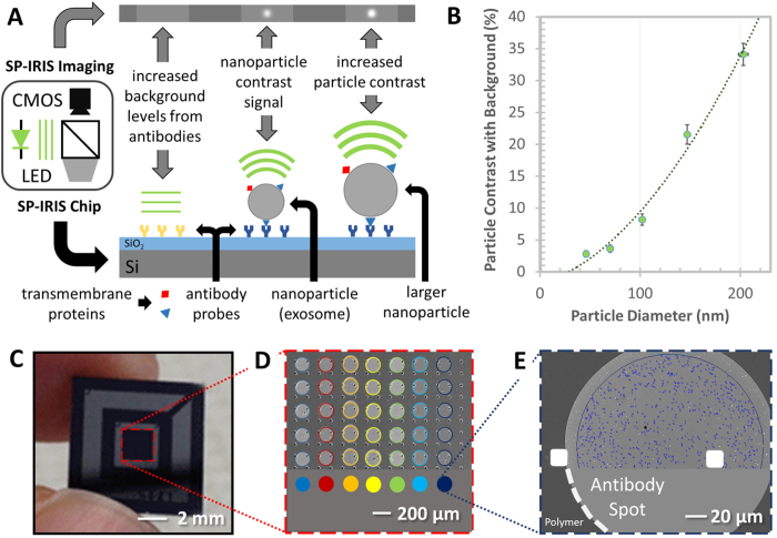
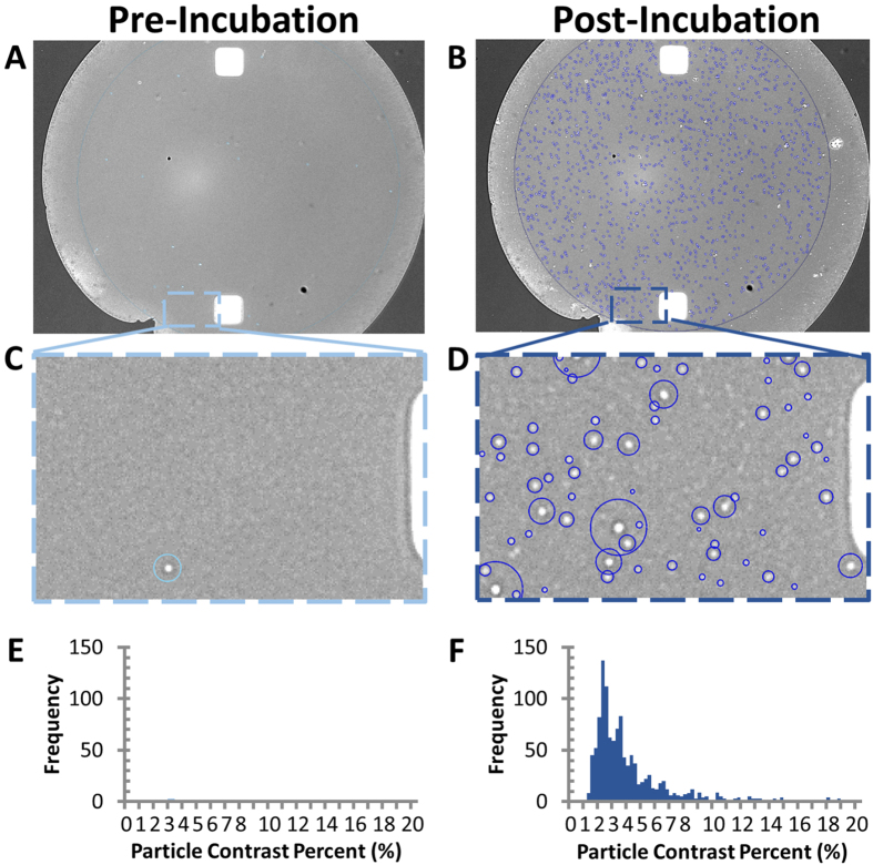
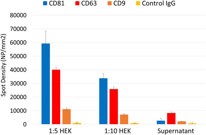
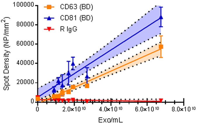
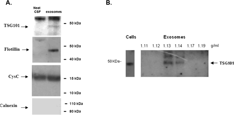
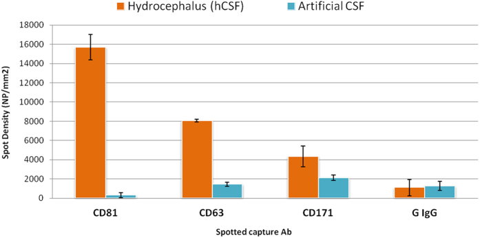
Exosomes, which are membranous nanovesicles, are actively released by cells and have been attributed to roles in cell-cell communication, cancer metastasis, and early disease diagnostics. The small size (30-100 nm) along with low refractive index contrast of exosomes makes direct characterization and phenotypical classification very difficult. In this work we present a method based on Single Particle Interferometric Reflectance Imaging Sensor (SP-IRIS) that allows multiplexed phenotyping and digital counting of various populations of individual exosomes (>50 nm) captured on a microarray-based solid phase chip. We demonstrate these characterization concepts using purified exosomes from a HEK 293 cell culture. As a demonstration of clinical utility, we characterize exosomes directly from human cerebrospinal fluid (hCSF). Our interferometric imaging method could capture, from a very small hCSF volume (20 uL), nanoparticles that have a size compatible with exosomes, using antibodies directed against tetraspanins. With this unprecedented capability, we foresee revolutionary implications in the clinical field with improvements in diagnosis and stratification of patients affected by different disorders.


Conflict of interest statement

The authors declare the following competing financial interest(s): George G. Daaboul is the chief scientific officer of Nexgenarrays LLC. David Freedman is the chief executive officer of NexgenArrays LLC. All remaining contributing authors declare no competing financial interests.

Figures

Figure 1
Figure 1. Schematic representation of the SP-IRIS detection process.
(A) SP-IRIS detection principle, monochromatic LED light illuminates the sensor surface and the interferometriclly enhanced nanoparticle scattering signature is captured on a CMOS camera. (B) Demonstrates SP-IRIS signal for polystyrene nanoparticles with a diameter from 50–200 nm which can be used to infer size of captured EVs. (C) Image of the SP-IRIS chip. (D) Low-magnification interferometric image showing microarray of immobilized capture probes. (E) SP-IRIS image of a capture probe. NVDX analysis software recognize capture spot and detects nanoparticles captured.
Figure 2
Figure 2. Exosome capture, digital counting, and relative sizing.
(A,B) Anti-CD81 capture probe image acquired before and after incubation with purified HEK293 cells derived exosomes. (C,D) Zoom-box of particles detected pre- and post-incubation. (E–F) Particle contrast histogram pre- and post-incubation.
Figure 3
Figure 3. Nanoparticle capture validation with SEM.
(A) SP-IRIS image of exosomes being captured by anti-CD81 antibody. (B) Exosomes visualized by SEM of the same field-of-view for comparison. Scale bar is 1 micron.
Figure 4
Figure 4
Exosomes purified from HEK cell line, captured with anti- CD81 antibody on silicon chip and detected by SP-IRIS (A) and AFM (B,C). (A) SP-IRIS Image of the anti-CD81 spot incubated with a suspension of exosomes (6.75E + 10 exosomes/mL), the red circles highlight the countable nanoparticles. (B) AFM image of the same spot area: the blue dots identify particles larger than 15 nm; the yellow circles show in image (B) perfectly match the particles detected by SP-IRIS in image (A). (C) Zoom in area of the anti-CD81 spot shown in the green frame highlights the particles larger than 15 nm in height detected by AFM and SP-IRIS. Particles smaller than 15–10 nm are detectable only with AFM as they are below SP-IRIS detection limit.
Figure 5
Figure 5. Exosome Phenotyping.
Exosomes, isolated from HEK fibroblast cells and EV depleted supernatant, captured with antibodies against CD81, CD63, CD9 and IgG negative control and detected by SP-IRIS.
Figure 6
Figure 6. Dilution curve of exosomes purified from HEK cell line and detected with SP-IRIS.
A good correlation can be observed for both capture antibodies CD63 (yellow line, R2 0.97) and CD81 (blue line, R2 0.93); no particles were detected on the negative control R IgG (red line). The bright area around the regression line corresponds to the 95% confidence interval. Based on line equations, limits of detection (LOD) were calculated, resulting in 5.07E + 09 particles/mL for CD63 antibody and 3.94E + 09 particles/mL for CD81 antibody.
Figure 7
Figure 7
(A) Western blot analysis of exosomes isolated from 1.8 ml of hCSF and neat hCSF: the exosomal markers TSG101 and flotillin marked the exosomes fraction and not neat CSF, cystatin C marked both fractions and calnexin was not detected. (B) Sucrose gradient fractions of exosomal preparations from hCSF were immunoblotted with the TSG101 monoclonal antibody. TSG101 marked the exosomes-positive fractions (corresponding to 1.13 and 1.14 g/mL sucrose). HEK cells lysate was used as positive control. Western blot gels have been cropped to show only the relevant protein bands. Complete figures can be found as Supplementary, Figure SI 7.
Figure 8
Figure 8. SP-IRIS label-free assay on human hydrocephalus CSF sample and artificial CSF.
The expression of the typical exosomal biomarkers CD81 CD63 as well as the neural adhesion protein CD171 is significantly different than in the artificial CSF, negative control. An additional negative control, a non correlated IgG, shows a low level of non-specific binding. Each multiplexed chip was run in duplicate independent replicate tests.

References

    1. Beaudoin A. R. & Grondin G. Shedding of vesicular material from the cell surface of eukaryotic cells: different cellular phenomena. Biochim. Biophys. Acta - Rev. Biomembr. 1071, 203–219 (1991). - PubMed
    1. Zhang J. et al. Exosome and exosomal microRNA: Trafficking, sorting, and function. Genomics, Proteomics and Bioinformatics 13, 17–24 (2015). - PMC - PubMed
    1. Orozco A. F. & Lewis D. E. Flow cytometric analysis of circulating microparticles in plasma. J. Int. Soc. Adv. Cytom. 77, 502–514 (2010). - PMC - PubMed
    1. Gonzales P. A. et al. Large-scale proteomics and phosphoproteomics of urinary exosomes. J. Am. Soc. Nephrol. 20, 363–379 (2009). - PMC - PubMed
    1. Sharma S., Gillespie B. M., Palanisamy V. & Gimzewski J. K. Quantitative nanostructural and single-molecule force spectroscopy biomolecular analysis of human-saliva-derived exosomes. Langmuir 27, 14394–14400 (2011). - PMC - PubMed

Publication types