

Magnetic resonance and ultrasound contrast imaging of polymer-shelled microbubbles loaded with iron oxide nanoparticles
- PMID: 27853587
- PMCID: PMC5108937
- DOI: 10.1098/rsos.160063
Magnetic resonance and ultrasound contrast imaging of polymer-shelled microbubbles loaded with iron oxide nanoparticles
Abstract
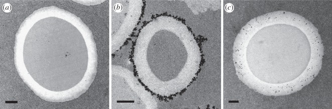
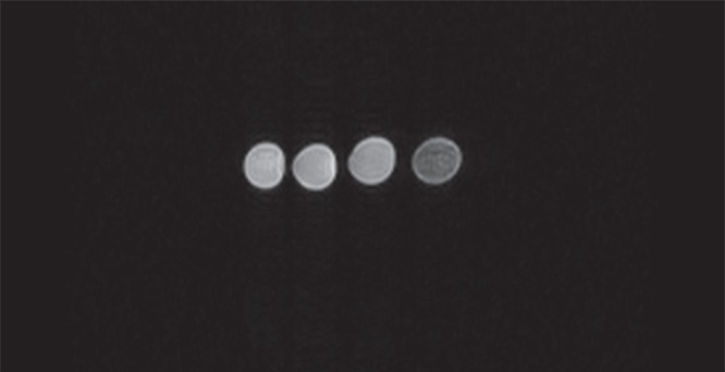
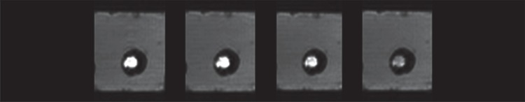
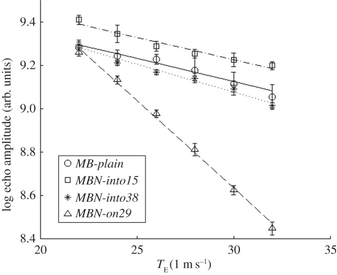
Dual-mode contrast agents (CAs) have great potential for improving diagnostics. However, the effectiveness of CAs is strictly related to both the solution adopted to merge the two agents into a single probe unit, and the ratio between the two agents. In this study, two dual-mode CAs for simultaneous magnetic resonance imaging (MRI) and ultrasound imaging (UI) were assessed. For this purpose, different densities of superparamagnetic iron oxide nanoparticles (SPIONs) were anchored to the external surface of polymer-shelled microbubbles (MBs) or were physically entrapped into the shell. In vitro static and dynamic experiments were carried out with a limited concentration of modified MBs (106 bubbles ml-1) by avoiding destruction during UI (performed at a peak pressure lower than 320 kPa) and by using a low-field MRI system (with a magnetic flux density equal to 0.25 T). Under these conditions, different imaging techniques, set-up parameters and SPION densities were used to achieve satisfactory detection of the CAs by using both UI and MRI. However, when the SPION density was increased, the MRI contrast improved, whereas the UI contrast worsened due to the reduced elasticity of the MB shell. For both UI and MRI, MBs with externally anchored SPIONs provided better performance than MBs with SPIONs entrapped into the shell. In particular, a SPION density of 29% with respect to the mass of the MBs was successfully tested.
Keywords: dual-mode contrast agents; magnetic resonance imaging; medical ultrasound; polymeric microbubbles; superparamagnetic iron oxide nanoparticles.
Figures
Similar articles
-
On the interplay of shell structure with low- and high-frequency mechanics of multifunctional magnetic microbubbles.Soft Matter. 2014 Jan 7;10(1):214-26. doi: 10.1039/c3sm51560e. Soft Matter. 2014. PMID: 24651844
-
Cellular Uptake of Plain and SPION-Modified Microbubbles for Potential Use in Molecular Imaging.Cell Mol Bioeng. 2017;10(6):537-548. doi: 10.1007/s12195-017-0504-9. Epub 2017 Aug 10. Cell Mol Bioeng. 2017. PMID: 29151981 Free PMC article.
-
Shell thickness determination of polymer-shelled microbubbles using transmission electron microscopy.Micron. 2016 Jun;85:39-43. doi: 10.1016/j.micron.2016.03.009. Epub 2016 Apr 1. Micron. 2016. PMID: 27077316
-
Iron-based superparamagnetic nanoparticle contrast agents for MRI of infection and inflammation.AJR Am J Roentgenol. 2015 Mar;204(3):W302-13. doi: 10.2214/AJR.14.12733. AJR Am J Roentgenol. 2015. PMID: 25714316 Free PMC article. Review.
-
Superparamagnetic Iron Oxide Nanoparticles-Current and Prospective Medical Applications.Materials (Basel). 2019 Feb 19;12(4):617. doi: 10.3390/ma12040617. Materials (Basel). 2019. PMID: 30791358 Free PMC article. Review.
Cited by
-
Functionalization of Metal and Carbon Nanoparticles with Potential in Cancer Theranostics.Molecules. 2021 May 21;26(11):3085. doi: 10.3390/molecules26113085. Molecules. 2021. PMID: 34064173 Free PMC article. Review.
-
Janus USPION modular platform (JUMP) for theranostic ultrasound-mediated targeted intratumoral microvascular imaging and DNA/miRNA delivery.Theranostics. 2022 Nov 7;12(18):7646-7667. doi: 10.7150/thno.78454. eCollection 2022. Theranostics. 2022. PMID: 36451861 Free PMC article.
-
Circulating Magnetic Microbubbles for Localized Real-Time Control of Drug Delivery by Ultrasonography-Guided Magnetic Targeting and Ultrasound.Theranostics. 2018 Jan 1;8(2):341-357. doi: 10.7150/thno.20781. eCollection 2018. Theranostics. 2018. PMID: 29290812 Free PMC article.
-
Hard-Shelled Glycol Chitosan Nanoparticles for Dual MRI/US Detection of Drug Delivery/Release: A Proof-of-Concept Study.Nanomaterials (Basel). 2023 Aug 1;13(15):2227. doi: 10.3390/nano13152227. Nanomaterials (Basel). 2023. PMID: 37570545 Free PMC article.
-
Nanotechnology strategies for hepatocellular carcinoma diagnosis and treatment.RSC Adv. 2022 Oct 31;12(48):31068-31082. doi: 10.1039/d2ra05127c. eCollection 2022 Oct 27. RSC Adv. 2022. PMID: 36349046 Free PMC article. Review.
References
-
- Townsend DW, Beyer T, Blodgett TM. 2003. PET/CT scanners: a hardware approach to image fusion. Semin. Nucl. Med. 33, 193–204. (doi:10.1053/snuc.2003.127314) - DOI - PubMed
-
- Cherry SR, Louie AY, Jacobs RE. 2008. The integration of positron emission tomography with magnetic resonance imaging. Proc. IEEE 96, 416–438. (doi:10.1109/JPROC.2007.913502) - DOI
-
- Louie A. 2010. Multimodality imaging probes: design and challenges. Chem. Rev. 110, 3146–3195. (doi:10.1021/cr9003538) - DOI - PMC - PubMed
-
- Yang F, Li Y, Chen Z, Zhang Y, Wu J, Gu N. 2009. Superparamagnetic iron oxide nanoparticle-embedded encapsulated microbubbles as dual contrast agents of magnetic resonance and ultrasound imaging. Biomaterials 30, 3882–3890. (doi:10.1016/j.biomaterials.2009.03.051) - DOI - PubMed
-
- Park JI, Dinesh J, Ross W, Wendy O, Siyon C, Greg JS, Kumacheva E. 2010. Microbubbles loaded with nanoparticles: a route to multiple imaging modalities. J. Am. Chem. Soc. Nano Lett. 4, 6579–6586. (doi:10.1021/nn102248g) - DOI - PubMed
LinkOut - more resources
Full Text Sources
Other Literature Sources
Miscellaneous
