

GUCY2C-directed CAR-T cells oppose colorectal cancer metastases without autoimmunity
- PMID: 27853651
- PMCID: PMC5087292
- DOI: 10.1080/2162402X.2016.1227897
GUCY2C-directed CAR-T cells oppose colorectal cancer metastases without autoimmunity
Abstract
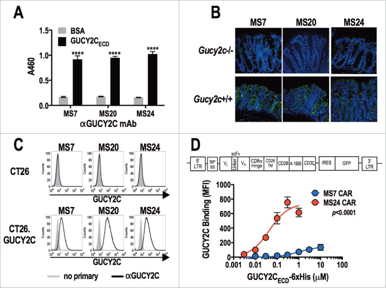
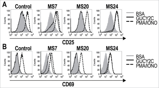
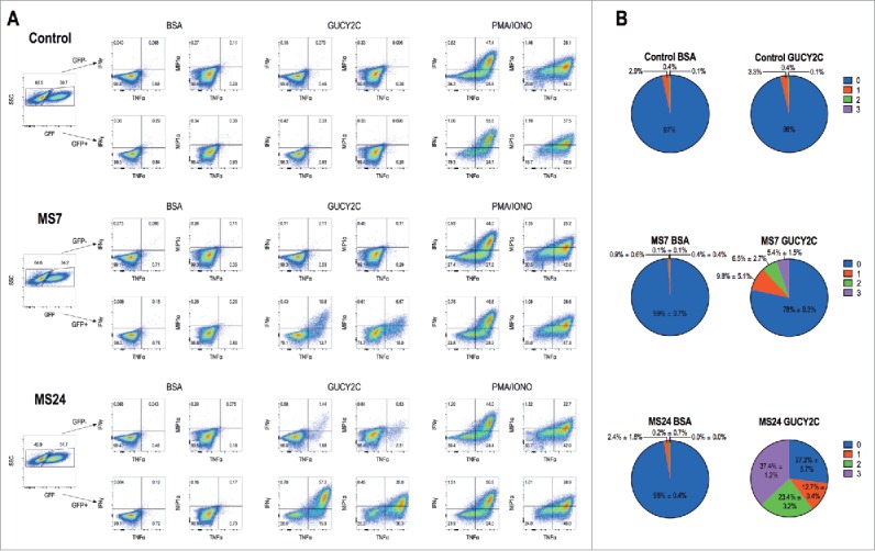
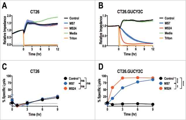
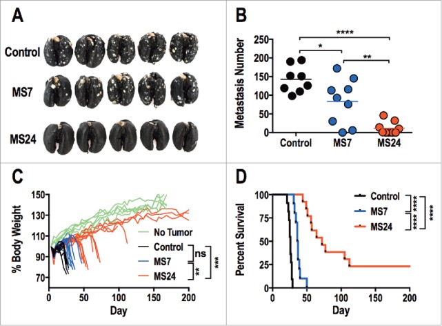
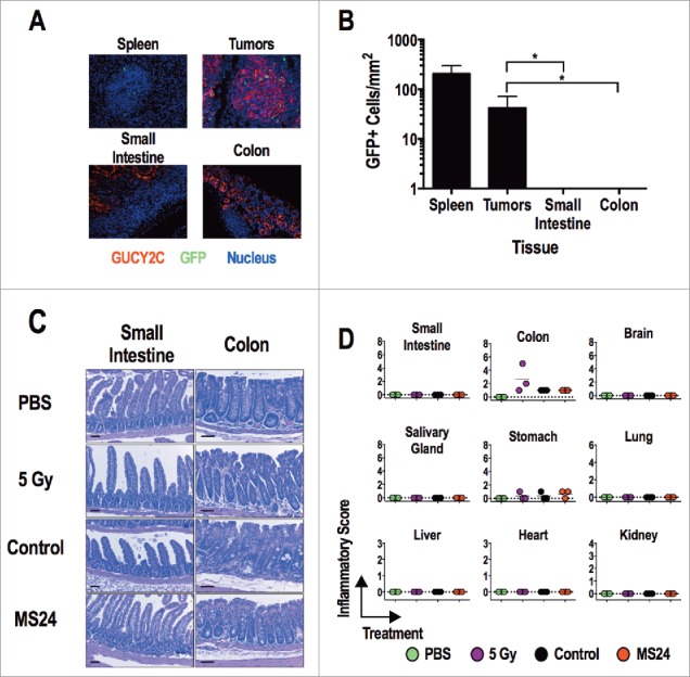
Adoptive T-cell therapy (ACT) is an emerging paradigm in which T cells are genetically modified to target cancer-associated antigens and eradicate tumors. However, challenges treating epithelial cancers with ACT reflect antigen targets that are not tumor-specific, permitting immune damage to normal tissues, and preclinical testing in artificial xenogeneic models, preventing prediction of toxicities in patients. In that context, mucosa-restricted antigens expressed by cancers exploit anatomical compartmentalization which shields mucosae from systemic antitumor immunity. This shielding may be amplified with ACT platforms employing antibody-based chimeric antigen receptors (CARs), which mediate MHC-independent recog-nition of antigens. GUCY2C is a cancer mucosa antigen expressed on the luminal surfaces of the intestinal mucosa in mice and humans, and universally overexpressed by colorectal tumors, suggesting its unique utility as an ACT target. T cells expressing CARs directed by a GUCY2C-specific antibody fragment recognized GUCY2C, quantified by expression of activation markers and cytokines. Further, GUCY2C CAR-T cells lysed GUCY2C-expressing, but not GUCY2C-deficient, mouse colorectal cancer cells. Moreover, GUCY2C CAR-T cells reduced tumor number and morbidity and improved survival in mice harboring GUCY2C-expressing colorectal cancer metastases. GUCY2C-directed T cell efficacy reflected CAR affinity and surface expression and was achieved without immune-mediated damage to normal tissues in syngeneic mice. These observations highlight the potential for therapeutic translation of GUCY2C-directed CAR-T cells to treat metastatic tumors, without collateral autoimmunity, in patients with metastatic colorectal cancer.
Keywords: Adoptive immunotherapy; chimeric antigen receptors; colorectal cancer; gene therapy; guanylyl cyclase C.
Figures
Similar articles
-
Human GUCY2C-Targeted Chimeric Antigen Receptor (CAR)-Expressing T Cells Eliminate Colorectal Cancer Metastases.Cancer Immunol Res. 2018 May;6(5):509-516. doi: 10.1158/2326-6066.CIR-16-0362. Epub 2018 Apr 3. Cancer Immunol Res. 2018. PMID: 29615399 Free PMC article.
-
Epitope-targeted cytotoxic T cells mediate lineage-specific antitumor efficacy induced by the cancer mucosa antigen GUCY2C.Cancer Immunol Immunother. 2012 May;61(5):713-23. doi: 10.1007/s00262-011-1133-0. Epub 2011 Nov 6. Cancer Immunol Immunother. 2012. PMID: 22057677 Free PMC article.
-
Antigen-independent activation is critical for the durable antitumor effect of GUCY2C-targeted CAR-T cells.J Immunother Cancer. 2024 Oct 4;12(10):e009960. doi: 10.1136/jitc-2024-009960. J Immunother Cancer. 2024. PMID: 39366753 Free PMC article.
-
GUCY2C-targeted cancer immunotherapy: past, present and future.Immunol Res. 2011 Dec;51(2-3):161-9. doi: 10.1007/s12026-011-8253-7. Immunol Res. 2011. PMID: 22038530 Free PMC article. Review.
-
An update on guanylyl cyclase C in the diagnosis, chemoprevention, and treatment of colorectal cancer.Expert Rev Clin Pharmacol. 2020 Oct;13(10):1125-1137. doi: 10.1080/17512433.2020.1826304. Epub 2020 Oct 6. Expert Rev Clin Pharmacol. 2020. PMID: 32945718 Free PMC article. Review.
Cited by
-
Novel antigens of CAR T cell therapy: New roads; old destination.Transl Oncol. 2021 Jul;14(7):101079. doi: 10.1016/j.tranon.2021.101079. Epub 2021 Apr 13. Transl Oncol. 2021. PMID: 33862524 Free PMC article. Review.
-
Bispecific c-Met/PD-L1 CAR-T Cells Have Enhanced Therapeutic Effects on Hepatocellular Carcinoma.Front Oncol. 2021 Mar 10;11:546586. doi: 10.3389/fonc.2021.546586. eCollection 2021. Front Oncol. 2021. PMID: 33777728 Free PMC article.
-
CAR-Based Immunotherapy of Solid Tumours-A Survey of the Emerging Targets.Cancers (Basel). 2023 Feb 11;15(4):1171. doi: 10.3390/cancers15041171. Cancers (Basel). 2023. PMID: 36831514 Free PMC article. Review.
-
Split tolerance permits safe Ad5-GUCY2C-PADRE vaccine-induced T-cell responses in colon cancer patients.J Immunother Cancer. 2019 Apr 23;7(1):104. doi: 10.1186/s40425-019-0576-2. J Immunother Cancer. 2019. PMID: 31010434 Free PMC article. Clinical Trial.
-
Human GUCY2C-Targeted Chimeric Antigen Receptor (CAR)-Expressing T Cells Eliminate Colorectal Cancer Metastases.Cancer Immunol Res. 2018 May;6(5):509-516. doi: 10.1158/2326-6066.CIR-16-0362. Epub 2018 Apr 3. Cancer Immunol Res. 2018. PMID: 29615399 Free PMC article.
References
-
- Howlader N, Noone AM, Krapcho M, Garshell J, Miller D, Altekruse SF, Kosary CL, Yu M, Ruhl J, Tatalovich Z,Mariotto A, Lewis DR, Chen HS, Feuer EJ, Cronin KA (eds). SEER Cancer Statistics Review, 1975-2012, National Cancer Institute. Bethesda, MD, http://seer.cancer.gov/csr/1975_2012/, based on November 2014 SEER data submission, posted to the SEER web site, April 2015.
-
- Couzin-Frankel J. Breakthrough of the year 2013. Cancer immunotherapy. Science 2013; 342:1432-3; PMID:24357284; http://dx.doi.org/10.1126/science.342.6165.1432 - DOI - PubMed
-
- Dudley ME, Wunderlich JR, Robbins PF, Yang JC, Hwu P, Schwartzentruber DJ, Topalian SL, Sherry R, Restifo NP, Hubicki AM et al.. Cancer regression and autoimmunity in patients after clonal repopulation with antitumor lymphocytes. Science 2002; 298:850-4; PMID:12242449; http://dx.doi.org/10.1126/science.1076514 - DOI - PMC - PubMed
-
- Grupp SA, Kalos M, Barrett D, Aplenc R, Porter DL, Rheingold SR, Teachey DT, Chew A, Hauck B, Wright JF et al.. Chimeric antigen receptor-modified T cells for acute lymphoid leukemia. N Engl J Med 2013; 368:1509-18; PMID:23527958; http://dx.doi.org/10.1056/NEJMoa12-15134 - DOI - PMC - PubMed
-
- Porter DL, Levine BL, Kalos M, Bagg A, June CH. Chimeric antigen receptor-modified T cells in chronic lymphoid leukemia. N Engl J Med 2011; 365:725-33; PMID:21830940; http://dx.doi.org/10.1056/NEJMoa1103849 - DOI - PMC - PubMed
Publication types
Grants and funding
LinkOut - more resources
Full Text Sources
Other Literature Sources
Research Materials