

Increased PD-L1 and T-cell infiltration in the presence of HLA class I expression in metastatic high-grade osteosarcoma: a rationale for T-cell-based immunotherapy
- PMID: 27853827
- PMCID: PMC5222929
- DOI: 10.1007/s00262-016-1925-3
Increased PD-L1 and T-cell infiltration in the presence of HLA class I expression in metastatic high-grade osteosarcoma: a rationale for T-cell-based immunotherapy
Abstract
Introduction: Immunotherapy may be an excellent choice for treating osteosarcoma given its exceptionally high genomic instability, potentially generating neoantigens. In this study, we aim to investigate the HLA class I expression, PD-L1 and tumour-infiltrating lymphocytes in primary osteosarcomas and relapses/metastases, as well as their changes during disease progression.
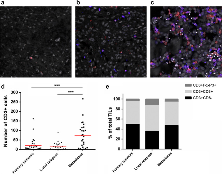
Materials and methods: Tumour samples from multiple stages of the disease (pretreatment biopsies, surgical resections of primary osteosarcomas, relapses and metastases) were collected and stained for HLA-A (HCA2), HLA-B/C (HC10), β2-microglobulin and PD-L1 using immunohistochemistry on whole sections. Density and type of T-cell infiltrate were characterised by a triple immunofluorescent staining CD3-CD8-FOXP3.
Results: Overall, 85 formalin-fixed, paraffin-embedded blocks from 25 osteosarcoma patients were included. HLA class I expression was detected in 94% of osteosarcomas (strongly positive in 56%, heterogeneous in 38%) and negative or weakly positive in 6%, without differences between the stages of the disease. HLA-A expression was more frequently negative than HLA-B/C. Tumour-infiltrating lymphocytes were highly heterogeneous and mainly observed in tumour areas with expression of HLA class I. Density of T cells was significantly higher in metastases than in primary tumours and local relapses (p = 0.0003). Positive PD-L1 expression was found in 13% of primary tumours, 25% of relapses and 48% of metastases and correlated with a high T-cell infiltrate (p = 0.002).
Conclusion: An increased number of tumour-infiltrating T cells and PD-L1 expression in metastases compared with primary tumours, suggesting accessibility for T cells, could imply that osteosarcoma patients with metastatic disease may benefit from T-cell-based immunotherapy.
Keywords: HLA class I; Immunotherapy; Osteosarcoma; PD-L1; Tumour-infiltrating lymphocytes.
Conflict of interest statement
The authors declare that they have no conflict of interest.
Figures


Similar articles
-
PD-L1 and IDO1 expression and tumor-infiltrating lymphocytes in osteosarcoma patients: comparative study of primary and metastatic lesions.J Cancer Res Clin Oncol. 2020 Oct;146(10):2607-2620. doi: 10.1007/s00432-020-03242-6. Epub 2020 May 9. J Cancer Res Clin Oncol. 2020. PMID: 32388585 Free PMC article.
-
Tumor mutational load, CD8+ T cells, expression of PD-L1 and HLA class I to guide immunotherapy decisions in NSCLC patients.Cancer Immunol Immunother. 2020 May;69(5):771-777. doi: 10.1007/s00262-020-02506-x. Epub 2020 Feb 12. Cancer Immunol Immunother. 2020. PMID: 32047958 Free PMC article.
-
A Combination of Positive Tumor HLA-I and Negative PD-L1 Expression Provides an Immune Rejection Mechanism in Bladder Cancer.Ann Surg Oncol. 2019 Aug;26(8):2631-2639. doi: 10.1245/s10434-019-07371-2. Epub 2019 Apr 22. Ann Surg Oncol. 2019. PMID: 31011905
-
Discordance of immunotherapy response predictive biomarkers between primary lesions and paired metastases in tumours: A systematic review and meta-analysis.EBioMedicine. 2021 Jan;63:103137. doi: 10.1016/j.ebiom.2020.103137. Epub 2020 Dec 11. EBioMedicine. 2021. PMID: 33310681 Free PMC article.
-
Immunotherapy in Breast Cancer: the Emerging Role of PD-1 and PD-L1.Curr Oncol Rep. 2017 Aug 10;19(10):64. doi: 10.1007/s11912-017-0627-0. Curr Oncol Rep. 2017. PMID: 28799073 Review.
Cited by
-
Evodiamine Induces Apoptosis, G2/M Cell Cycle Arrest, and Inhibition of Cell Migration and Invasion in Human Osteosarcoma Cells via Raf/MEK/ERK Signalling Pathway.Med Sci Monit. 2018 Aug 23;24:5874-5880. doi: 10.12659/MSM.909682. Med Sci Monit. 2018. Retraction in: Med Sci Monit. 2021 Mar 25;27:e932330. doi: 10.12659/MSM.932330. PMID: 30135419 Free PMC article. Retracted.
-
Clinicopathological and prognostic significance of PD-L1 expression in sarcoma: A systematic review and meta-analysis.Medicine (Baltimore). 2018 Jun;97(25):e11004. doi: 10.1097/MD.0000000000011004. Medicine (Baltimore). 2018. PMID: 29923984 Free PMC article.
-
The Updated Status and Future Direction of Immunotherapy Targeting B7-H1/PD-1 in Osteosarcoma.Cancer Manag Res. 2021 Jan 27;13:757-764. doi: 10.2147/CMAR.S285560. eCollection 2021. Cancer Manag Res. 2021. PMID: 33536783 Free PMC article. Review.
-
Cytokine screening identifies TNF to potentially enhance immunogenicity of pediatric sarcomas.Front Immunol. 2024 Dec 11;15:1347404. doi: 10.3389/fimmu.2024.1347404. eCollection 2024. Front Immunol. 2024. PMID: 39723214 Free PMC article.
-
Regulatory Role of N6-methyladenosine (m6A) Modification in Osteosarcoma.Front Oncol. 2021 May 19;11:683768. doi: 10.3389/fonc.2021.683768. eCollection 2021. Front Oncol. 2021. PMID: 34094986 Free PMC article. Review.
References
-
- Rosenberg A, Cleton-Jansen A, de Pinieux G. Conventional osteosarcoma. In: Fletcher CDM, Bridge JA, Hogendoorn PC, Mertens F, editors. WHO classification of tumours of soft tissue and bone. 4. Lyon: IARC Press; 2013. pp. 282–288.
Publication types
MeSH terms
Substances
LinkOut - more resources
Full Text Sources
Other Literature Sources
Medical
Research Materials
