. 2016 Nov 16;17(11):1907.
doi: 10.3390/ijms17111907.

Visualization of Oxidative Stress Induced by Experimental Periodontitis in Keap1-Dependent Oxidative Stress Detector-Luciferase Mice

Affiliations

Visualization of Oxidative Stress Induced by Experimental Periodontitis in Keap1-Dependent Oxidative Stress Detector-Luciferase Mice

Kota Kataoka et al. Int J Mol Sci. .

Abstract

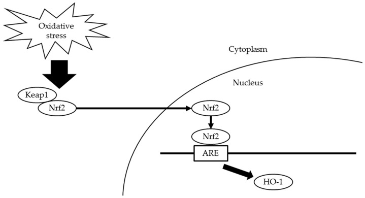
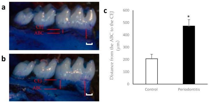
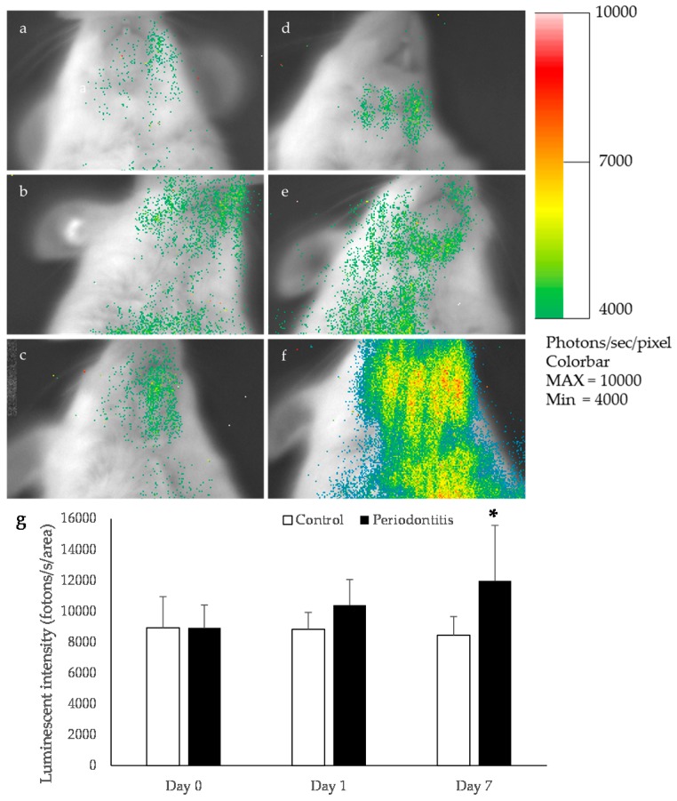
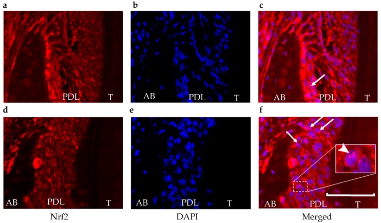
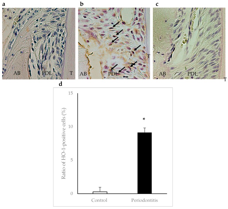
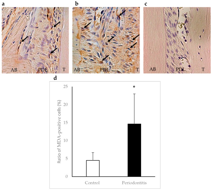
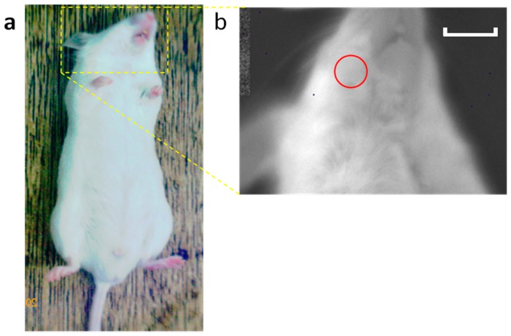
The aim of this study was to investigate whether a Keap1-dependent oxidative stress detector-luciferase (OKD-LUC) mouse model would be useful for the visualization of oxidative stress induced by experimental periodontitis. A ligature was placed around the mandibular first molars for seven days to induce periodontitis. Luciferase activity was measured with an intraperitoneal injection of d-luciferin on days 0, 1, and 7. The luciferase activity in the periodontitis group was significantly greater than that in the control group at seven days. The expressions of heme oxygenase-1 (HO-1) and malondialdehyde in periodontal tissue were significantly higher in the periodontitis group than in the control group. Immunofluorescent analysis confirmed that the nuclear translocation of nuclear factor erythroid 2-related factor 2 (Nrf2) occurred more frequently in the periodontitis group than in the control group. This study found that under oxidative stress induced by experimental periodontitis, the Nrf2/antioxidant defense pathway was activated and could be visualized from the luciferase activity in the OKD-LUC model. Thus, the OKD-LUC mouse model may be useful for exploring the mechanism underlying the relationship between the Nrf2/antioxidant defense pathway and periodontitis by enabling the visualization of oxidative stress over time.

Keywords: Nrf2; heme oxygenase-1; luciferase activity; oxidative stress; periodontitis.


Conflict of interest statement

The authors declare no conflict of interest.

Figures

Figure 1
Figure 1
Scheme for Keap1-Nrf2 defense pathway. Under oxidative stress, the Keap1-Nrf2 complex dissociates and Nrf2 translocates into nuclei to bind AREs. Nrf2 activates HO-1 to attenuate cellular oxidative stress. Keap1, Kelch-like ECH-associated protein 1; Nrf2, nuclear factor erythroid 2-related factor 2; AREs, antioxidant response elements; HO-1, heme oxygenase-1.
Figure 2
Figure 2
Alveolar bone loss at day 7. Representative photographs of mandibular bone in the control (a) and periodontitis (b) groups are shown. Red lines indicate the distance from the alveolar bone crest (ABC) to the cemento-enamel junction (CEJ). The distance was significantly higher in the periodontitis group than in the control group at day 7 (c) (* p < 0.05, t-test) (n = 6/group). White scale bar = 200 µm.
Figure 3
Figure 3
Bioluminescence imaging in oxidative stress detector-luciferase (OKD-LUC) mice. The color scale bar shows the photon counts. Representative photographs taken at baseline (a); the control group at day 1 (b) and day 7 (c); the periodontitis group just after ligation (d); and the periodontitis group at day 1 (e) and day 7 (f). In the periodontitis group, a high luminescence intensity was observed in the mandibular area at day 7 (d). This luminescence intensity was significantly higher in the periodontitis group than in the control group (* p < 0.05, t-test) (n = 6/group) (g).
Figure 4
Figure 4
Translocation of Nrf2. For the Nrf2 nuclear translocation assay, the nuclear level of Nrf2 was determined using immunofluorescence staining (red) (a,d). The cell nuclei were visualized by DAPI staining (blue) (b,e). In the control group, Nrf2 expression was observed (a), but there was less translocation (c) than in the periodontitis group (f). Arrows show cells that are positive for both Nrf2 and DAPI in the nucleus. The arrowhead in white box shows a magnification of an area of cells with positive staining for both Nrf2 and DAPI in the nucleus. These double-positive cells mainly existed adjacent to the alveolar bone surface within the periodontal ligament. 4′,6-diamidino-2-phenylindole, DAPI; AB, alveolar bone; PDL, periodontal ligament; T, tooth. Scale bar = 50 μm.
Figure 5
Figure 5
Immunohistochemical staining of heme oxygenase-1 (HO-1) in periodontal tissue at day 7. Arrows show HO-1-positive cells. A higher number of HO-1-positive cells was observed in areas adjacent to the alveolar bone surface within the periodontal ligament in the periodontitis group (b) than in the control group (a) (×400). Panel (c) shows a negative control. The ratio of HO-1-positive cells was significantly higher in the periodontitis group than in the control group (* p < 0.05, t-test) (n = 6/group) (d). AB, alveolar bone; PDL, periodontal ligament; T, tooth.
Figure 6
Figure 6
Immunohistochemical staining of malondialdehyde (MDA) in periodontal tissue at day 7. Arrows show MDA-positive cells. A higher number of MDA-positive cells was observed in areas adjacent to the alveolar bone surface within the periodontal ligament in the periodontitis group (b) than in the control group (a) (×400). Panel (c) shows a negative control. The ratio of MDA-positive cells was significantly higher in the periodontitisgroup than in the control group (* p < 0.01, t-test) (n = 6/group) (d). AB, alveolar bone; PDL, periodontal ligament; T, tooth.
Figure 7
Figure 7
Scheme of bioluminescence imaging. Imaging of the head area was performed (a). The red circle shows the region of interest (ROI) (b). Luciferase activity was quantified from images displaying surface radiance using circular ROIs. Scale bar = 1 cm.

References

    1. Pihlstrom B., Michalowicz B., Johnson N. Periodontal diseases. Lancet. 2005;366:1809–1820. doi: 10.1016/S0140-6736(05)67728-8. - DOI - PubMed
    1. Chapple I.L., Matthews J.B. The role of reactive oxygen and antioxidant species in periodontal tissue destruction. Periodontol 2000. 2007;43:160–232. doi: 10.1111/j.1600-0757.2006.00178.x. - DOI - PubMed
    1. Circu M.L., Aw T.Y. Reactive oxygen species, cellular redox systems, and apoptosis. Free Radic. Biol. Med. 2010;48:749–762. doi: 10.1016/j.freeradbiomed.2009.12.022. - DOI - PMC - PubMed
    1. Tomofuji T., Azuma T., Kusano H., Sanbe T., Ekuni D., Tamaki N., Yamamoto T., Watanabe T. Oxidative damage of periodontal tissue in the rat periodontitis model: Effects of a high-cholesterol diet. FEBS Lett. 2006;580:3601–3604. doi: 10.1016/j.febslet.2006.05.041. - DOI - PubMed
    1. Ekuni D., Tomofuji T., Tamaki N., Sanbe T., Azuma T., Yamanaka R., Yamamoto T., Watanabe T. Mechanical stimulation of gingiva reduces plasma 8-OHdG level in rat periodontitis. Arch. Oral Biol. 2008;53:324–329. doi: 10.1016/j.archoralbio.2007.10.005. - DOI - PubMed

LinkOut - more resources