. 2016 Nov 16;8(11):729.
doi: 10.3390/nu8110729.

Parenchymal and Stromal Cells Contribute to Pro-Inflammatory Myocardial Environment at Early Stages of Diabetes: Protective Role of Resveratrol

Affiliations

Parenchymal and Stromal Cells Contribute to Pro-Inflammatory Myocardial Environment at Early Stages of Diabetes: Protective Role of Resveratrol

Monia Savi et al. Nutrients. .

Abstract

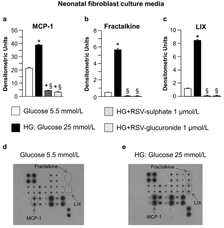
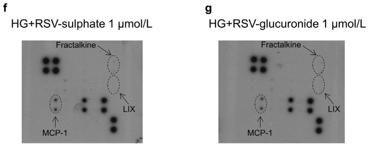
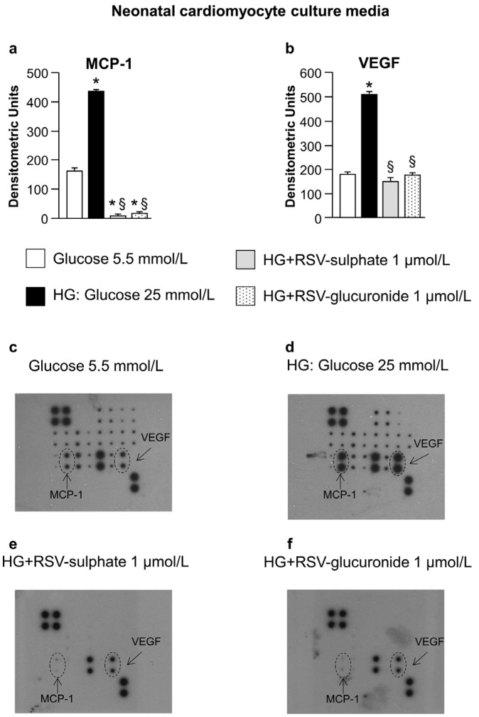
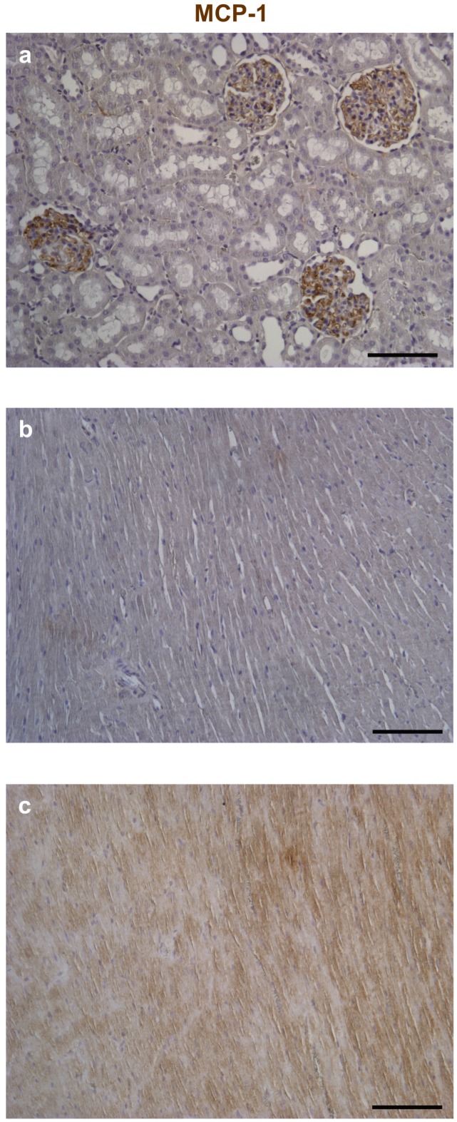
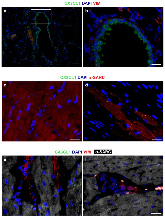
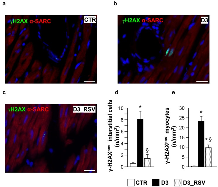
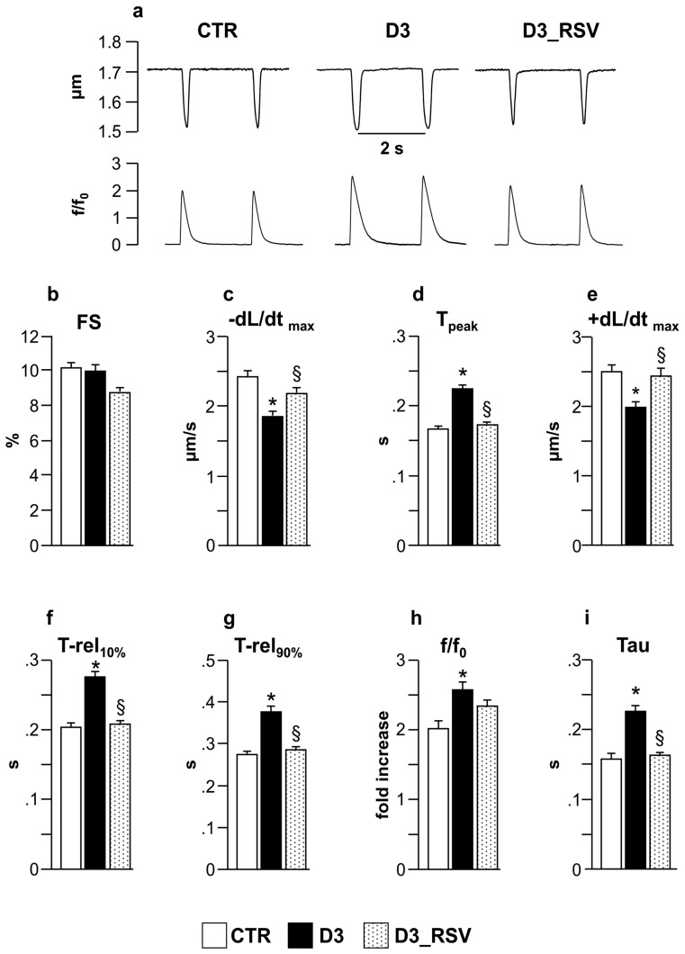
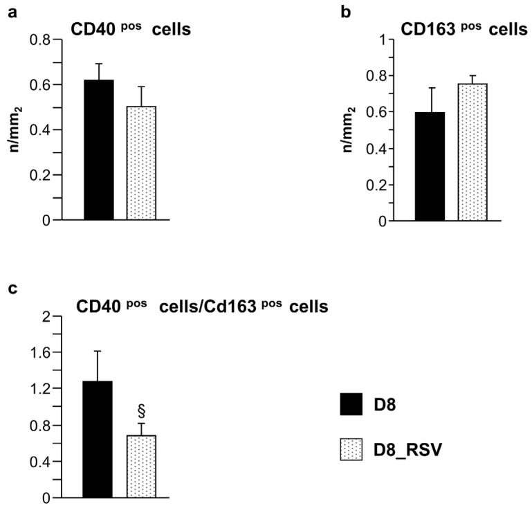
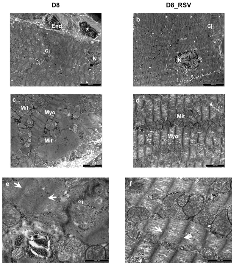
Background: Little information is currently available concerning the relative contribution of cardiac parenchymal and stromal cells in the activation of the pro-inflammatory signal cascade, at the initial stages of diabetes. Similarly, the effects of early resveratrol (RSV) treatment on the negative impact of diabetes on the different myocardial cell compartments remain to be defined. Methods: In vitro challenge of neonatal cardiomyocytes and fibroblasts to high glucose and in vivo/ex vivo experiments on a rat model of Streptozotocin-induced diabetes were used to specifically address these issues. Results: In vitro data indicated that, besides cardiomyocytes, neonatal fibroblasts contribute to generating initial changes in the myocardial environment, in terms of pro-inflammatory cytokine expression. These findings were mostly confirmed at the myocardial tissue level in diabetic rats, after three weeks of hyperglycemia. Specifically, monocyte chemoattractant protein-1 and Fractalkine were up-regulated and initial abnormalities in cardiomyocyte contractility occurred. At later stages of diabetes, a selective enhancement of pro-inflammatory macrophage M1 phenotype and a parallel reduction of anti-inflammatory macrophage M2 phenotype were associated with a marked disorganization of cardiomyocyte ultrastructural properties. RSV treatment inhibited pro-inflammatory cytokine production, leading to a recovery of cardiomyocyte contractile efficiency and a reduced inflammatory cell recruitment. Conclusion: Early RSV administration could inhibit the pro-inflammatory diabetic milieu sustained by different cardiac cell types.

Keywords: cardiac cell compartments; cardiomyocyte mechanics; cytokines; diabetes; intracellular calcium dynamics; polyphenols.


Conflict of interest statement

The authors declare no conflict of interest.

Figures

Figure 1
Figure 1
Analysis of neonatal fibroblast culture media. Average values ± standard error of the mean SEM of pro-inflammatory cytokines expressed by neonatal cardiac fibroblasts (ac) cultured in normoglucidic (Glucose 5.5 mmol/L), hyperglucidic (HG, High Glucose: 25 mmol/L) or hyperglucidic + RSV metabolites medium. * p < 0.05 vs. normoglucidic conditions. § p < 0.05 vs. HG conditions (one-way ANOVA, Holm-Šídák post test); Panels (dg) typical images obtained with RayBio Rat Cytokine Antibody Array 1. The membranes were probed with conditioned media from neonatal cardiac fibroblasts cultured in different conditions. Membranes were exposed to Kodak X-Omat AR film. MCP-1: Monocyte chemotactic protein-1; LIX: Lipopolysaccharide-inducible CXC chemokine.
Figure 1
Figure 1
Analysis of neonatal fibroblast culture media. Average values ± standard error of the mean SEM of pro-inflammatory cytokines expressed by neonatal cardiac fibroblasts (ac) cultured in normoglucidic (Glucose 5.5 mmol/L), hyperglucidic (HG, High Glucose: 25 mmol/L) or hyperglucidic + RSV metabolites medium. * p < 0.05 vs. normoglucidic conditions. § p < 0.05 vs. HG conditions (one-way ANOVA, Holm-Šídák post test); Panels (dg) typical images obtained with RayBio Rat Cytokine Antibody Array 1. The membranes were probed with conditioned media from neonatal cardiac fibroblasts cultured in different conditions. Membranes were exposed to Kodak X-Omat AR film. MCP-1: Monocyte chemotactic protein-1; LIX: Lipopolysaccharide-inducible CXC chemokine.
Figure 2
Figure 2
Analysis of neonatal cardiomyocyte culture media. Average values ± SEM of pro-inflammatory cytokines expressed by neonatal cardiomyocytes (a,b) cultured in normoglucidic (Glucose 5.5 mmol/L), hyperglucidic (HG, High Glucose: 25 mmol/L) or hyperglucidic + RSV metabolites medium. * p < 0.05 vs. normoglucidic conditions. § p < 0.05 vs. HG conditions (one-way ANOVA, Holm-Šídák post test); Panels (cf) typical images obtained with RayBio Rat Cytokine Antibody Array 1. VEGF: Vascular endothelial growth factor. Other explanations as in Figure 1.
Figure 3
Figure 3
RSV-treatment reduced diabetes-induced tissue inflammation. Mean values ± SEM of MCP-1 (a), Fractalkine (b) and VEGF (c) expression levels in LV myocardium of control (CTR), diabetic (D3) and RSV-treated diabetic rats (D3_RSV), after three weeks of hyperglycemia (Western blot assay). Quantitative comparisons were performed on data derived from different gels processed in parallel (see Methods). Sets of bands related to cytokine expression of three animals representative of the average behavior observed in each group are reported at the bottom of the corresponding graph. Lanes were not adjacent in the gel, as indicated by the white space between them. The visualization of all proteins in one membrane stained with Ponceau Red is reported in (d), together with the molecular weights. * p < 0.05 vs. CTR, § p < 0.05 significant differences between D3 and D3_RSV (Kruskal-Wallis analysis of variance and Mann Whitney U-test).
Figure 4
Figure 4
Immunohistochemical analysis of Monocyte Chemoattractant Protein-1 (MCP-1). Reference tissues for the detection of MCP-1 by immunoperoxidase (brownish) were represented by the rat kidney (a); Compared to control (b); Higher expression of the chemokine is apparent in both cardiomyocytes and interstitial cells of the diabetic left ventricular (LV) myocardium (c). Nuclei were counterstained by Hematoxylin. Scale bars: 100 µm.
Figure 5
Figure 5
Immunofluorescence analysis of CX3CL1 (Fractalkine). The granular and diffuse cytoplasmic expression of CX3CL1 (green) is apparent in bronchiolar epithelial cells of the rat lung, used as a positive control tissue; (a,b) The white square includes an area shown at higher magnification in b where interstitial cells are depicted by the red fluorescence of vimentin (VIM); (c) Internal negative control. Absence of immunofluorescence signals in a section of the rat diabetic heart exposed to FITC secondary antibodies in the absence of primary anti-CX3CL1 antibodies. Cardiomyocytes are recognized by the red fluorescence of alpha-sarcomeric actin (α-SARC); (d) A rather dense granular, dot-like pattern of CX3CL1 (green) expression in α-SARC (red) positive cardiomyocytes is illustrated in a section of the rat infarcted myocardium; (e,f) A granular, dot-like expression of Fractalkine (green) is apparent in VIM (red) positive fibroblasts and interstitial cells surrounding α-SARC (withe) positive cardiomyocytes. The pro-inflammatory cytokine is also detectable in cardiomyocytes. (af): Blue fluorescence corresponds to DAPI staining of nuclei. Scale Bars: (a) = 50 µm, (bf) = 20 µm.
Figure 6
Figure 6
RSV treatment reduced diabetes-induced DNA damage. Panels a–c: detection of DNA double strand breaks in sections of the LV myocardium from control (a), untreated (b) and RSV-treated (c) diabetic rats, by immunofluorescence. Nuclear labeling by antibodies against gamma-Histone2 AX (green, γH2AX) is documented in cardiomyocytes recognized by the red fluorescence of α-sarcomeric actin (red). Two cardiomyocytes show diffuse nuclear fluorescence in the D3 heart while dot-like signals are present in a cardiomyocyte from RSV-treated myocardium. Blue fluorescence corresponds to DAPI staining of nuclei. Scale bars: 20 µm; In (de), bar graphs illustrating the density of γH2AX positive interstitial cells (d) and cardiomyocytes (e), in control (CTR), untreated (D3) and RSV-treated (D3_RSV) diabetic myocardium. Data are reported as mean ± SEM * p < 0.05 vs. CTR; § p < 0.05 significant differences between D3 and D3_RSV (one-way ANOVA, Games-Howell post-hoc test).
Figure 7
Figure 7
RSV treatment significantly restored cell mechanics and Ca2+ handling. (a) Representative examples of sarcomere shortening and corresponding Ca2+ transients (normalized traces: fold increase) recorded from CTR, D3 and D3_RSV ventricular myocytes, after three weeks of hyperglycemia; In bar graphs (bi): mean values ± SEM of cardiomyocyte mechanical properties. FS: fraction of shortening (b); −dL/dtmax: maximal rate of shortening (c); Tpeak: duration of cell contraction (d); +dL/dtmax: maximal rate of relengthening (e); T-rel10%: time to 10% of relengthening (f); T-rel90%: time to 90% of relengthening (g); f/f0: Ca2+ transient amplitude expressed as peak fluorescence normalized to baseline fluorescence (h); and Tau: time constant of the intracellular Ca2+ decay (i). * p < 0.05: significant differences vs. CTR; § p < 0.05: significant differences between D3 and D3_RSV (one-way ANOVA, Games-Howell post-hoc test).
Figure 8
Figure 8
Positive effects of RSV treatment on macrophage recruitment. Mean values ± SEM of CD40pos and CD163pos cell density (a,b), and CD40pos/CD163pos cell ratio (c), in the LV myocardium of D8 and D8_RSV rats (immunohistochemistry). § p < 0.05, significant differences between D8 and D8_RSV groups (Student’s t-test).
Figure 9
Figure 9
Positive effects of RSV treatment on ultrastructural damage of diabetic LV myocardium. Ultra-thin longitudinal sections of the LV myocardium from untreated (D8) and RSV-treated (D8_RSV) diabetic rats. (a,b) Low magnification images illustrating cardiomyocytes connected by gap junctions (Gj). An inflammatory cell (*) showing electron-dense granules near a capillary endothelial cell (End) is also documented in the diabetic rat myocardium (a). N: cardiomyocyte nuclei. (c,d) Disorganization and severe alteration of contractile myofibrils (Myo) are illustrated in the diabetic myocardium (c) whereas a more preserved sarcomere striation is present in RSV-treated heart (d). Mitochondria (Mit) are running parallel to myofibrils in RSV-treated rat myocardium (d) which are swollen and clustering between disarrayed contractile filaments in diabetic rats (c). At higher magnification, a marked ultrastructural effacement of sarcomeres (white arrows) at the junctional interface (Gj) of diabetic cardiomyocytes (e) compared to RSV-treated myocardium (f) is apparent. Scale bars: (a,b): 5 µm; (c,d): 2 µm; (e,f): 1 µm.

Similar articles

Cited by

References

    1. Rubler S., Dlugash J., Yuceoglu Y.Z., Kumral T., Branwood A.W., Grishman A. New type of cardiomyopathy associated with diabetic glomerulosclerosis. Am. J. Cardiol. 1972;30:595–602. doi: 10.1016/0002-9149(72)90595-4. - DOI - PubMed
    1. Bugger H., Abel E.D. Molecular mechanisms of diabetic cardiomyopathy. Diabetologia. 2014;57:660–671. doi: 10.1007/s00125-014-3171-6. - DOI - PMC - PubMed
    1. Becher P.M., Lindner D., Fröhlich M., Savvatis K., Westermann D., Tschöpe C. Assessment of cardiac inflammation and remodeling during the development of streptozotocin-induced diabetic cardiomyopathy in vivo: A time course analysis. Int. J. Mol. Med. 2013;32:158–164. - PubMed
    1. Lorenzo O., Picatoste B., Ares-Carrasco S., Ramirez E., Egido J., Tunon J. Potential role of nuclear factor κB in diabetic cardiomyopathy. Mediat. Inflamm. 2011;2011:652097. doi: 10.1155/2011/652097. - DOI - PMC - PubMed
    1. Wang W.K., Wang B., Lu Q.H., Zhang W., Qin W.D., Liu X.J., Liu X.Q., An F.S., Zhang Y., Zhang M.X. Inhibition of high-mobility group box 1 improves myocardial fibrosis and dysfunction in diabetic cardiomyopathy. Int. J. Cardiol. 2014;172:202–212. doi: 10.1016/j.ijcard.2014.01.011. - DOI - PubMed