. 2017 Jan 24;61(2):e02130-16.
doi: 10.1128/AAC.02130-16. Print 2017 Feb.

Long-Chain Fatty Acyl Coenzyme A Ligase FadD2 Mediates Intrinsic Pyrazinamide Resistance in Mycobacterium tuberculosis

Affiliations

Long-Chain Fatty Acyl Coenzyme A Ligase FadD2 Mediates Intrinsic Pyrazinamide Resistance in Mycobacterium tuberculosis

Brandon C Rosen et al. Antimicrob Agents Chemother. .

Abstract

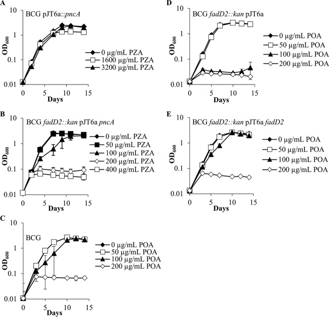
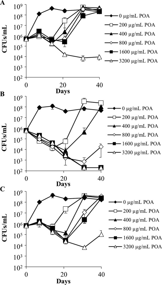
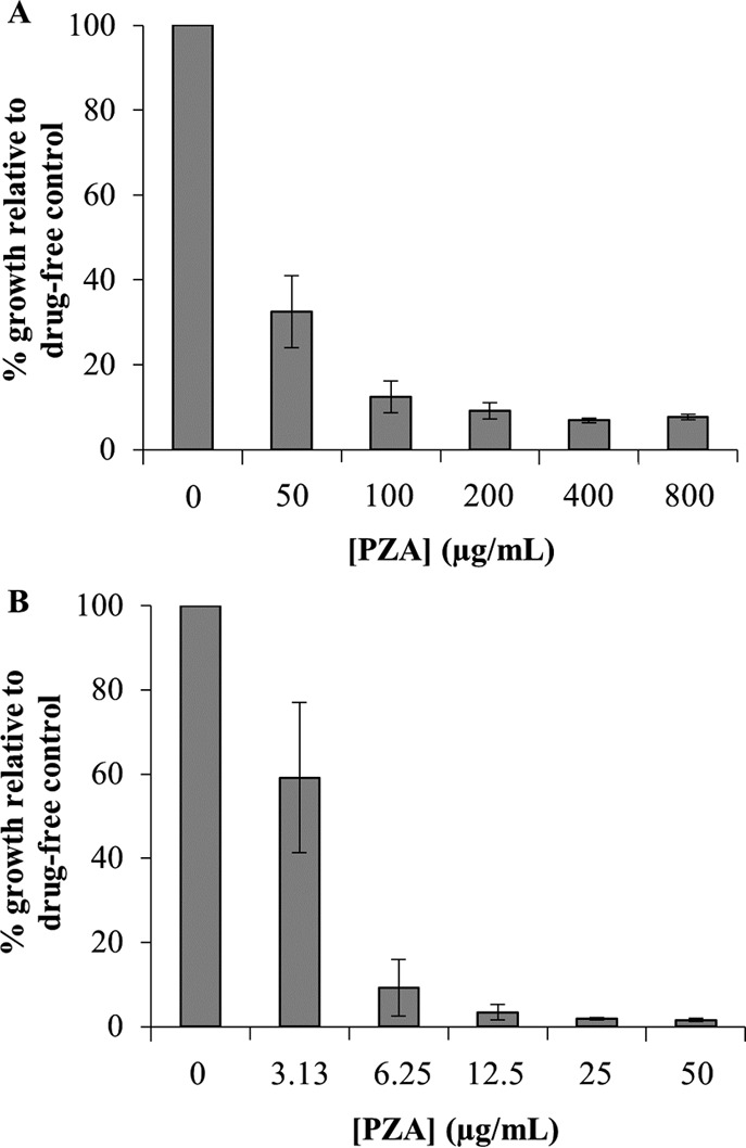
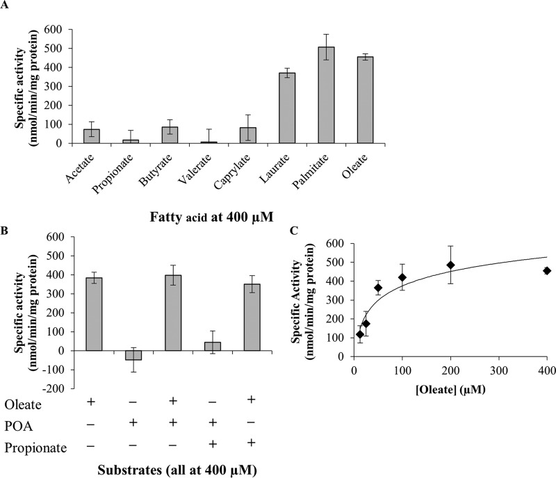
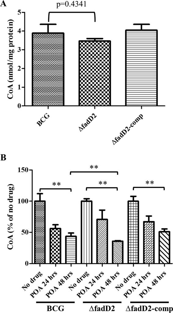
Pyrazinamide (PZA) is a first-line tuberculosis (TB) drug that has been in clinical use for 60 years yet still has an unresolved mechanism of action. Based upon the observation that the minimum concentration of PZA required to inhibit the growth of Mycobacterium tuberculosis is approximately 1,000-fold higher than that of other first-line drugs, we hypothesized that M. tuberculosis expresses factors that mediate intrinsic resistance to PZA. To identify genes associated with intrinsic PZA resistance, a library of transposon-mutagenized Mycobacterium bovis BCG strains was screened for strains showing hypersusceptibility to the active form of PZA, pyrazinoic acid (POA). Disruption of the long-chain fatty acyl coenzyme A (CoA) ligase FadD2 enhanced POA susceptibility by 16-fold on agar medium, and the wild-type level of susceptibility was restored upon expression of fadD2 from an integrating mycobacterial vector. Consistent with the recent observation that POA perturbs mycobacterial CoA metabolism, the fadD2 mutant strain was more vulnerable to POA-mediated CoA depletion than the wild-type strain. Ectopic expression of the M. tuberculosis pyrazinamidase PncA, necessary for conversion of PZA to POA, in the fadD2 transposon insertion mutant conferred at least a 16-fold increase in PZA susceptibility under active growth conditions in liquid culture at neutral pH. Importantly, deletion of fadD2 in M. tuberculosis strain H37Rv also resulted in enhanced susceptibility to POA. These results indicate that FadD2 is associated with intrinsic PZA and POA resistance and provide a proof of concept for the target-based potentiation of PZA activity in M. tuberculosis.

Keywords: Mycobacterium tuberculosis; antimicrobial agents; coenzyme A; fatty acids; metabolism; pyrazinamide.


Figures

FIG 1
FIG 1
Growth kinetics of Mycobacterium bovis BCG strains in the presence of PZA or POA at pH 6.8. M. bovis BCG strains in mid-logarithmic phase were subcultured into 7H9 medium supplemented with OADC (10%), glycerol (0.2%), and tyloxapol (0.05%) at neutral pH (6.8). Strains ectopically expressing the M. tuberculosis PncA in a wild-type background (A) and a fadD2 disruption background (B) were grown in the presence of various PZA concentrations. Wild-type M. bovis BCG (C), the M. bovis BCG fadD2 disruption mutant (D), and the complemented fadD2 disruption mutant (E) were grown in the presence of a range of POA concentrations.
FIG 2
FIG 2
Bactericidal activity of pyrazinoic acid against Mycobacterium bovis BCG strains at pH 6.8. Wild-type M. bovis BCG (A), M. bovis BCG fadD2::kan/pJT6a (B), and M. bovis BCG fadD2::kan/pJT6a::fadD2 (C) cells in mid-logarithmic phase were subcultured into supplemented 7H9 medium at neutral pH (6.8) and treated with a range of POA concentrations. Aliquots were drawn from each culture at the indicated time points, serially diluted, and plated on drug-free supplemented 7H9 agar to permit CFU enumeration. The limit of detection was 200 CFU/ml. Error bars depict the standard deviations of the cell concentrations at a particular time point from three independent experiments.
FIG 3
FIG 3
Pyrazinamide susceptibility of Mycobacterium bovis BCG strains ectopically expressing the Mycobacterium tuberculosis PncA at acidic pH. M. bovis BCG/pJT6a::pncA (A) and M. bovis BCG fadD2::kan/pJT6a::pncA (B) were diluted to an OD600 of 0.01 in 7H9 medium supplemented with OADC (10%, vol/vol), glycerol (0.2%, vol/vol), and tyloxapol (0.05%, vol/vol) at pH 5.8. Cells were aliquoted into 30-ml square bottles, and PZA was added to a range of concentrations. Cultures were incubated by shaking at 37°C for 7 days, after which OD600 values were acquired. The average OD600 values for the drug-free cultures for M. bovis BCG/pJT6a::pncA and M. bovis BCG fadD2::kan/pJT6a::pncA after 7 days of incubation were 1.21 ± 0.10 and 2.18 ± 0.18, respectively. The data depicted represent the averages from three independent experiments; error bars depict standard deviations.
FIG 4
FIG 4
Enzymatic characterization of the M. tuberculosis FadD2. (A) Fatty acid substrate specificity of FadD2; (B) additional assays conducted to assess the allosteric modulation of FadD2 and use of POA as the substrate; (C) kinetic profile of FadD2 with oleate as the substrate. Recombinant histidine-tagged FadD2 was propagated in E. coli and purified by nickel affinity chromatography. Enzymatic assays were conducted using 5,5′-dithiobis-(2-nitrobenzoic acid) (DTNB; or Ellman's reagent) to monitor CoA-SH consumption at 412 nm. Specific activities were computed using the previously determined extinction coefficient for DTNB of 13,600 M−1 cm−1. All specific activity values shown are equivalent to the average reaction velocity over the initial 5-min incubation period. Error bars depict the standard deviations from three assays.
FIG 5
FIG 5
Intracellular CoA levels of Mycobacterium bovis BCG strains. (A) Intracellular CoA levels were measured from wild-type M. bovis BCG (BCG), M. bovis BCG fadD2::hyg (ΔfadD2), and M. bovis BCG fadD2::hyg/pTIC6a::fadD2 (ΔfadD2-comp) that were grown in supplemented 7H9 medium at neutral pH (pH 6.8) until early logarithmic phase (OD600 = 0.2 to 0.3). (B) Intracellular CoA levels were measured 24 h (POA 24 hrs) and 48 h (POA 48 hrs) after 1 mg/ml POA was added to M. bovis BCG strains grown at early logarithmic phase. Intracellular CoA levels were measured as described in Materials and Methods. CoA levels were normalized by the protein concentrations. These results represent the means and standard deviations from three biological replicates. P values of pairwise comparisons (marked by the ends of the brackets) were calculated by using the Student t test. **, P < 0.05.

References

    1. Ormerod LP, Horsfield N. 1987. Short-course antituberculous chemotherapy for pulmonary and pleural disease: 5 years' experience in clinical practice. Br J Dis Chest 81:268–271. doi: 10.1016/0007-0971(87)90160-4. - DOI - PubMed
    1. Konno K, Feldmann FM, McDermott W. 1967. Pyrazinamide susceptibility and amidase activity of tubercle bacilli. Am Rev Respir Dis 95:461–469. - PubMed
    1. Scorpio A, Zhang Y. 1996. Mutations in pncA, a gene encoding pyrazinamidase/nicotinamidase, cause resistance to the antituberculous drug pyrazinamide in tubercle bacillus. Nat Med 2:662–667. doi: 10.1038/nm0696-662. - DOI - PubMed
    1. Scorpio A, Lindholm-Levy P, Heifets L, Gilman R, Siddiqi S, Cynamon M, Zhang Y. 1997. Characterization of pncA mutations in pyrazinamide-resistant Mycobacterium tuberculosis. Antimicrob Agents Chemother 41:540–543. - PMC - PubMed
    1. Vilchèze C, Weinrick B, Wong K-W, Chen B, Jacobs WR Jr. 2010. NAD(+) auxotrophy is bacteriocidal for the tubercle bacilli. Mol Microbiol 76:365–377. doi: 10.1111/j.1365-2958.2010.07099.x. - DOI - PMC - PubMed

Publication types

MeSH terms

LinkOut - more resources