. 2016 Nov 17;11(11):e0166621.
doi: 10.1371/journal.pone.0166621. eCollection 2016.

Characterization of a vraG Mutant in a Genetically Stable Staphylococcus aureus Small-Colony Variant and Preliminary Assessment for Use as a Live-Attenuated Vaccine against Intrammamary Infections

Affiliations

Characterization of a vraG Mutant in a Genetically Stable Staphylococcus aureus Small-Colony Variant and Preliminary Assessment for Use as a Live-Attenuated Vaccine against Intrammamary Infections

Julie Côté-Gravel et al. PLoS One. .

Abstract

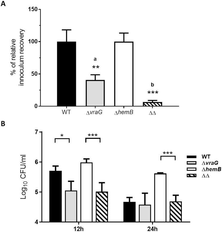
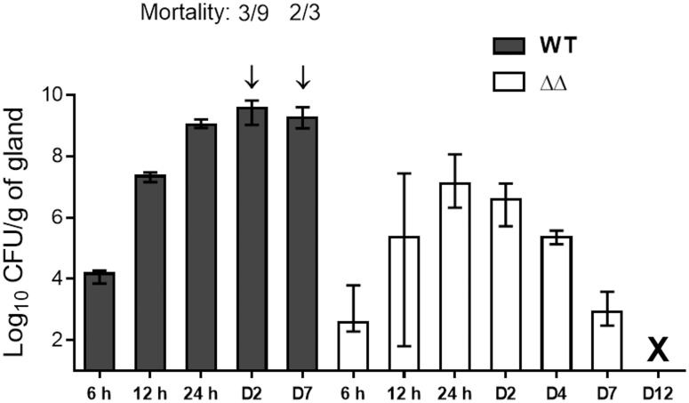
Staphylococcus aureus is a leading cause of bovine intramammary infections (IMIs) that can evolve into difficult-to-treat chronic mastitis. To date, no vaccine formulation has shown high protective efficacy against S. aureus IMI, partly because this bacterium can efficiently evade the immune system. For instance, S. aureus small colony variants (SCVs) have intracellular abilities and can persist without producing invasive infections. As a first step towards the development of a live vaccine, this study describes the elaboration of a novel attenuated mutant of S. aureus taking advantage of the SCV phenotype. A genetically stable SCV was created through the deletion of the hemB gene, impairing its ability to adapt and revert to the invasive phenotype. Further attenuation was obtained through inactivation of gene vraG (SACOL0720) which we previously showed to be important for full virulence during bovine IMIs. After infection of bovine mammary epithelial cells (MAC-T), the double mutant (ΔvraGΔhemB) was less internalized and caused less cell destruction than that seen with ΔhemB and ΔvraG, respectively. In a murine IMI model, the ΔvraGΔhemB mutant was strongly attenuated, with a reduction of viable counts of up to 5-log10 CFU/g of mammary gland when compared to the parental strain. A complete clearance of ΔvraGΔhemB from glands was observed whereas mortality rapidly (48h) occurred with the wild-type strain. Immunization of mice using subcutaneous injections of live ΔvraGΔhemB raised a strong immune response as judged by the high total IgG titers measured against bacterial cell extracts and by the high IgG2a/IgG1 ratio observed against the IsdH protein. Also, ΔvraGΔhemB had sufficient common features with bovine mastitis strains so that the antibody response also strongly recognized strains from a variety of mastitis associated spa types. This double mutant could serve as a live-attenuated component in vaccines to improve cell-mediated immune responses against S. aureus IMIs.


Conflict of interest statement

The authors have declared that no competing interests exist.

Figures

Fig 1
Fig 1. Influence of S. aureus ΔhemB, ΔvraG, and ΔvraGΔhemB mutations on MAC-T cell infectivity.
MAC-T cells were infected with each of the four strains for 3h, then were incubated with lysostaphin an additional 30 min (t = 3h), 12h or 24h and lysed for measurement of intracellular bacteria (CFUs). (A) Relative recovery of the initial inoculum found within cells at 3h for the ΔvraG and ΔvraGΔhemB (ΔΔ) mutants. Results are normalized according to that obtained with ATCC 29213 (WT) for comparison to ΔvraG, or with ΔhemB for comparison to ΔvraGΔhemB (ΔΔ), and are expressed as means with SD (**, P ≤ 0.01; ***, P ≤ 0.001; unpaired t test). (B) Means and SD of intracellular CFUs for WT and mutants at 12h (left) and 24h (right). A two-way ANOVA and Tukey's multiple comparisons test was used (*: P ≤ 0.05; ***: P ≤ 0.001). All values indicate the mean of three independent experiments, each performed in triplicate.
Fig 2
Fig 2. Persistence of S. aureus ATCC 29213 (WT) and isogenic mutants within MAC-T cells over time.
MAC-T cells were infected with each of the four strains for 3h, then were incubated with lysostaphin an additional 30 min, 12h or 24h and lysed for measurement of intracellular bacteria (CFU). Intracellular bacterial CFUs are expressed as the percentage of the initial inoculum after being transformed in base 10 logarithmic values (Log10 CFU/ml). The full lines relate to strains of the normal phenotypes (WT and ΔvraG), whereas the dotted lines represent the strains having the SCV phenotype (ΔhemB and ΔvraGΔhemB [ΔΔ]). Values indicate the mean of three independent experiments, each done in triplicate, with standard deviations.
Fig 3
Fig 3. Viability of MAC-T cells infected by S. aureus ATCC 29213 (WT) and isogenic mutants.
MAC-T cells were infected with each of the four strains (WT, ΔvraG, ΔhemB and ΔvraG,ΔhemB [ΔΔ]) for 3h, then were incubated with lysostaphin for 12 h (A) or 24 h (B). MTT viability assays were then performed as described in materials and methods. The results are reported as percent viability relative to uninfected cells and are expressed as the mean with SD of three independent experiments done in triplicate. Statistical significance with the “Φ” symbols are compared to the WT, and the “*” symbols compare the indicated strains (Two-way ANOVA and Tukey's multiple comparisons test: * or Φ, P ≤ 0.05; **, P≤ 0.01; ***, P ≤ 0.001; ΦΦΦΦ, P ≤ 0.0001).
Fig 4
Fig 4. Murine IMIs with the parental (WT) and ΔvraGΔhemB (ΔΔ) strains.
Mice were infected as previously described and glands were harvested at the indicated hour (h) or day (D) after infection. Each column represents the median value of bacterial CFU counts for a group of glands, and ranges are indicated by bars. A minimum of six glands per group were used except for the WT strain at D7 (2 glands: only one mouse survived). Mortality of mice at specific time points is indicated by arrows. The X indicates the clearance of ΔvraGΔhemB from glands (below the detection limit of 10 CFU/gland).
Fig 5
Fig 5. Double mutant ΔvraGΔhemB (ΔΔ) stimulates neutrophil influx in mammary glands at levels comparable to S. aureus ATCC 29213 (WT) in the first 24 h of infection.
Mice were infected as described in materials and methods, and a non-infected control group of mice received a sterile PBS injection (PBS). Glands were harvested at the indicated times, homogenized and kinetically assayed for MPO activity as described in materials and methods. Each dot represents MPO Units for one gland and is shown as a raw value adjusted by gram of gland. Means are represented by thick lines.
Fig 6
Fig 6. Visual inflammation of the large R4 and L4 mouse mammary glands 24 h after IMI with S. aureus ATCC 29213 (WT) and the double mutant ΔvraGΔhemB (ΔΔ).
Mice were infected as described in materials and methods, and the non-infected control group of mice received a sterile PBS injection (PBS). Pictures show the pairs of glands (R4, left, and L4, right) for each mouse in each group (PBS, n = 2 mice; WT, n = 3 mice; ΔΔ, n = 3 mice) that were harvested after 24 h.
Fig 7
Fig 7. Neutrophil infiltration goes back to normal levels after clearance of the double mutant ΔvraGΔhemB (ΔΔ) from the mammary glands.
Mice were infected as described in materials and methods, and a non-infected control group of mice received a sterile PBS injection (PBS). Glands were harvested at the indicated times, homogenized and kinetically assayed for MPO activity as described in materials and methods. Columns represent means of MPO Units of a group of 6 glands (4 for the PBS control) adjusted by gram of gland, and error bars illustrate standard deviation. Statistical significance between the Day 4 and 12 groups post infection is shown by the “Φ” symbols. One-Way ANOVA and Tukey’s multiple comparison tests were used (ΦΦ, P≤ 0.01; NS, no significant difference between groups).
Fig 8
Fig 8. Immunization of mice with the live-attenuated double mutant (Δ720ΔhemB) induces a strong humoral response against S. aureus bovine mastitis isolates and against a specific cell-wall associated antigen (IsdH).
Mice were immunized as previously described: serums were collected before priming immunization (preimmune, open circles) and ten days after the boost immunization (immune, blue squares). A. IgG titers rise with increasing immunization doses (106, 107, 108 CFU) of the live-attenuated mutant ΔvraGΔhemB: each dot represents the total IgG titer of one mouse against a ΔvraGΔhemB whole cell extract. Medians are represented by thick lines for immune titers and dashed lines for preimmune titers. Titers were compared to their corresponding preimmune titers (Two-way ANOVA and Tukey’s multiple comparisons test: ****: P ≤ 0.0001). B. Immunization with the live-attenuated mutant ΔvraGΔhemB confers high IgG titers against components that are shared by mastitis strains of commonly found spa types. Each dot represents the total IgG titer of one mouse against the whole cell extract of the indicated strain. Medians are represented by thick lines for immune titers and dashed lines for preimmune titers. All immune titers were compared to their corresponding preimmune titers (Two-way ANOVA and Tukey’s multiple comparisons test: P ≤ 0.0001 for all groups). C. Immunization with the live-attenuated mutant ΔvraGΔhemB confers specific IgG titers against the cell-wall associated protein IsdH. Each dot represents the total IgG titer of one mouse against recombinant IsdH. Compared groups were immunized with the 108 CFU of the live-attenuated ΔvraGΔhemB (ΔΔ) or 25 μg of the purified recombinant IsdH protein (IsdH). D. IgG isotype ratios (IgG2a/IgG1) of mice immunized with the live-attenuated mutant ΔvraGΔhemB (open diamonds) or immunized with the recombinant IsdH (black diamonds), against whole-cell extracts of strain ΔvraGΔhemB (vs ΔΔ) or against the recombinant IsdH protein (vs IsdH). Each diamond represents the IgG2a/IgG1 titer ratio for one mouse. Medians are represented by thick lines (One-way ANOVA and Dunn’s multiple comparison test: *: P ≤ 0.05).

References

    1. Foster TJ. Colonization and infection of the human host by staphylococci: adhesion, survival and immune evasion. Vet Dermatol. 2009;20: 456–70. 10.1111/j.1365-3164.2009.00825.x - DOI - PubMed
    1. Novick RP, Geisinger E. Quorum Sensing in Staphylococci. Annu Rev Genet. 2008;42: 541–564. 10.1146/annurev.genet.42.110807.091640 - DOI - PubMed
    1. Otto M. Staphylococcal Infections: Mechanisms of Biofilm Maturation and Detachment as Critical Determinants of Pathogenicity. Annu Rev Med. 2013;64: 175–88. 10.1146/annurev-med-042711-140023 - DOI - PubMed
    1. Brouillette E, Martinez A, Boyll BJ, Allen NE, Malouin F. Persistence of a Staphylococcus aureus small-colony variant under antibiotic pressure in vivo. FEMS Immunol Med Microbiol. 2004;41: 35–41. 10.1016/j.femsim.2003.12.007 - DOI - PubMed
    1. Chambers HF, Deleo FR. Waves of Resistance: Staphylococcus aureus in the Antibiotic Era. Nat Rev Microbiol. 2009;7: 629–641. - PMC - PubMed

MeSH terms