

Disk displacement, eccentric condylar position, osteoarthrosis - misnomers for variations of normality? Results and interpretations from an MRI study in two age cohorts
- PMID: 27855674
- PMCID: PMC5114831
- DOI: 10.1186/s12903-016-0319-4
Disk displacement, eccentric condylar position, osteoarthrosis - misnomers for variations of normality? Results and interpretations from an MRI study in two age cohorts
Abstract
Background: Clinical decision-making and prognostic statements in individuals with manifest or suspected temporomandibular disorders (TMDs) may involve assessment of (a) the position of articular disc relative to the mandibular condyle, (b) the location of the condyle relative to the temporal joint surfaces, and (c) the depth of the glenoid fossa of the temporomandibular joints (TMJs). The aim of this study was twofold: (1) Determination of the prevalence of these variables in two representative population-based birth cohorts. (2) Reinterpretation of the clinical significance of the findings.
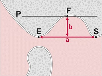
Methods: From existing magnetic resonance imaging (MRI) scans of the TMJs that had been taken in 2005 and 2006 from 72 subjects born between 1930 and 1932 and between 1950 and 1952, respectively, the condylar position at closed jaw was calculated as percentage displacement of the condyle from absolute centricity. By using the criteria introduced by Orsini et al. (Oral Surg Oral Med Oral Pathol Oral Radiol Endod 86:489-97, 1998), a textbook-like disc position at closed jaw was distinguished from an anterior location. TMJ morphology of the temporal joint surfaces was assessed at open jaw by measuring the depth of the glenoid fossa, using the method proposed by Muto et al. (J Oral Maxillofac Surg 52:1269-72, 1994). Frequency distributions were recorded for the condylar and disc positions at closed jaw. Student's t-test with independent samples was used as test of significance to detect differences of condylar positions between the age cohorts (1930 vs. 1950) and the sexes. The significance levels were set at 5%. First, the results from the measurement of the age cohorts were compared without differentiation of sexes, i.e., age cohort 1930-1932 versus age cohort 1950-1952. Subsequently, the age cohorts were compared by sex, i.e., men in cohort 1930-1932 versus men in cohort 1950-1952, and women in cohort 1930-1932 women men in cohort 1950-1952.
Results: In both cohorts, condylar position was characterized by great variability. About 50% of the condyles were located centrically, while the other half was either in an anterior or in a posterior position. In both female cohorts, a posterior position predominated, whereas a centric position prevailed among men. Around 75% of the discs were positioned textbook-like, while the remaining forth was located anteriorly. Age had no statistically significant influence on condylar or on disc position. Conversely, comparison between the age groups revealed a statistically significant decrease of the depth of the glenoid fossa in both older cohorts. This age-dependent changes may be interpreted as flattening of the temporal joint surfaces.
Conclusions: We call for a re-interpretation of imaging findings because they may insinuate pathology which usually is not present. Instead, anterior or posterior positions of the mandibular condyle as well as an anterior location of the articular disc should be construed as a variation of normalcy. Likewise, flattening of articular surfaces of the TMJs may be considered as normal adaptive responses to increased loading, rather than pathological degenerative changes.
Trial registration: Not applicable.
Keywords: Image interpretation; Mandibular condyle; Medical overuse; Medicalization; Osteoarthritis; Osteoarthrosis; Overdiagnosis; Temporomandibular disorders; Temporomandibular joint disc; Terminology.
Figures

References
-
- Howard JA. Temporomandibular joint disorders, facial pain, and dental problems in performing artists. In: Sataloff RT, Brandfonbrener AG, Lederman RJ, editors. Textbook of performing arts medicine. New York: Raven; 1991. pp. 111–69.
-
- Al-Jundi MA, John MT, Setz JM, Szentpétery A, Kuss O. Meta-analysis of treatment need for temporomandibular disorders in adult nonpatients. J Orofacial Pain. 2008;22:97–107. - PubMed
-
- Kim TY, Shin JS, Lee J, Lee YJ, Kim MR, Ahn YJ, Park KB, Hwang DS, Ha IH. Gender difference in associations between chronic temporomandibular disorders and general quality of life in Koreans: a cross-sectional study. PLoS One. 2015;10:e0145002. doi: 10.1371/journal.pone.0145002. - DOI - PMC - PubMed
-
- Kuttila M, Niemi PM, Kuttila S, Alanen P, Le Bell Y. TMD treatment need in relation to age, gender, stress, and diagnostic subgroup. J Orofac Pain. 1998;12:67–74. - PubMed
MeSH terms
LinkOut - more resources
Full Text Sources
Other Literature Sources
Medical
Miscellaneous
