. 2017 Jan 8;482(2):346-351.
doi: 10.1016/j.bbrc.2016.11.066. Epub 2016 Nov 14.

Aspirin increases mitochondrial fatty acid oxidation

Affiliations

Aspirin increases mitochondrial fatty acid oxidation

Radha Uppala et al. Biochem Biophys Res Commun. .

Abstract

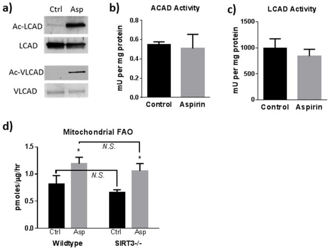
The metabolic effects of salicylates are poorly understood. This study investigated the effects of aspirin on fatty acid oxidation. Aspirin increased mitochondrial long-chain fatty acid oxidation, but inhibited peroxisomal fatty acid oxidation, in two different cell lines. Aspirin increased mitochondrial protein acetylation and was found to be a stronger acetylating agent in vitro than acetyl-CoA. However, aspirin-induced acetylation did not alter the activity of fatty acid oxidation proteins, and knocking out the mitochondrial deacetylase SIRT3 did not affect the induction of long-chain fatty acid oxidation by aspirin. Aspirin did not change oxidation of medium-chain fatty acids, which can freely traverse the mitochondrial membrane. Together, these data indicate that aspirin does not directly alter mitochondrial matrix fatty acid oxidation enzymes, but most likely exerts its effects at the level of long-chain fatty acid transport into mitochondria. The drive on mitochondrial fatty acid oxidation may be a compensatory response to altered mitochondrial morphology and inhibited electron transport chain function, both of which were observed after 24 h incubation of cells with aspirin. These studies provide insight into the pathophysiology of Reye Syndrome, which is known to be triggered by aspirin ingestion in patients with fatty acid oxidation disorders.

Keywords: Aspirin; Fatty acid oxidation; Lysine acetylation; Mitochondria; Peroxisomes; SIRT3.


Figures

Figure 1
Figure 1. Aspirin increases mitochondrial long-chain FAO
(a) HEK293 cells were treated with 5 mM aspirin for either 3 hr or 24 hr and total fatty acid oxidation was measured with 3H-labeled palmitate. (b) 3H-palmitate oxidation was measured in HEK293 cells ± 5 mM aspirin for 24 hrs, with or without etomoxir, an irreversible inhibitor of CPT-1. Etomoxir-insensitive fatty acid oxidation represents the peroxisomal pathway. (c) Aspirin (0 to 5 mM for 24 hr) did not affect protein levels of the acyl-CoA dehydrogenases or their electron acceptor ETF. Note that ETF is a heterodimer with two different-sized subunits. (d,e) The experiment from panel A was repeated with side-by-side comparison to 5 mM salicylic acid (SA) for either 3 hr (d) or 24 hr (e). All palmitate oxidation assays were conducted in quadruplicate wells of cells on a 24-well plate; bars represent means and standard deviations of the four wells. *P < 0.01 versus untreated control cells.
Figure 2
Figure 2. Aspirin chemically acetylates mitochondrial proteins
(a) Anti-acetyllysine western-blotting of HEK293 whole-cell lysates after 24 hr of 5 mM aspirin versus 5mM salicylic acid (SA). (b) Anti-acetyllysine western-blotting of mitochondria isolated from HEK293 cells treated with aspirin from 0 to 48 hr. (c) Anti-acetyllysine western-blotting of recombinant LCAD after incubation with no aspirin (Ctr) or 5 mM aspirin for 1 hr at each indicated pH. (d) Anti-acetyllysine western-blotting of recombinant LCAD after 1 hr incubation with either 5 mM acetyl-CoA or 5 mM aspirin.
Figure 3
Figure 3. Reversible acetylation of matrix proteins does not regulate FAO flux in aspirin-treated cells
(a) Anti-acetyllysine western-blotting of LCAD-Flag and VLCAD-Flag recovered from HEK293 cell lysates by immunoprecipitation ± 24 hr of 5 mM aspirin. (b) Acyl-CoA dehydrogenase activity against palmitoyl-CoA in HEK293 cell lysates ± 24 hr of 5 mM aspirin. This reflects combined activities of LCAD and VLCAD. (c) LCAD activity against palmitoyl-CoA ± 1 hr incubation with 5 mM aspirin. (d) Etomoxir-sensitive (mitochondrial) 3H-palmitate oxidation in wild-type versus SIRT3−/− MEFs ± 24 hr of 5 mM aspirin. All enzyme activity assays and palmitate oxidation assays were conducted on quadruplicate samples of aspirin-treated cells or LCAD protein. Bars represent means and standard deviations. *P < 0.01 of aspirin versus untreated control cells; NS= difference not statistically significant.
Figure 4
Figure 4. Aspirin fragments the mitochondrial network and decreases respiration on pyruvate and succinate
(a–c) Wild-type MEFs treated with vehicle (a), 1 mM aspirin for 24 hr (b), or 5 mM aspirin for 24 hr (c). The mitochondrial network (red) was visualized with anti-ATP synthase antibody and peroxisomes (green) with anti-PMP70 antibody. MEFs were used rather than HEK293 cells because of better adherence. The experiment was repeated with a separate plate of cells yielding similar images.(d,e) Oxygen consumption traces for digitonin-permeabilized wild-type MEFs (d) and HEK293 cells (e) ± 24 hr of 5 mM aspirin. Both cell lines show inhibited malate/pyruvate/succinate-driven mitochondrial respiration. These experiments were repeated three times witch each cell line, yielding similar oxygen traces. (f) Oxidation of 14C-octanoate, a medium-chain fatty acid which can enter mitochondria independently of CPT-1, in HEK293 cells ± 24 hr of 5 mM aspirin treatment. Cells were assayed in suspension with released 14CO2 captured in hanging baskets. Bars represent means and standard deviations of four separate preparations of cells.

References

    1. Smith T, et al. Aspirin in the 21st century-common mechanisms of disease and their modulation by aspirin: a report from the 2015 scientific conference of the international aspirin foundation, 28 August, London, UK. Ecancermedicalscience. 2015;9:581. - PMC - PubMed
    1. Shirakawa K, et al. Salicylate, diflunisal and their metabolites inhibit CBP/p300 and exhibit anticancer activity. Elife. 2016:5. - PMC - PubMed
    1. Mohlig M, et al. Acetylsalicylic acid improves lipid-induced insulin resistance in healthy men. J Clin Endocrinol Metab. 2006;91(3):964–7. - PubMed
    1. Ford RJ, et al. Metformin and salicylate synergistically activate liver AMPK, inhibit lipogenesis and improve insulin sensitivity. Biochem J. 2015;468(1):125–32. - PMC - PubMed
    1. He Z, et al. Aspirin regulates hepatocellular lipid metabolism by activating AMPK signaling pathway. J Toxicol Sci. 2015;40(1):127–36. - PubMed

MeSH terms

LinkOut - more resources