. 2017 Feb;38(2):270-275.
doi: 10.3174/ajnr.A5003. Epub 2016 Nov 17.

Impact of Pial Collaterals on Infarct Growth Rate in Experimental Acute Ischemic Stroke

Affiliations

Impact of Pial Collaterals on Infarct Growth Rate in Experimental Acute Ischemic Stroke

G A Christoforidis et al. AJNR Am J Neuroradiol. 2017 Feb.

Abstract

Background and purpose: Cerebral infarction evolves at different rates depending on available blood flow suggesting that treatment time windows vary depending on the degree of pial collateral recruitment. This work sought to mathematically model infarct growth and determine whether infarct volume growth can be predicted by angiographic assessment of pial collateral recruitment in an experimental MCA occlusion animal model.

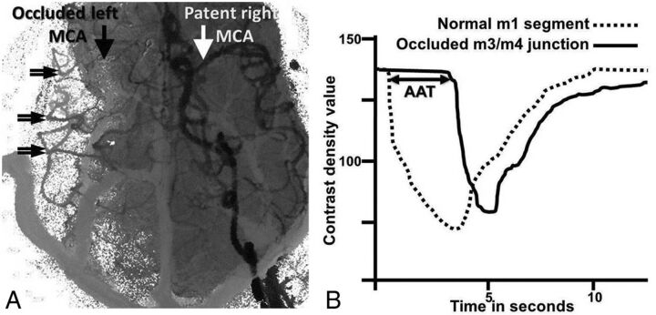
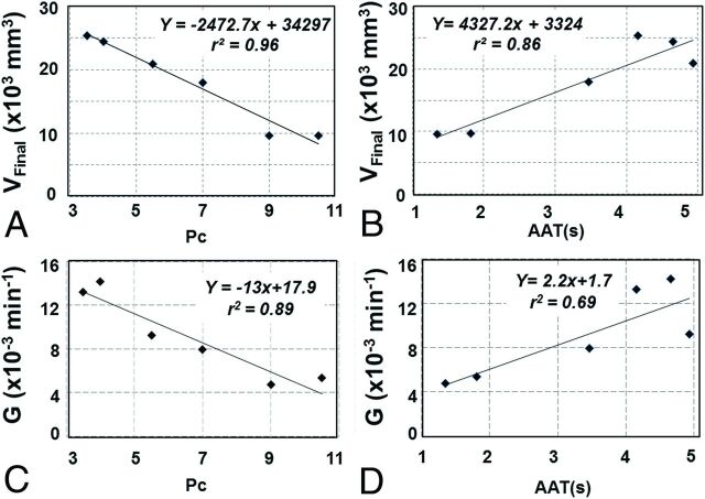
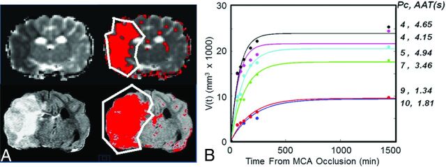
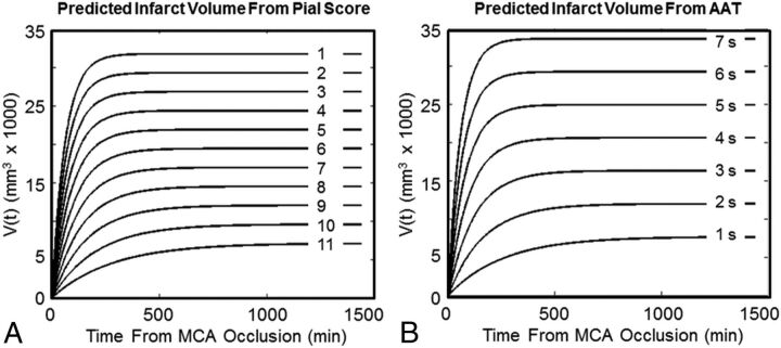
Materials and methods: Pial collateral recruitment was quantified by using DSA, acquired 15 minutes following permanent MCA occlusion in 6 canines based on a scoring system (average pial collateral score) and arterial arrival time. MR imaging-based infarct volumes were measured 60, 90, 120, 180, 240 and 1440 minutes following MCA occlusion and were parameterized in terms of the growth rate index and final infarct volume (VFinal) as V(t) = VFinal [1 - e(-G × t)] (t = time). Correlations of the growth rate index and final infarct volume to the average pial collateral score and arterial arrival time were assessed by linear bivariate analysis. Correlations were used to generate asymptotic models of infarct growth for average pial collateral score or arterial arrival time values. Average pial collateral score- and arterial arrival time-based models were assessed by F tests and residual errors.

Results: Evaluation of pial collateral recruitment at 15 minutes postocclusion was strongly correlated with 24-hour infarct volumes (average pial collateral score: r2 = 0.96, P < .003; arterial arrival time: r2 = 0.86, P < .008). Infarct growth and the growth rate index had strong and moderate linear relationships to the average pial collateral score (r2 = 0.89; P < .0033) and arterial arrival time (r2 = 0.69; P < .0419), respectively. Final infarct volume and the growth rate index were algebraically replaced by angiographically based collateral assessments to model infarct growth. The F test demonstrated no statistical advantage to using the average pial collateral score- over arterial arrival time-based predictive models, despite lower residual errors in the average pial collateral score-based model (P < .03).

Conclusions: In an experimental permanent MCA occlusion model, assessment of pial collaterals correlates with the infarct growth rate index and has the potential to predict asymptotic infarct volume growth.


Figures

Fig 1.
Fig 1.
Arterial arrival times measured from angiographic time-density curves. ROIs within the normal MCA proximal M1 segment (white arrow, A) and from collateralized MCA branches (double arrows, A) are identified on composite angiographic images. ROIs were used to calculate time-density curves (B). “Average arterial time” (in seconds) was defined as the time interval between contrast arrival at the normal M1 segment (interrupted curve, B) and the average of 3 ROIs at the M3/4 junction of the MCA corresponding to the occluded MCA (continuous curve, B). AAT is graphically depicted by the horizontal double arrow line.
Fig 2.
Fig 2.
Final infarct volumes by T2 FLAIR images acquired 24 hours post-MCA occlusion are compared with baseline (ie, 15 minutes postocclusion) angiographic measures of pial collateral recruitment. A, Angiographic scoring of pial collateral score and arterial arrival time (B) strongly correlates with final infarct volume. Both pial collateral score (C) and AAT (D) are predictive the infarct growth rate index (derived from the fits shown in Fig 3B) on the basis of a linear function.
Fig 3.
Fig 3.
Single-section 120-minute mean diffusivity (upper part) and 24-hour FLAIR (lower part) images (A) were used to estimate and plot the growth of the infarct volume with time (B). A semiautomated algorithm was used to estimate the volume of the infarct on the basis of signal intensity within the affected hemisphere (red area), varying 1.5 times the SD from the mean of the signal in the contralateral normal-appearing hemisphere exclusive of spinal fluid. B, Infarct volume growth with time follows a predicable trend. Each curve corresponds to 1 experiment. The pial collateral scores and AAT measured immediately after occlusion (ie, at t = 15 minutes) for each curve are listed on the right.
Fig 4.
Fig 4.
Families of infarct volume growth curves over 24 hours predicted by the pial collateral score and arterial arrival time.

References

    1. Olivot JM, Mlynash M, Thijs VN, et al. . Geography, structure and evolution of diffusion and perfusion lesions in DEFUSE. Stroke 2009;40:3245–51 10.1161/STROKEAHA.109.558635 - DOI - PMC - PubMed
    1. Hacke W, Albers G, Al Rawi Y, et al. ; DIAS Study Group. The Desmoteplase in Acute Ischemic Stroke Trial (DIAS): a phase II MRI-based 9-hour window acute stroke thrombolysis trial with intravenous desmoteplase. Stroke 2005;36:66–73 10.1161/01.STR.0000149938.08731.2c - DOI - PubMed
    1. Schlaug G, Benfield A, Baird AE, et al. . The ischemic penumbra: operationally defined by diffusion and perfusion MRI. Neurology 1999;53:1528–37 10.1212/WNL.53.7.1528 - DOI - PubMed
    1. Fitzek S, Fitzek C, Urban PP, et al. . Time course of lesion development in patients with acute brain stem infarction and correlation with NIHSS score. Eur J Radiol 2001;39:180–85 10.1016/S0720-048X(01)00372-2 - DOI - PubMed
    1. Christoforidis GA, Mohammad Y, Kehagias D, et al. . Angiographic assessment of pial collaterals as a prognostic indicator following intra-arterial thrombolysis for acute ischemic stroke. AJNR Am J Neuroradiol 2005;26:1789–97 - PMC - PubMed

LinkOut - more resources