. 2016 Dec;6(12):1342-1351.
doi: 10.1158/2159-8290.CD-16-0607. Epub 2016 Oct 7.

Genomics of Ovarian Cancer Progression Reveals Diverse Metastatic Trajectories Including Intraepithelial Metastasis to the Fallopian Tube

Affiliations

Genomics of Ovarian Cancer Progression Reveals Diverse Metastatic Trajectories Including Intraepithelial Metastasis to the Fallopian Tube

Mark A Eckert et al. Cancer Discov. 2016 Dec.

Abstract

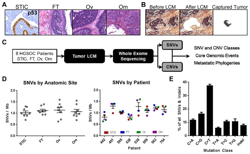
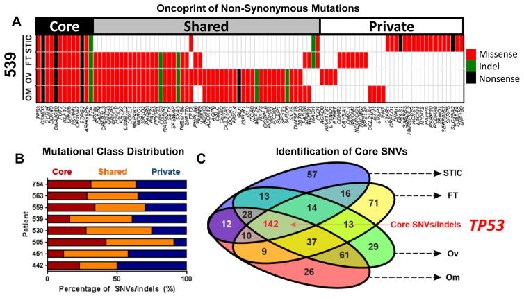
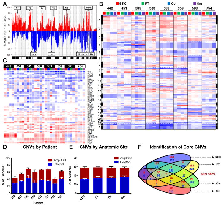
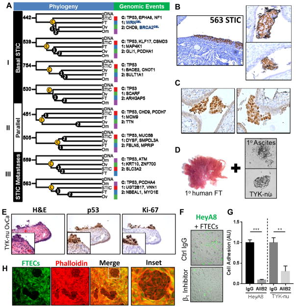
Accumulating evidence has supported the fallopian tube rather than the ovary as the origin for high-grade serous ovarian cancer (HGSOC). To understand the relationship between putative precursor lesions and metastatic tumors, we performed whole-exome sequencing on specimens from eight HGSOC patient progression series consisting of serous tubal intraepithelial carcinomas (STIC), invasive fallopian tube lesions, invasive ovarian lesions, and omental metastases. Integration of copy number and somatic mutations revealed patient-specific patterns with similar mutational signatures and copy-number variation profiles across all anatomic sites, suggesting that genomic instability is an early event in HGSOC. Phylogenetic analyses supported STIC as precursor lesions in half of our patient cohort, but also identified STIC as metastases in 2 patients. Ex vivo assays revealed that HGSOC spheroids can implant in the fallopian tube epithelium and mimic STIC lesions. That STIC may represent metastases calls into question the assumption that STIC are always indicative of primary fallopian tube cancers.

Significance: We find that the putative precursor lesions for HGSOC, STIC, possess most of the genomic aberrations present in advanced cancers. In addition, a proportion of STIC represent intraepithelial metastases to the fallopian tube rather than the origin of HGSOC. Cancer Discov; 6(12); 1342-51. ©2016 AACR.See related commentary by Swisher et al., p. 1309This article is highlighted in the In This Issue feature, p. 1293.


Conflict of interest statement

The authors disclose no potential conflicts of interest.

Figures

Figure 1
Figure 1. High grade serous ovarian cancer (HGSOC) mutational processes are established early and are patient specific
A) Representative p53 IHC and H&E images of the hypothetical progression series of HGSOC in a patient with STIC, and invasive lesions in the fallopian tube, ovary, and abdominal omentum. B) Laser capture microdissection of the tumor compartment from omentum. C) Workflow to elucidate the spatiotemporal pattern of genomic alterations in HGSOC to capture tumor phylogenies and core events. D) Frequency of SNV by anatomic site and patient reveal that mutational burden is patient-specific rather than determined by anatomic tumor location. Average mutational burden across the patient cohort is approximately 1 somatic mutation per megabase (50 mutations total per tumor sample). E) Identification of an age-related mutational signature class characterized by high rates of C>T substitutions in all samples of the patient cohort, reveals a comparable mutational process underlying disease progression in all patients examined. STIC = serous tubal intraepithelial carcinoma; FT = invasive fallopian tube tumor; OV = invasive ovarian tumor; OM = omental metastasis.; SNV = single nucleotide variants.
Figure 2
Figure 2. Core, recurrent SNVs in HGSOC are restricted to TP53 mutations
A) Oncoprint of all non-synonymous mutations in patient 539 reveals mutation of TP53 and distribution of mutations into core, shared, and private classes. B) A minority of SNVs and insertions/deletions (indels) are present in all samples (“core”). The majority of mutations are shared between 2–3 anatomic sites (“shared”), or present in only one site (“private”) C) Euler diagram of all non-synonymous SNVs and indels. Core SNVs/indels present in all anatomic sites are highlighted in red.
Figure 3
Figure 3. Genomic instability is a core feature of ovarian cancer that frequently involves DNA-damage repair genes
A) Frequency plot of copy number variations (CNVs) across all patients and all four anatomic sites. Annotated arm level events were also identified as significant in the TCGA analysis of HGSOC. For all plots in Figure 3, amplifications are red and deletions are blue. B) Genomic aberration plot of CNVs across all anatomic sites and patients imply high degree of genomic instability in all anatomic sites. Chromosome numbers are indicated on margin. Amplifications are red; deletions are blue. C) CNV status of significantly altered DNA repair pathway genes (from REPAIRtoire) reveals common deletion of DNA damage response and repair genes known to be involved in HGSOC. Amplifications are red; deletions are blue. D/E) Extent of genomic instability is patient-specific (D) and does not vary by anatomic site (E). F) GISTIC identification and analysis of significantly altered genes identifies conserved core amplifications and deletions. Number of amplifications in red and deletions in blue.
Figure 4
Figure 4. Phylogenetic analyses of ovarian cancer progression reveal diverse metastatic processes and evidence of intraepithelial metastasis
A) Phylogenetic trees of each patient with key genomic events (SNVs, Indels, CNVs) that characterize each branching event annotated (C (“core”), 1, or 2). Deletions are in blue. B) Representative p53 staining of a “STIC Metastasis” in patient #563 (left), as well as intraluminal HGSOC spheroids within the same fallopian tube (right). C) P53 staining of intraluminal HGSOC spheroids adhering to the epithelium of the fallopian tube (patient 530) D) Human fallopian tube fimbriae and spheroids that were co-cultured. E) Co-culture of human fallopian tube explants with TYK-nu ovarian cancer spheroids mimic STICs with clearance of normal epithelium and implantation of tumor cells expressing Ki-67 and nuclear p53. F/G) Adhesion of fluorescently-labeled HGSOC cells (green) to primary FTEC monolayer (brightfield) after 15 minutes. Pretreatment with β1-integrin blocking antibody (AIIB2) attenuated adhesion of HeyA8 and TYK-nu cells to FTEC monolayer. H) TYK-nu ovarian cancer spheroids (phalloidin only) clear a primary human fallopian tube epithelial monolayer (phalloidin and CellTracker Green) after 12 hours.

Comment in

References

    1. Lengyel E. Ovarian cancer development and metastasis. Am J Pathol. 2010;177(3):1053–64. - PMC - PubMed
    1. Norquist BM, Harrell MI, Brady MF, Walsh T, Lee MK, Gulsuner S, et al. Inherited Mutations in Women With Ovarian Carcinoma. JAMA oncology. 2015:1–9. - PMC - PubMed
    1. Patch AM, Christie EL, Etemadmoghadam D, Garsed DW, George J, Fereday S, et al. Whole-genome characterization of chemoresistant ovarian cancer. Nature. 2015;521(7553):489–94. - PubMed
    1. Piek JM, van Diest PJ, Zweemer RP, Jansen JW, Poort-Keesom RJ, Menko FH, et al. Dysplastic changes in prophylactically removed fallopian tubes of women predisposed to developing ovarian cancer. Journal of Pathology. 2001;195(4):451–6. - PubMed
    1. Kuhn E, Kurman RJ, Vang R, Sehdev AS, Han G, Soslow R, et al. TP53 mutations in serous tubal intraepithelial carcinoma and concurrent pelvic high-grade serous carcinoma-evidence supporting the clonal relationship of the two lesions. Journal of Pathology. 2012;226:421–6. - PMC - PubMed

MeSH terms