. 2017 Jan;37(1):98-107.
doi: 10.1161/ATVBAHA.116.307922. Epub 2016 Nov 17.

RasGRF Couples Nox4-Dependent Endoplasmic Reticulum Signaling to Ras

Affiliations

RasGRF Couples Nox4-Dependent Endoplasmic Reticulum Signaling to Ras

Ru Feng Wu et al. Arterioscler Thromb Vasc Biol. 2017 Jan.

Abstract

Objectives: In response to endoplasmic reticulum (ER) stress, endothelial cells initiate corrective pathways such as the unfolded protein response. Recent studies suggest that reactive oxygen species produced on the ER may participate in homeostatic signaling through Ras in response to ER stress. We sought to identify mechanisms responsible for this focal signaling pathway.

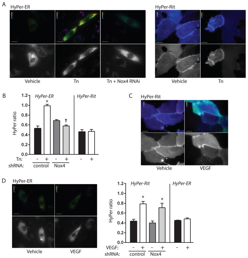
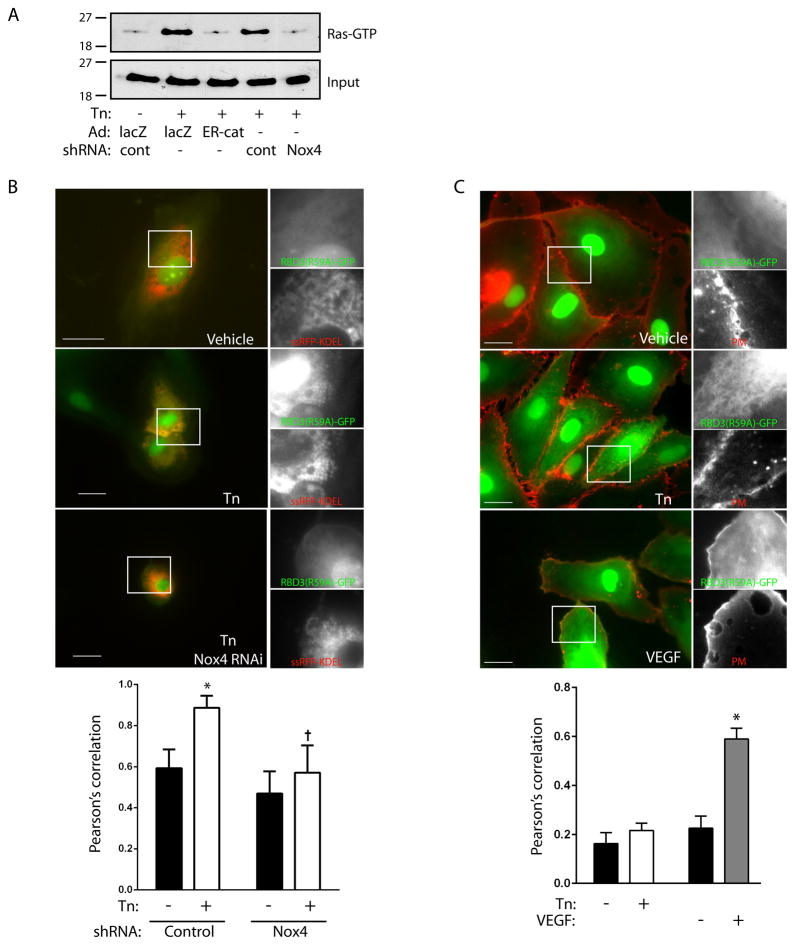
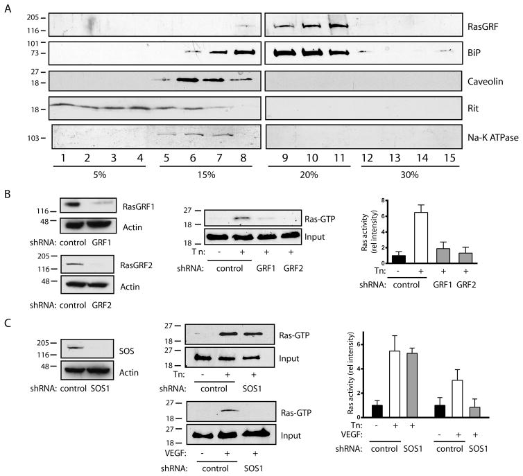
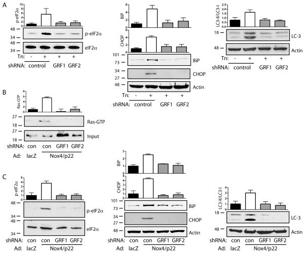
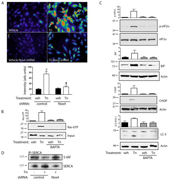
Approach and results: In endothelial cells, we found that ER stress induced by tunicamycin activates the NADPH (nicotinamide adenine dinucleotide phosphate) oxidase Nox4 focally on the ER surface but not on the plasma membrane. Ras activation is also restricted to the ER, occurs downstream of Nox4, and is required for activation of the unfolded protein response. In contrast, treatment with the growth factor VEGF (vascular endothelial growth factor) results in Ras activation and reactive oxygen species production confined instead to the plasma membrane and not to the ER, demonstrating local coupling of reactive oxygen species and Ras signals. We further identify the calcium-responsive, ER-resident guanyl exchange factors RasGRF1 and RasGRF2 as novel upstream mediators linking Nox4 with Ras activation in response to ER stress. Oxidation of the sarcoendoplasmic reticulum calcium ATPase and increases in cytosolic calcium caused by ER stress are blocked by Nox4 knockdown, and reduction in cytosolic free calcium prevents both Ras activation and the unfolded protein response.

Conclusions: ER stress triggers a localized signaling module on the ER surface involving Nox4-dependent calcium mobilization, which directs local Ras activation through ER-associated, calcium-responsive RasGRF.

Keywords: RasGRF1; apoptosis; autophagy; calcium; tunicamycin.


Figures

Figure 1
Figure 1. Focal production of ROS by tunicamycin and VEGF
A. HUVEC expressing control or Nox4 shRNA were transduced with HyPer-ER and exposed to vehicle (DMSO) or tunicamycin (Tn, 10 μg/ml) for 16 h. Pseudocolored ratiometric images for excitation wavelengths 492/405 nm are shown in top panels. Grayscale images at bottom show 405 nm excitation images to document HyPer-ER expression. B. Quantification of HyPer intensity ratios for indicated conditions. *P<0.001 compared to vehicle with control shRNA, † P<0.001 compared to Tn with control shRNA, mean ± SEM of 18–39 determinations. C, D. Ratiometric images of HUVEC expressing HyPer-Rit (C) or HyPer-ER (D) and stimulated with saline or VEGF (50 U/ml) for 3–10 min. Bar graphs in D show quantification of HyPer intensity ratios for indicated conditions. *P<0.01 compared to vehicle with corresponding control or Nox4 shRNA, mean ± SEM of 11–20 determinations. All scale bars are 20 μm.
Figure 2
Figure 2. Ras activation by tunicamycin is restricted to the ER
A. HUVEC expressing control or Nox4-targeted shRNA were transduced with lacZ or ER-targeted catalase (ER-cat) and treated with tunicamycin (10 μg/ml) for 16 h. Ras activity was assessed by pulldown, with input shown in lower panel. Representative of two separate experiments. B. Representative photomicrographs of cells cotransfected with RBD3(R59A)-GFP and the ER marker ssRFP-KDEL, under the conditions as labeled. Inset shows magnification of green and red channels separately. Quantification of colocalization for the red and green channels for whole-cell regions of interest are shown in bar graph below. *P<0.001 compared to vehicle with control shRNA, † P<0.001 compared to Tn with control shRNA, mean ± SEM of 9 fields. C. Cells were transduced with RBD3(R59A)-GFP (green channel), stimulated with Tn (10 μg/ml, 16 h) or VEGF (50 U/ml, 5 min), and plasma membranes stained (PM, red channel). Bar graph shows quantification of red/green colocalization for membrane regions of interest. *P<0.001 compared to vehicle. Mean ± SEM of 15–20 determinations. All scale bars are 20 μm.
Figure 3
Figure 3. K-Ras mediates the UPR
A. Cells expressing control or K-Ras shRNA were stimulated with tunicamycin (Tn, 10 μg/ml for 16 h). Ras and BiP protein expression are shown. K-Ras knockdown was 85% efficient. Bar graph shows BiP levels, mean ± SEM of 3 determinations. B–D. Cells expressing control or K-Ras shRNAs were stimulated with Tn and eIF2α phosphorylation (B), CHOP (C), and LC3-I (upper band, D) to LC3-II (lower band, D) conversion were determined. Bar graphs show mean ± SEM of 3 determinations.
Figure 4
Figure 4. RasGRF is required for tunicamycin-induced Ras activation
A. HUVEC were subjected to discontinuous iodixanol gradient fractionation. RasGRF cosedimented with ER fractions (9–11, marked by BiP), and not plasma membrane fractions (5–7, marked by caveolin, Rit, and Na-K ATPase). B. Effect of shRNAs against RasGRF1 and RasGRF2 on protein expression is shown in left panels. Cells expressing control or RasGRF shRNAs were stimulated with Tn (10 μg/ml for 16 h) and assessed for Ras activity. Representative pulldowns in center panel, graph at right shows Ras activity, mean ± SEM of 3 determinations. Knockdowns were 77% (RasGRF1) and 93% (RasGRF2) efficient. C. Effect of shRNA against SOS1 on protein expression shown in left panel. Cells expressing control or SOS1 shRNA were stimulated with Tn (10 μg/ml for 16 h) or VEGF (50 U/ml, 5 min) and assessed for Ras activity. Representative pulldowns in center panel, graph at right shows Ras activity mean ± SEM of 3 determinations. SOS1 knockdown was 98% efficient.
Figure 5
Figure 5. RasGRF mediates Nox4-dependent ER stress signaling
A. Cells expressing control, RasGRF1, or RasGRF2 shRNAs were stimulated with Tn (10 μg/ml for 16 h) and eIF2α phosphorylation (left), BiP and CHOP expression (center), and LC3-I to LC3-II conversion (right) were determined. Bar graphs are mean ± SEM of 3 determinations. B. Cells expressing control, RasGRF1, or RasGRF2 shRNAs were transduced with lacZ or Nox4/p22phox and Ras activity was assessed by pulldown. Bar graph is mean ± SEM of 3 determinations. C. Cells were treated as in (B) and eIF2α phosphorylation (left), BiP and CHOP expression (center), and LC3-I to LC3II conversion (right) were determined. Bar graphs are mean ± SEM of 3–4 determinations.
Figure 6
Figure 6. Calcium mediates Ras-dependent ER signaling
A. Fluo-3-AM fluorescence intensities were measured following treatment of control or Nox4 shRNA-expressing cells with Tn (10 μg/ml, 16 h). Top panels show representative pseudocolored Fluo-3-AM images, bar graph below shows mean ± SEM of 50 determinations; *P<0.001 compared to vehicle with control shRNA, † P<0.001 compared to Tn with control shRNA. B. Cells were preloaded with BAPTA-AM and treated with Tn. Ras activity was assessed by pulldown. Bar graph is mean ± SEM of 3 determinations. C. Cells were treated as in (B) and eIF2α phosphorylation, BiP and CHOP expression, and LC3-I to LC3-II conversion were determined. Bar graphs are mean ± SEM of 3 determinations. D. Cells expressing control or Nox4 shRNA were treated with Tn and reactive cysteines were alkylated with 5-iodoacetamidofluorescein (5-IAF). Lysates were immunoprecipitated for SERCA and immunoblotted for either SERCA or fluorescein.

References

    1. Avivar-Valderas A, Salas E, Bobrovnikova-Marjon E, Diehl JA, Nagi C, Debnath J, Aguirre-Ghiso JA. PERK integrates autophagy and oxidative stress responses to promote survival during extracellular matrix detachment. Mol Cell Biol. 2011;31:3616–29. - PMC - PubMed
    1. Civelek M, Manduchi E, Riley RJ, Stoeckert CJ, Jr, Davies PF. Chronic endoplasmic reticulum stress activates unfolded protein response in arterial endothelium in regions of susceptibility to atherosclerosis. Circ Res. 2009;105:453–61. - PMC - PubMed
    1. Hotamisligil GS. Endoplasmic reticulum stress and the inflammatory basis of metabolic disease. Cell. 2010;140:900–17. - PMC - PubMed
    1. Walter P, Ron D. The unfolded protein response: from stress pathway to homeostatic regulation. Science. 2011;334:1081–6. - PubMed
    1. Onken B, Wiener H, Philips MR, Chang EC. Compartmentalized signaling of Ras in fission yeast. Proc Natl Acad Sci USA. 2006;103:9045–50. - PMC - PubMed

Publication types

MeSH terms

LinkOut - more resources