

doi: 10.3324/haematol.2016.153197.
Epub 2016 Nov 17.
Extracellular vesicles released from chronic lymphocytic leukemia cells exhibit a disease relevant mRNA signature and transfer mRNA to bystander cells
Affiliations
- PMID: 27856511
- PMCID: PMC5394970
- DOI: 10.3324/haematol.2016.153197
Item in Clipboard
Extracellular vesicles released from chronic lymphocytic leukemia cells exhibit a disease relevant mRNA signature and transfer mRNA to bystander cells
Haematologica.
2017 Mar.
No abstract available
Figures
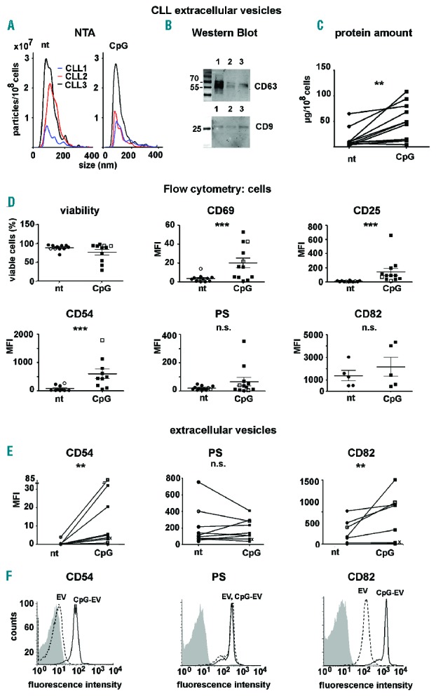
Characterization of extracellular vesicles. (A) Size distribution of CLL-derived vesicles (untreated, left panel, or treated with a TLR9 agonist (CpG, right panel) analyzed by NanoSight Technology (NS300, Malvern). Mean size diameter of vesicles from untreated/treated samples was 106/124 nm for CLL1, 115/99 nm for CLL2 and 97/98 nm for CLL3. All samples were similarly diluted and measured with a sample flow rate of 10 (n=6). (B) Western blot analysis of the exosome-enriched proteins CD63 and CD9 (both antibodies from BioLegend). EV released by 5×10⁹ CLL cells within 38–45 h were resuspended in 40 μL non-reducing lysis buffer; sodium dodecyl sulfate (SDS) gel electrophoresis was performed using 10 μL EV/lane in 12.5% SDS gels and subsequently proteins were transferred to a nitrocellulose membrane. (C) The amount of purified EV protein released by 108 cells was measured with Nanodrop1000 (Thermo Scientific) (n=11). (D) Flow cytometric analysis of CLL cells after 38–45 h culture in Panserin medium with or without 200 nM CpG. Most patients had IgHV mutations (see black circles or squares). Samples without mutations are represented by non-filled circles or non-filled squares. Cell viability was assessed with an apoptosis assay, staining with annexin V-PE and 7-AAD (BioLegend) (PS = phosphatidylserine). Cells were stained with CD69-FITC (BD Biosciences), CD25-FITC (BD Biosciences), purified CD54 and CD82 (both BioLegend), which were detected with goat-anti-mouse-PE (BioLegend). The analysis was performed with a FACS Calibur (BD Biosciences) or FACS Gallios (BeckmanCoulter). Dead cells were excluded by gating for 7AAD (BD Biosciences) negative cells (n=5–14). (E) Flow cytometric analysis of EV: EV were bound to polystyrene beads (Polysciences) and subsequently stained and analyzed as described in (D). Only EV derived from cell samples with a viability of more than 90% were used for further analysis (n=5–9). The IgHV status of one patient analyzed for CD54 expression (left panel) is unknown (labeled with an x). (F) One representative histogram for FACS data depicted in (E).
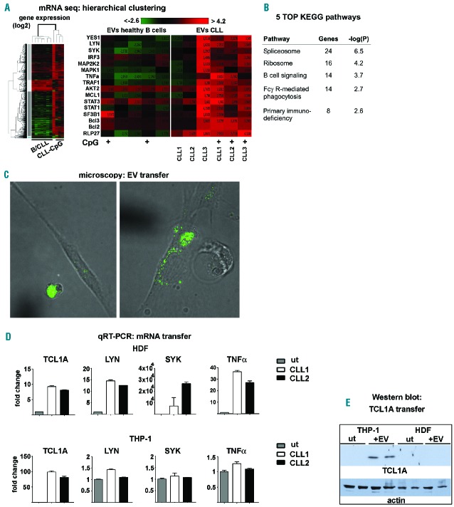
mRNA signature of CLL-derived vesicles. (A) Hierarchical clustering of mRNAseq data (left: all; right: selected for CLL-relevant genes) obtained using next-generation sequencing of RNA isolated from vesicles of normal B cells (obtained from 3 healthy donors) and CLL cells (obtained from 3 patients) cultured in Panserin medium with or without 200 nM CpG (maximal cell density of 2.5×10⁷ cells/mL). Vesicles were purified after 38–45 h from supernatant by serial ultracentrifugation to extract RNA using RNAeasy Mini Kit (Qiagen). B: B-cell derived vesicles, CLL: CLL-derived vesicles, CpG: stimulated with CpG. (B) KEGG pathway analysis of sequencing data to identify top canonical pathways was performed with DAVID 6.7. The functional annotation clustering was performed with the highest classification stringency. The KEGG-pathway function included in DAVID 6.7 was used to interpret the data in the context of biological processes, pathways and networks. Both up- and down-regulated identifiers were defined as value parameters for the analysis. (C–E) Uptake of CLL-vesicles results in TCL1A, LYN and SYK expression in fibroblasts and macrophages. (C) CLL cells were labeled with 5 mM DiO for 10 min and incubated together with HDFn fibroblasts for 16 h. Live cell imaging was performed using a Nikon spinning disc. (D–E) HDFn fibroblasts or THP-1 cells (monocytic cell line) were incubated with purified CLL-EV from two different patients (43 and 64) for 48 h to exclude any contribution or interference with CLL debris. (D) Expression of TCL1A, SYK and LYN in untreated HDFn fibroblasts and THP-1 and in cells treated with CLL-vesicles was measured using quantitative real-time polymerase chain reaction (see also Online Supplementary Table S4). (E) Western blot of cell lysates from THP-1 and HDFn cells untreated (ut) or cultured with CLL-EV (n=2, two independent patients) detecting TCL1A protein.
References
Publication types
MeSH terms
Substances
LinkOut - more resources
Full Text Sources
Other Literature Sources
Miscellaneous
