Review
. 2017 Jan;30(1):233-276.
doi: 10.1128/CMR.00032-16.

Chromoblastomycosis

Affiliations
Review

Chromoblastomycosis

Flavio Queiroz-Telles et al. Clin Microbiol Rev. 2017 Jan.

Abstract

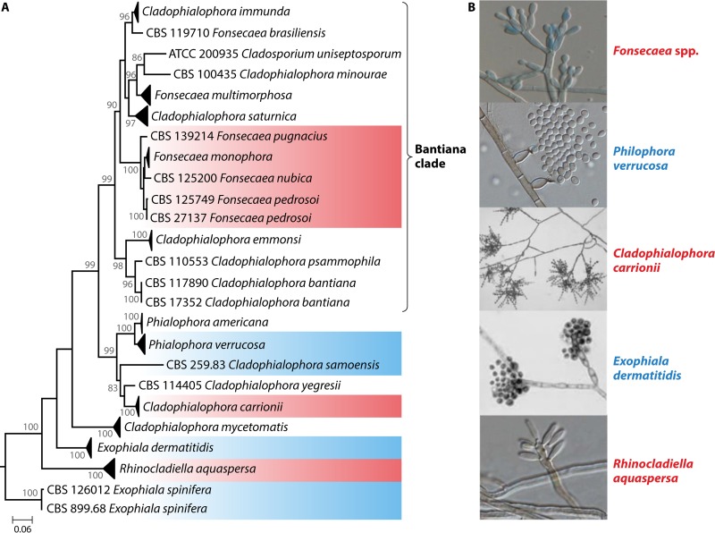
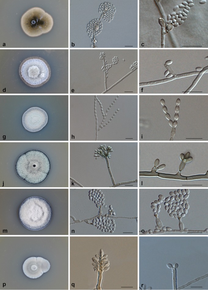
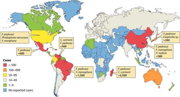
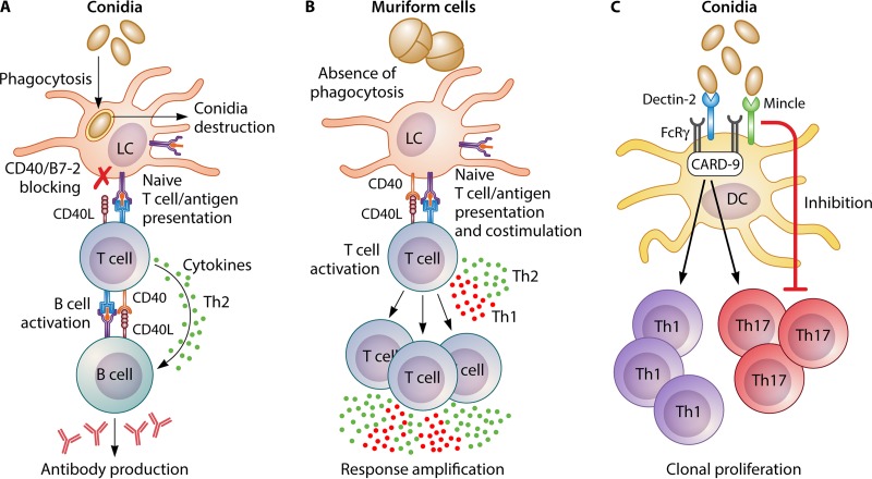
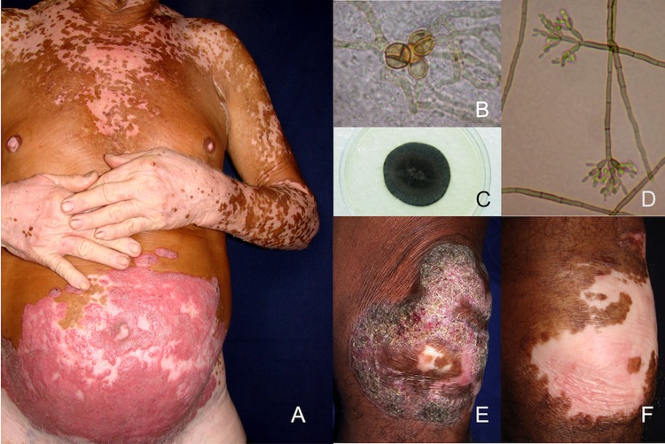
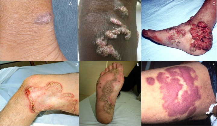
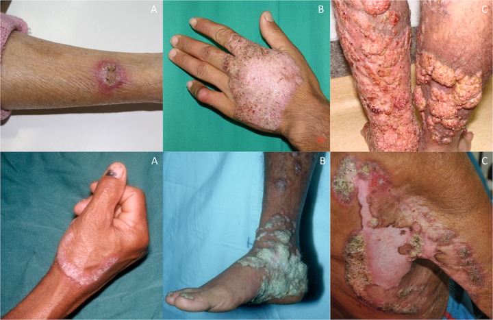
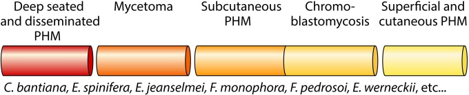
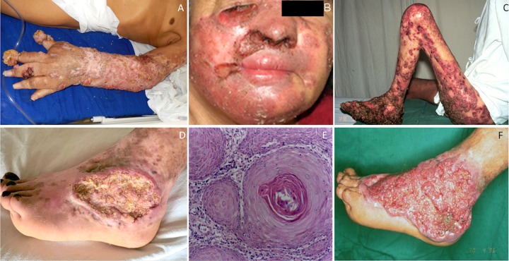
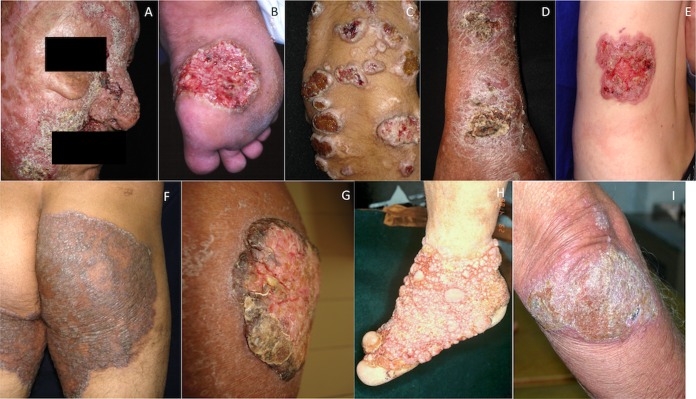
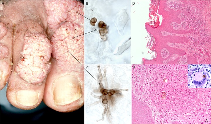
Chromoblastomycosis (CBM), also known as chromomycosis, is one of the most prevalent implantation fungal infections, being the most common of the gamut of mycoses caused by melanized or brown-pigmented fungi. CBM is mainly a tropical or subtropical disease that may affect individuals with certain risk factors around the world. The following characteristics are associated with this disease: (i) traumatic inoculation by implantation from an environmental source, leading to an initial cutaneous lesion at the inoculation site; (ii) chronic and progressive cutaneous and subcutaneous tissular involvement associated with fibrotic and granulomatous reactions associated with microabscesses and often with tissue proliferation; (iii) a nonprotective T helper type 2 (Th2) immune response with ineffective humoral involvement; and (iv) the presence of muriform (sclerotic) cells embedded in the affected tissue. CBM lesions are clinically polymorphic and are commonly misdiagnosed as various other infectious and noninfectious diseases. In its more severe clinical forms, CBM may cause an incapacity for labor due to fibrotic sequelae and also due to a series of clinical complications, and if not recognized at an early stage, this disease can be refractory to antifungal therapy.

Keywords: black fungi; chromoblastomycosis; chromomycosis; melanized fungi; muriform (sclerotic) cells; neglected disease.


Figures

FIG 1
FIG 1
Phylogeny of a representative selection of species of the Chaetothyriales, based on the confidently aligned LSU rDNA D1-D2 domains of LSU sequences constructed by the maximum likelihood method implemented in MEGA 5.10. Bootstrap values of >80% from 500 resampled data sets are shown with branches. Morphologies of the species concerned are shown at the right. Exophiala spinifera was used as the outgroup.
FIG 2
FIG 2
(a to c) Exophiala dermatitidis CBS 748.88. (a) Colony on malt extract agar (MEA) after 3 and 4 weeks of incubation at 30°C; (b) conidial head; (c) conidiophore and conidia. (d to f) Exophiala spinifera CBS 899.68. (d) Colony on MEA after 3 weeks of incubation; (e) conidiophore and conidia clustered at the apex of the conidiophore; (f) conidiophore and liberated conidia. (g to i) Cladophialophora carrionii CBS 166.54. (g) Colony on MEA after 3 weeks of incubation; (h) branching conidial system and conidial chains; (i) conidial chains. (j to l) Fonsecaea pedrosoi CBS 273.66. (j) Colony on MEA after 3 weeks of incubation; (k) conidiophores and conidia; (l) phialides and conidia. (m to o) Phialophora verrucosa BMU 07506. (m) Colony on MEA; (n) phialides and conidia; (o) flask-shaped phialides and conidia. (p to r) Rhinocladiella aquaspersa CBS 122635. (p) Colony on MEA after 3 weeks of incubation; (q) conidiophore and conidia; (r) young conidia and conidiophore. All cultures were incubated at 30°C.
FIG 3
FIG 3
Global distribution of chromoblastomycosis based on reported case series.
FIG 4
FIG 4
CBM immunology. Epidermal Langerhans cells (LCs) are at the front line of defense and may respond differently to conidia or meristematic cells. (A) After recognition of conidia, engulfment and destruction occur, followed by inhibition of costimulatory molecules. Conidial antigens may be presented to T cells, which activates B cells to produce antibodies against CBM pathogens. (B) If muriform cells are present, although there is no phagocytosis by LCs, naive T cells may be activated and proliferate, amplifying the immune response. (C) Recognition of conidia by dendritic cells (DCs) can be made by the C-type lectin dectin-2 or Mincle. However, the dectin-2 pathway leads to T-cell activation with proliferation of Th1 and Th17 cells, while Mincle signaling inhibits the same process, in a clear demonstration of PRR antagonism of the immune response following recognition of CBM fungi by DCs.
FIG 5
FIG 5
Vitiligo and chromoblastomycosis. (A) A 62-year-old Brazilian man presented with a 10-year history of a slow-growing plaque lesion, which started as a small nodule near his navel and spread centrifugally until it reached 30 cm in diameter. Concomitantly, he noted the presence of interwoven achromic patches, which extended to other regions of his skin after 5 years of having the disease restricted to the abdominal area. (B) A skin scraping from the plaque lesion revealed the presence of muriform cells. (C and D) After 3 weeks of culture in Mycosel, a grayish colony grew (C), and micromorphology depicted Fonsecaea pedrosoi structures (D). (E and F) An association of CBM and vitiligo has not been reported, but the presence of antimelanin antibodies in CBM patients is known. Most of the cases that were treated with itraconazole were healed, leaving an achromic patch (F) in place of the previous verrucous/plaque lesions (E). This patient developed vitiligo after CBM, which could be related to the presence of antimelanin antibodies.
FIG 6
FIG 6
Clinical types of lesions observed in patients with chromoblastomycosis. (A) Initial lesion with a 3-month duration in the lower leg. (B) Confluent nodular lesions on the knee. (C) Tumoral (cauliflower-like) lesion on the posterior part of the foot. (D) Cicatricial lesion with verruca showing serpiginous and verrucous contours. (E) Hyperkeratotic verrucous lesion on the sole of the foot. (F) Soft violaceous plaque lesion in the root of the thigh.
FIG 7
FIG 7
Lesions of chromoblastomycosis with different severity grades. (A) Mild forms; (B) moderate forms; (C) severe forms.
FIG 8
FIG 8
Spectrum of fungal diseases caused by melanized fungi. PHM, phaeohyphomycosis.
FIG 9
FIG 9
Complications and sequelae related to severe forms of chromoblastomycosis. (A) Chronic lymphedema hyperkeratotic lesions in the upper limb. (B) Ectropium, secondary bacterial infection, and facial lymphedema. (C) Ankylosis of the knee. (D) Neoplastic transformation of a foot lesion. Shown is an ulcerative lesion with chronic bacterial infection. (E) Skin biopsy specimen taken 80 months later showing a well-differentiated epidermoid carcinoma with typical nuclear atypias and “corn pearls” in the middle of neoplastic cell blocks. Shown is an HE-stained section at a ×400 magnification. (F) Vegetant and papillomatous lesions resulting from the association of chromoblastomycosis with neoplastic lesions.
FIG 10
FIG 10
Differential diagnosis of chromoblastomycosis. (A) Coccidioidomycosis; (B) paracoccidioidomycosis; (C) phaeohyphomycosis; (D) sporotrichosis; (E) deep dermatophytosis associated with CARD9 homozygous mutation; (F) verrucous tuberculosis; (G) Bowen disease; (H) mossy foot; (I) mycosis fungoid.
FIG 11
FIG 11
Laboratory diagnosis of chromoblastomycosis. (A) Skin scrapes and biopsy specimens should be taken from the “black dot” area (arrows). (B and C) Direct examination shows muriform cells (B), which may germinate and form filaments near the cutaneous surface (C). (D to F) In HE-stained sections, tissue reaction with hyperkeratosis, pseudoepitheliomatous hyperplasia (D), may be observed, associated with neutrophilic microabscesses containing muriform cells (E), which may also be found in Langerhans cells (F).
None
None
None
None
None
None
None
None
None
None
None
None
None

Similar articles

Cited by

References

    1. Savioli L, Daumerie D, World Health Organization Department of Control of Neglected Tropical Diseases . 2013. Sustaining the drive to overcome the global impact of neglected tropical diseases: second WHO report on neglected tropical diseases. World Health Organization, Geneva, Switzerland.
    1. van de Sande WWJ, Maghoub ES, Fahal AH, Goodfellow M, Welsh O, Zijlstra ED. 2014. The mycetoma knowledge gap: identification of research priorities. PLoS Negl Trop Dis 8:e2667. doi: 10.1371/journal.pntd.0002667. - DOI - PMC - PubMed
    1. Queiroz-Telles F, Nucci M, Colombo AL, Tobón A, Restrepo A. 2011. Mycoses of implantation in Latin America: an overview of epidemiology, clinical manifestations, diagnosis and treatment. Med Mycol 49:225–236. doi: 10.3109/13693786.2010.539631. - DOI - PubMed
    1. Reiss H, Shadomy HJ, Lyon GM III. 2012. Fundamental medical mycology. Wiley-Blackwell, Hoboken, NJ.
    1. La Hoz RM, Baddley JW. 19 July 2012. Subcutaneous fungal infections. Curr Infect Dis Rep doi: 10.1007/s11908-012-0275-3. - DOI - PubMed

MeSH terms

Substances

LinkOut - more resources