. 2016 Nov 1;30(21):2433-2442.
doi: 10.1101/gad.288035.116. Epub 2016 Nov 17.

Epigenetic regulation of intestinal stem cells by Tet1-mediated DNA hydroxymethylation

Affiliations

Epigenetic regulation of intestinal stem cells by Tet1-mediated DNA hydroxymethylation

Rinho Kim et al. Genes Dev. .

Abstract

Methylated cytosines are associated with gene silencing. The ten-eleven translocation (TET) hydroxylases, which oxidize methylated cytosines to 5-hydroxymethylcytosine (5hmC), are essential for cytosine demethylation. Gene silencing and activation are critical for intestinal stem cell (ISC) maintenance and differentiation, but the potential role of TET hydroxylases in these processes has not yet been examined. Here, we generated genome-wide maps of the 5hmC mark in ISCs and their differentiated progeny. Genes with high levels of hydroxymethylation in ISCs are strongly associated with Wnt signaling and developmental processes. We found Tet1 to be the most abundantly expressed Tet gene in ISCs; therefore, we analyzed intestinal development in Tet1-deficient mice and determined that these mice are growth-retarded, exhibit partial postnatal lethality, and have significantly reduced numbers of proliferative cells in the intestinal epithelium. In addition, the Tet1-deficient intestine displays reduced organoid-forming capacity. In the Tet1-deficient crypt, decreased expression of Wnt target genes such as Axin2 and Lgr5 correlates with lower 5hmC levels at their promoters. These data demonstrate that Tet1-mediated DNA hydroxymethylation plays a critical role in the epigenetic regulation of the Wnt pathway in intestinal stem and progenitor cells and consequently in the self-renewal of the intestinal epithelium.

Keywords: Tet1; epigenomics; hydroxymethylation; intestinal differentiation; intestinal stem cell.


Figures

Figure 1.
Figure 1.
Differential 5hmC distribution in intestinal stem and differentiated cells. (A) The dot blot shows global 5hmC levels in the mouse adult intestinal epithelium compared with the brain and ESCs. (B,C) Immunofluorescence staining of mouse jejunum with 5mC (B), 5hmC (C), and Ki67 antibodies, as indicated. Bars, 50 µm. (D) FACS plot to isolate Lgr5-EGFP+ cells and RT-qPCR validation for Lgr5 expression in GFP+ cells. (E) H&E staining image of scraped differentiated villus cells and RT-qPCR validation for cell-specific marker genes. (Creb3l3) Enterocyte; (vimentin) mesenchymal cells; (hemoglobin) red blood cells.
Figure 2.
Figure 2.
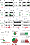
Genome-wide distribution of 5hmC and its dynamics. (AC) Screenshots of bisulfite sequencing (which measures the sum of 5mC and 5hmC) and hMeDIP-seq (which determines 5hmC abundance only) at marker genes in Lgr5+ ISCs (A), Wnt target genes (B), and marker genes of differentiated villus cells (C). Data are presented as percent methylation for the bisulfite sequencing data and as reads per million mapped reads (RPM) for the 5hmC data. (D) The relative 5hmC signal of hMedIP-seq in Lgr5+ stem and differentiated cells. The ratio of hMeDIP/input reads numbers in differentiated epithelial cells (DIFF) was set to 100%. (E) The total number of 5hmC-enriched regions (peaks) and genomic distribution in Lgr5+ stem and differentiated cells (fourfold or more enrichment over local tag count). P-value < 0.001. (F) Genomic distribution of differentially hydroxymethylated regions (DhMRs). (Top pie chart) regions with 5hmC levels that are higher in Lgr5+ stem cells than in differentiated epithelial cells (DIFF) (at least fivefold). P-value < 0.0001. (Bottom pie chart) Regions with 5hmC levels that are higher in differentiated epithelial cells (DIFF) than in Lgr5+ stem cells (at least fivefold). P-value < 0.0001. Circle size represents the total number of DhMRs in each group. (G) Gene ontology search results for genes closest to DhMRs.
Figure 3.
Figure 3.
Positive correlation between 5hmC-enriched gene expression levels. (AC) RNA-seq results for ISC marker genes (A), Wnt target genes (B), and differentiated villus cell marker genes (C). RNA-seq data for both cell populations are represented as reads per kilobase per million mapped read (RPKM). (D) Heat map for differential gene expression of differentially hydroxymethylated genes (≥1.5-fold change) during intestinal epithelial differentiation. (E) Box plot for differential gene expression of differentially hydroxymethylated genes in each cell population. (***) P-value < 0.001 by t-test.
Figure 4.
Figure 4.
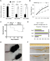
Postnatal developmental defects in the intestines of Tet1-null mice. (A) Relative Tet1/2/3 mRNA expression levels in Lgr5+ intestinal stem and differentiated epithelial cells of adult mice. mRNA levels were normalized to those of Tbp. (B) Tet1-null mice are growth-retarded. Weight curve during the postnatal period. (**) P-value < 0.01 by t-test. (C) Survival curve during the postnatal period. (***) P-value < 0.001 by a log rank (Mantel-Cox) test. (D) Reduced perinatal viability of Tet1 homozygous mutants. Mice were quantified on P3. (***) P-value <0.0005, χ2 test. (E) Representative Tet1+/+ and Tet1−/− mice on P11. (F) Representative images of intestinal tracts of Tet1+/+ and Tet1−/− mice at 1 wk of age.
Figure 5.
Figure 5.
Decreased Wnt target gene expression in the postnatal Tet1-null intestine. (A) Histological analysis of Tet1+/+ and Tet1−/− jejunum of 1-wk-old mice. Proliferating cells are marked by immunohistochemistry for Ki67 (dark-brown signal). Bars, 50 µm. (B) Villus length in Tet1+/+ and Tet1−/− 1-wk-old jejunum. (***) P-value < 0.001. (C) Ki67+ cell number per crypt is reduced in 1-wk-old Tet1−/− jejunum compared with littermate controls. (***) P-value < 0.001. (D) Immunostaining results of Wnt target gene expression, including Axin2, c-Myc, and Sox9 in P7 Tet1+/+ and Tet1−/− mice. Bars, 50 µm.
Figure 6.
Figure 6.
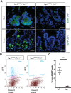
Reduction of Lgr5 expression in Tet1-null mouse intestines. (A) Decreased Lgr5-EGFP expression levels in 2-wk-old and 2-mo-old Tet1-null jejunum as detected by immunofluorescence staining of GFP (green). Bars, 20 µm. (B) FACS analysis demonstrates loss of Lgr5-EGFP+ cells in 2-mo-old Tet1−/− mice. (C) Quantification of Lgr5-EGFP+ cell number in 2-mo-old Tet1+/+ and Tet1−/− mice. (***) P-value < 0.001. n = 4.
Figure 7.
Figure 7.
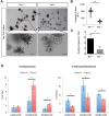
Budding organoid-forming potential is reduced in the absence of Tet1. (A) Images of intestinal organoid formation from 2-wk-old Tet1+/+ and Tet1−/− jejunal crypts. Bars, 10 µm. (B) Quantification of intestinal organoid volume after 9 d of culture. (**) P-value < 0.01. n = 3. (C) Ratio of budding organoids over total organoids and spheroids. (*) P-value < 0.05. n = 3. (D) Differential 5mC and 5hmC levels at the promoters of Lgr5, Axin2, and Sox9 in 2-wk-old Tet1+/+ and Tet1−/− crypts. (*) P-value < 0.05. n = 3.

References

    1. Avrahami D, Li C, Zhang J, Schug J, Avrahami R, Rao S, Stadler MB, Burger L, Schübeler D, Glaser B, et al. 2015. Aging-dependent demethylation of regulatory elements correlates with chromatin state and improved β cell function. Cell Metab 22: 619–632. - PMC - PubMed
    1. Barker N, van Es JH, Kuipers J, Kujala P, van den Born M, Cozijnsen M, Haegebarth A, Korving J, Begthel H, Peters PJ, et al. 2007. Identification of stem cells in small intestine and colon by marker gene Lgr5. Nature 449: 1003–1007. - PubMed
    1. Baylin SB, Esteller M, Rountree MR, Bachman KE, Schuebel K, Herman JG. 2001. Aberrant patterns of DNA methylation, chromatin formation and gene expression in cancer. Hum Mol Genet 10: 687–692. - PubMed
    1. Choi I, Kim R, Lim H-WW, Kaestner KH, Won K-JJ. 2014. 5-hydroxymethylcytosine represses the activity of enhancers in embryonic stem cells: a new epigenetic signature for gene regulation. BMC Genomics 15: 670. - PMC - PubMed
    1. Cimmino L, Dawlaty MM, Ndiaye-Lobry D, Yap Y, Bakogianni S, Yu Y, Bhattacharyya S, Shaknovich R, Geng H, Lobry C, et al. 2015. TET1 is a tumor suppressor of hematopoietic malignancy. Nat Immunol 16: 653–662. - PMC - PubMed

Publication types

MeSH terms