. 2016 Nov 29;113(48):13732-13737.
doi: 10.1073/pnas.1611904113. Epub 2016 Nov 15.

Global transformation of erythrocyte properties via engagement of an SH2-like sequence in band 3

Affiliations

Global transformation of erythrocyte properties via engagement of an SH2-like sequence in band 3

Estela Puchulu-Campanella et al. Proc Natl Acad Sci U S A. .

Abstract

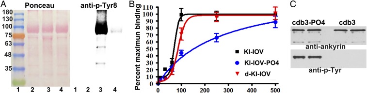
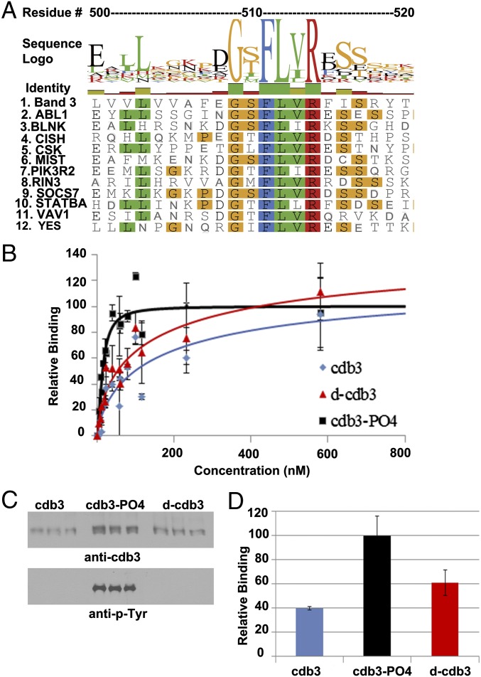
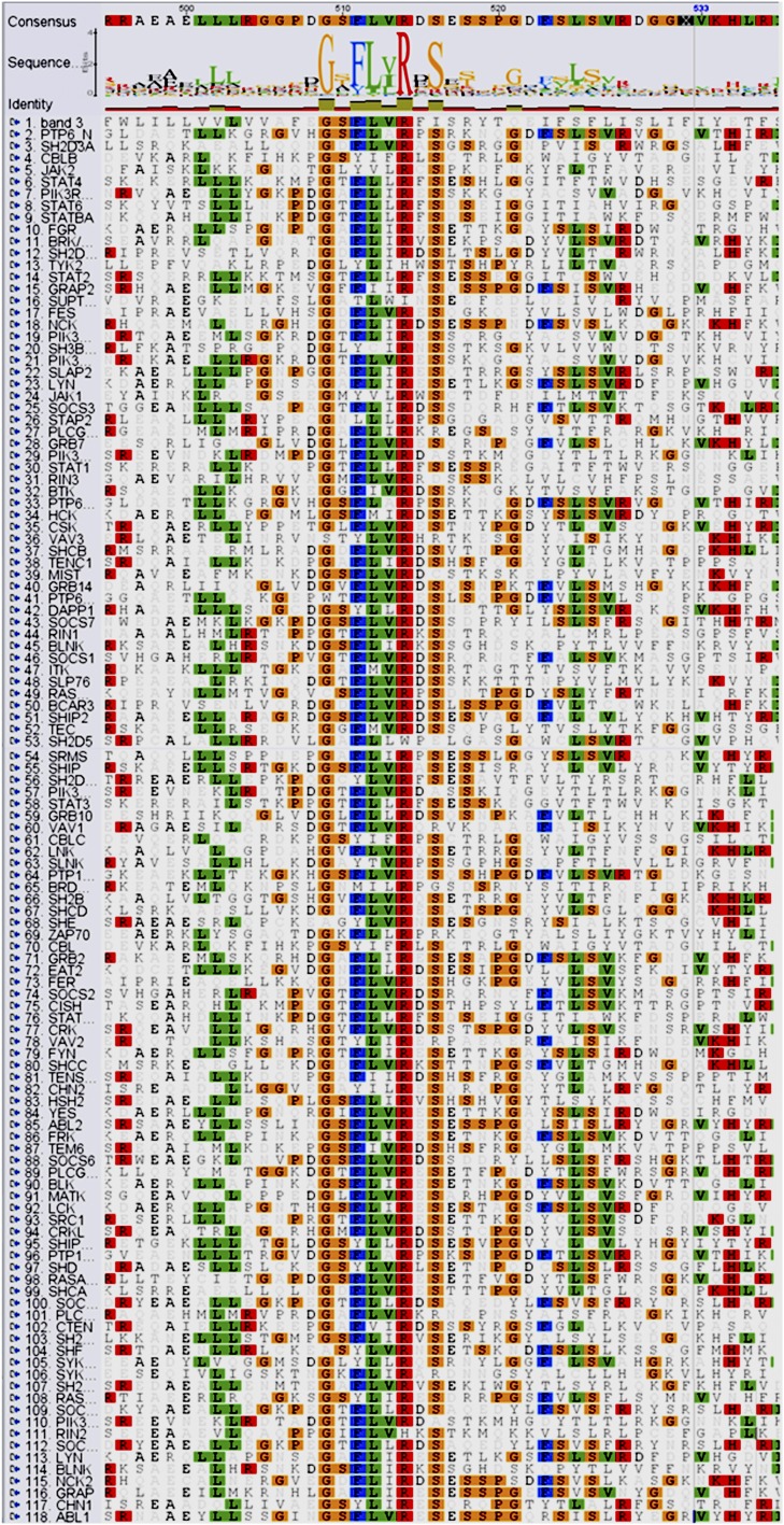
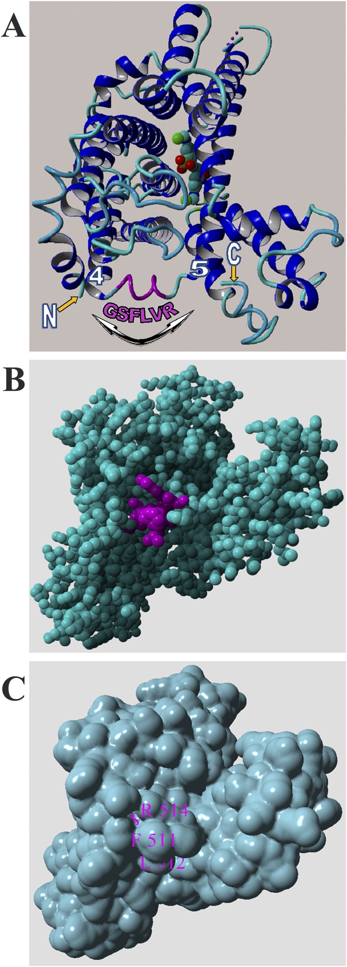
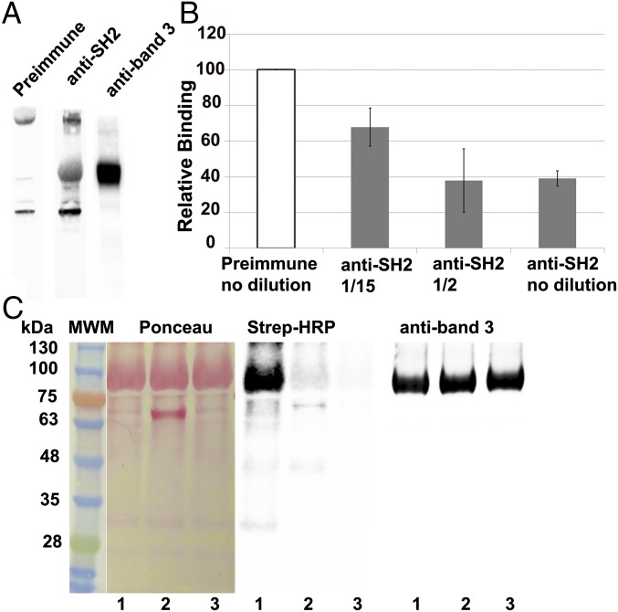
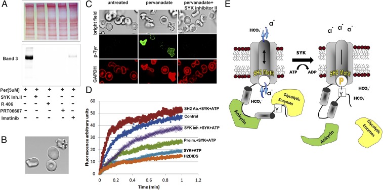
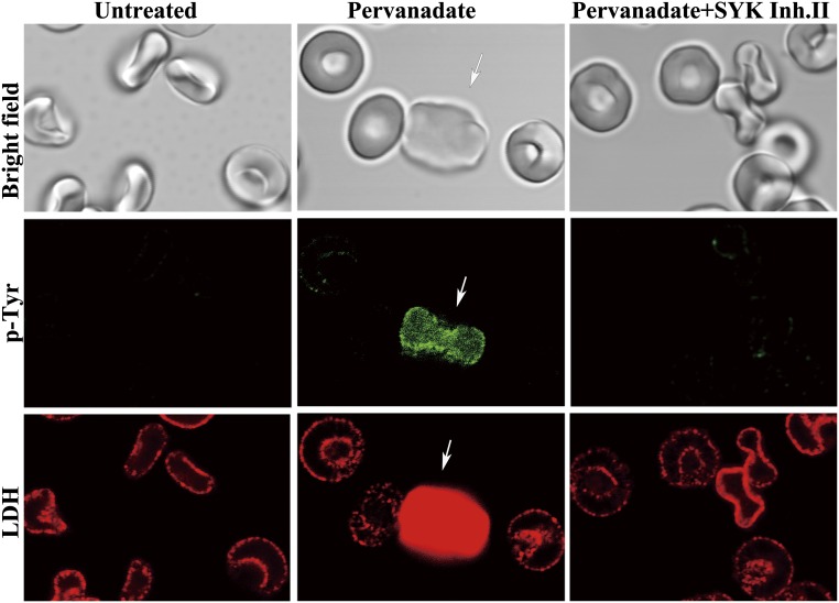
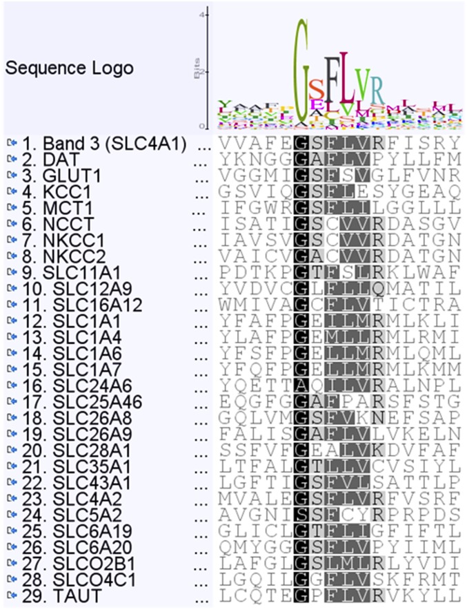
Src homology 2 (SH2) domains are composed of weakly conserved sequences of ∼100 aa that bind phosphotyrosines in signaling proteins and thereby mediate intra- and intermolecular protein-protein interactions. In exploring the mechanism whereby tyrosine phosphorylation of the erythrocyte anion transporter, band 3, triggers membrane destabilization, vesiculation, and fragmentation, we discovered a SH2 signature motif positioned between membrane-spanning helices 4 and 5. Evidence that this exposed cytoplasmic sequence contributes to a functional SH2-like domain is provided by observations that: (i) it contains the most conserved sequence of SH2 domains, GSFLVR; (ii) it binds the tyrosine phosphorylated cytoplasmic domain of band 3 (cdb3-PO4) with Kd = 14 nM; (iii) binding of cdb3-PO4 to erythrocyte membranes is inhibited both by antibodies against the SH2 signature sequence and dephosphorylation of cdb3-PO4; (iv) label transfer experiments demonstrate the covalent transfer of photoactivatable biotin from isolated cdb3-PO4 (but not cdb3) to band 3 in erythrocyte membranes; and (v) phosphorylation-induced binding of cdb3-PO4 to the membrane-spanning domain of band 3 in intact cells causes global changes in membrane properties, including (i) displacement of a glycolytic enzyme complex from the membrane, (ii) inhibition of anion transport, and (iii) rupture of the band 3-ankyrin bridge connecting the spectrin-based cytoskeleton to the membrane. Because SH2-like motifs are not retrieved by normal homology searches for SH2 domains, but can be found in many tyrosine kinase-regulated transport proteins using modified search programs, we suggest that related cases of membrane transport proteins containing similar motifs are widespread in nature where they participate in regulation of cell properties.

Keywords: SH2 domain motif; anion exchanger 1; erythrocyte glycolysis; regulation of transport proteins; tyrosine phosphorylation.


Conflict of interest statement

The authors declare no conflict of interest.

Figures

Fig. 1.
Fig. 1.
Effect of band 3 tyrosine phosphorylation on the binding of ankyrin to erythrocyte KI-IOVs and cdb3. (A) KI-IOVs were imaged with Ponceau stain (Left) and then immunoblotted with antibody to phosphotyrosine 8 on band 3 (Right). Lanes contain: 1, molecular weight standards; 2, control KI-IOVs; 3, KI-IOVs from o-vanadate-treated RBCs isolated in the presence of phosphatase inhibitors (phosphorylated;KI-IOV-PO4); 4, same as 3 except phosphatase inhibitors were excluded (dephosphorylated; d-KI-IOV). (B) Binding of increasing concentrations of GST-D3D4-ankyrin to each of the above KI-IOV preparations. (C) Western blotting of the pelleted proteins shows that similar amounts of ankyrin are collected in the pellet (Upper) regardless of the phosphorylation state of cdb3 (Lower).
Fig. 2.
Fig. 2.
Evidence for an SH2 signature sequence (GSFLVR) in a cytoplasmic loop of the membrane-spanning domain of band 3 (msdb3). (A) Alignment of sequences surrounding the second cytoplasmic loop of the msdb3 with 11 prominent SH2 domains in the human sequence database. Full alignment of band 3 with all known human SH2 domains is shown in Fig. S1. Colors used to reveal sequence alignments constitute the default colors used by the ClustalX program: where amino acids labeled with the respective colors are orange (G, P, S, and T), red (H, K, and R), blue (F, W, and T), and green (I, L, M, and V). The sequence logo represents the conservation of the residues. (B) Binding of tyrosine phosphorylated cdb3 (cdb3-PO4), dephosphorylated cdb3 (d-cdb3), or untreated cdb3 (cdb3) to KI-stripped and trypsin-digested IOVs containing solely the membrane-spanning proteins of the erythrocyte membrane (n = 6; mean ± SD). (C) Copelleting of either nonphosphorylated cdb3 (cdb3), cdb3-PO4, or phosphorylated and then dephosphorylated cdb3 (d-cdb3) with stripped digested IOVs. Blots were stained with an anti-band 3 antibody or anti–p-tyrosine antibody. (D) Densitometric analysis of the blots shown in C (n = 3).
Fig. S1.
Fig. S1.
Alignment of residues surrounding the GSFLVR sequence in band 3 with established SH2 domains present in enzymes, adaptors, scaffolds, and signal regulators in the human genome. Sequence alignment was performed with Geneious version 5.5.7 (www.geneious.com, using an identity matrix global alignment with free end gaps. Colors used to reveal aligned sequences constitute the default colors used by the ClustalX program, where amino acids labeled with the respective colors are orange (G, P, S, and T), red (H, K, and R), blue (F, W, and T), and green (I, L, M, and V).
Fig. S2.
Fig. S2.
Structure of the membrane-spanning domain of band 3 showing the location of the conserved SH2 domain sequence (GSFLVR; colored in magenta in A) that forms part of the cytoplasmic loop connecting membrane-spanning helices 4 and 5. This cytoplasmic loop also connects the “core” and “gate” domains of band 3 that must pivot to open and close the anion channel (double-pointed arrow). The most NH2-terminal (G381) and COOH-terminal residues (D887) resolved in the crystal structure (26) are marked N and C, respectively. The highly specific covalent inhibitor of anion transport, DIDS, is shown with space-filling atoms. The figure was constructed using YASARA version 11.10.18, 1993–2015 (www.yasara.org/). (B) Space filling model of the cytoplasmic view of A with the GSLVR residues shown in magenta. (C) Surface presentation of B with GSLVR labeled.
Fig. 3.
Fig. 3.
Confirmation of the role of the GSFLVR sequence in binding of cdb3-PO4. (A) Western blots of whole erythrocyte membranes demonstrating that the anti-SH2 signature sequence antibody recognizes band 3. Lane 1, preimmune serum; lane 2, anti-SH2 antiserum from the same rabbit used in lane 1; and lane 3, anti-band 3 antibody. (B) Inhibition of cdb3-PO4 binding to SD-IOVs by increasing concentrations of anti-SH2 antiserum. SD-IOVs were incubated with different dilutions of anti-SH2 preimmune serum (white bar) or immune serum (gray bars) before incubation with phosphorylated cdb3. Copelleted cdb3-PO4 was determined by Western blotting with anti-cdb3 antibody. (C) Identification of the polypeptide in the erythrocyte membrane that binds sulfo-SBED-cdb3-PO4. Sulfo-SBED–labeled cdb3-PO4 was incubated with SD-IOVs in the absence of any competing protein (lanes 1) or in the presence of anti-SH2 antiserum (lanes 2) or excess underivatized cdb3-PO4 (lanes 3). Biotin-labeled proteins were visualized by streptavidin-HRP staining.
Fig. 4.
Fig. 4.
Tyrosine phosphorylation of band 3 induces shape changes in erythrocytes, dissociation of glycolytic enzymes from the membrane, and inhibition of anion exchange across the membrane. (A) Western blot showing induction of band 3 tyrosine phosphorylation by 5 μM pervanadate (lane 1) and its prevention by preincubation with 10 μM SYK inhibitor II (lane 2), 10 μM R406 (lane 3), 10 μM PRT 062607 (lane 4), or 20 μM imatinib (lane 5). (B) Erythrocyte shape changes induced by 5 µM pervanadate include echinocytes and spherocytes. (C) Erythrocyte shape changes (Top row) and displacement of glyceraldehyde-3-phosphate dehydrogenase from the membrane (Bottom row) only occur in erythrocytes in which pervanadate has induced tyrosine phosphorylation of the membrane (Middle row). Analogous findings with lactate dehydrogenase are shown in Fig. S3. (D) Tracings of anion exchange in lysed erythrocytes following resealing in the presence of SYK kinase + ATP, SYK kinase + SYK kinase inhibitor II, and/or SYK kinase + anti-SH2 antiserum, as indicated. Preimmune serum is used as a control for treatment with anti-SH2 domain antiserum. DIDS blocks all band 3-mediated anion transport. (E) Proposed mechanism of regulation of erythrocyte properties by tyrosine phosphorylation of band 3 and engagement of an SH2-like motif in msdb3.
Fig. S3.
Fig. S3.
Tyrosine phosphorylation of band 3 induces both cell shape changes and dissociation of glycolytic enzymes from the erythrocyte membrane. Displacement of the glycolytic enzyme, lactate dehydrogenase, from the membrane (Bottom row) only occurs in erythrocytes in which pervanadate has induced tyrosine phosphorylation of the membrane (Middle row). Moreover, comparison of pervanadate-treated cells in the absence (Middle column) and presence (Right column) of SYK inhibitor II demonstrates that the enzyme displacement is mediated by SYK tyrosine kinase.
Fig. S4.
Fig. S4.
Alignment of SH2 domain signature sequences in tyrosine kinase-regulated membrane transporters. Alignment of individual proteins is centered on the most conserved SH2 domain motif, GSFLVR, using Geneious version 5.5.7 (www.geneious.com, and using identity matrix global alignment with free end gaps. Proteins are named as in Sprowl et al. (49).

Similar articles

Cited by

References

    1. Hunter T. Tyrosine phosphorylation: Thirty years and counting. Curr Opin Cell Biol. 2009;21(2):140–146. - PMC - PubMed
    1. Pawson T, Kofler M. Kinome signaling through regulated protein-protein interactions in normal and cancer cells. Curr Opin Cell Biol. 2009;21(2):147–153. - PubMed
    1. Liu BA, Engelmann BW, Nash PD. The language of SH2 domain interactions defines phosphotyrosine-mediated signal transduction. FEBS Lett. 2012;586(17):2597–2605. - PubMed
    1. van den Akker E, Satchwell TJ, Williamson RC, Toye AM. Band 3 multiprotein complexes in the red cell membrane; of mice and men. Blood Cells Mol Dis. 2010;45(1):1–8. - PubMed
    1. Anong WA, et al. Adducin forms a bridge between the erythrocyte membrane and its cytoskeleton and regulates membrane cohesion. Blood. 2009;114(9):1904–1912. - PMC - PubMed

Publication types

MeSH terms

Substances

LinkOut - more resources