. 2016 Nov 29;113(48):13756-13761.
doi: 10.1073/pnas.1609718113. Epub 2016 Nov 16.

Neutron structures of the Helicobacter pylori 5'-methylthioadenosine nucleosidase highlight proton sharing and protonation states

Affiliations

Neutron structures of the Helicobacter pylori 5'-methylthioadenosine nucleosidase highlight proton sharing and protonation states

Michael T Banco et al. Proc Natl Acad Sci U S A. .

Abstract

MTAN (5'-methylthioadenosine nucleosidase) catalyzes the hydrolysis of the N-ribosidic bond of a variety of adenosine-containing metabolites. The Helicobacter pylori MTAN (HpMTAN) hydrolyzes 6-amino-6-deoxyfutalosine in the second step of the alternative menaquinone biosynthetic pathway. Substrate binding of the adenine moiety is mediated almost exclusively by hydrogen bonds, and the proposed catalytic mechanism requires multiple proton-transfer events. Of particular interest is the protonation state of residue D198, which possesses a pKa above 8 and functions as a general acid to initiate the enzymatic reaction. In this study we present three corefined neutron/X-ray crystal structures of wild-type HpMTAN cocrystallized with S-adenosylhomocysteine (SAH), Formycin A (FMA), and (3R,4S)-4-(4-Chlorophenylthiomethyl)-1-[(9-deaza-adenin-9-yl)methyl]-3-hydroxypyrrolidine (p-ClPh-Thio-DADMe-ImmA) as well as one neutron/X-ray crystal structure of an inactive variant (HpMTAN-D198N) cocrystallized with SAH. These results support a mechanism of D198 pKa elevation through the unexpected sharing of a proton with atom N7 of the adenine moiety possessing unconventional hydrogen-bond geometry. Additionally, the neutron structures also highlight active site features that promote the stabilization of the transition state and slight variations in these interactions that result in 100-fold difference in binding affinities between the DADMe-ImmA and ImmA analogs.

Keywords: Helicobacter; enzyme mechanism; neutron diffraction; nucleosidase; proton transfer.


Conflict of interest statement

The authors declare no conflict of interest.

Figures

Fig. 1.
Fig. 1.
(A) A surface model of HpMTAN in complex with SAH. The enzyme is an obligatory homodimer in which the two chains of the dimer are represented by the white and the green surfaces. The red box highlights the several hydrogen-bond interactions that contribute to substrate binding in the HpMTAN active site. (B) A structural representation of the HpMTAN catalytic reaction (PDB ID codes 4OY3 and 4OTJ). (Left) Substrate and the nucleophilic water-bound molecule. After formation of the oxocarbenium ion intermediate, a bound water molecule in the active site attacks the C1′ of the ribosyl moiety, resulting in the hydrolysis of the substrate. (Right) The product complex.
Fig. 2.
Fig. 2.
The observed positions of the deuterium atom in the adenine-binding pocket of HpMTAN for the product complex presented. In both panels, the 2FO-FC for the nuclear density is contoured to 1σ and is represented in light blue. The FO-FC nuclear and X-ray density are represented in dark blue and light green, respectively. (A) The 2FO-FC density illustrating the hydrogen-bond network that aids in positioning D198. (B) Difference omit FO-FC nuclear density of the shared D+ ion and of the N9 nitrogen and deuteron atoms contoured to 3.5σ. The FO-FC X-ray map of the adenine molecule and D198, contoured to 3.0σ, demonstrates the lack of density for the shared D+ ion. Hydrogen-bonding interactions and the respective distances are indicated.
Fig. 3.
Fig. 3.
The proposed catalytic mechanism of HpMTAN. The colored hydrogen atoms represent the protons that are involved in the enzymatic reaction determined by the neutron structures. Briefly, the catalytic reaction is initiated by D198 protonating the N7 of the adenyl moiety. Delocalization of electrons in the adenyl moiety leads to bond elongation of the N-ribosidic bond, consequently forming an oxocarbenium ion intermediate. A bound water molecule then will attack the C1′ of the ribosyl moiety and will donate a proton to the N9 of adenine after the N7 proton is shared.
Fig. 4.
Fig. 4.
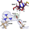
Positioning of the nucleophilic water molecule in the substrate complex. The nuclear and X-ray 2Fo-Fc maps are contoured to 1σ. The nuclear 2FO-FC map is presented in light blue, and the X-ray 2FO-FC map is presented in green. The positioning of the nucleophilic water molecule is afforded by hydrogen-bonding interactions with the carboxylate moiety of E13 and the guanidinium moiety of R194. The hydrogen-bonding interactions with the two residues allow one lone pair of electrons of the water molecule to be positioned near the oxocarbenium ion intermediate to allow nucleophilic attack at the C1′ position of the ribose moiety as indicated by the arrow.
Fig. 5.
Fig. 5.
The observed positions of deuterium atoms in the adenine-binding pocket of HpMTAN-D198N for the inactive binary substrate complex. In both panels, the 2FO-FC for the nuclear and X-ray density is contoured to 1σ and is represented in light blue and green, respectively. (A) Due to the D198N variant, the hydrogen of the hydroxyl group of S197 is reoriented to interact with a bound water molecule. (B) The interactions between the water molecule and the conserved F208 residue.
Fig. 6.
Fig. 6.
The hydrogen-bonding interactions observed for the nucleophile in the presence of the fully and early dissociative transition-state analogs. The 2FO-FC nuclear and X-ray maps are contoured to 1σ and represented in light blue and green, respectively. (A) The deuteron positions of the nucleophile and the various hydrogen-bond interactions with p-ClPh-Thio-DADMe-ImmA are shown. Additionally, the 2FO-FC nuclear maps show a protonated ammonium in the pyrrolidine moiety of the DADMe-ImmA analog. (B) Positioning of the observed deuterium atoms for FMA and the unusual orientation of the nucleophile in the HpMTAN/FMA neutron structure. In the FMA complex the deuterium atom of the 3′ hydroxyl of the ribose moiety is in an unexpected position that disrupts the hydrogen-bond interaction with Oδ2 of E175 that was observed in all the other neutron structures.
Fig. S1.
Fig. S1.
The high-resolution X-ray structure of the p-ClPh-Thio-DADMe-ImmA analog demonstrates the tetrahedral geometry of the N1′ atom. The ImmA compound and active site residues coordinating the nucleophilic water molecule are shown by yellow and green carbon atoms, respectively. The FO-FC omit density in which the ImmA compound, each residue, and the water molecule were omitted from the map calculation is shown at 3σ.

Similar articles

Cited by

References

    1. Dairi T. An alternative menaquinone biosynthetic pathway operating in microorganisms: An attractive target for drug discovery to pathogenic Helicobacter and Chlamydia strains. J Antibiot (Tokyo) 2009;62(7):347–352. - PubMed
    1. Thomas K, et al. Femtomolar inhibitors bind to 5′-methylthioadenosine nucleosidases with favorable enthalpy and entropy. Biochemistry. 2012;51(38):7541–7550. - PubMed
    1. Wang S, et al. A picomolar transition state analogue inhibitor of MTAN as a specific antibiotic for Helicobacter pylori. Biochemistry. 2012;51(35):6892–6894. - PMC - PubMed
    1. Wang S, et al. New antibiotic candidates against Helicobacter pylori. J Am Chem Soc. 2015;137(45):14275–14280. - PMC - PubMed
    1. Mishra V, Ronning DR. Crystal structures of the Helicobacter pylori MTAN enzyme reveal specific interactions between S-adenosylhomocysteine and the 5′-alkylthio binding subsite. Biochemistry. 2012;51(48):9763–9772. - PMC - PubMed

Publication types

MeSH terms