. 2016 Nov 16;8(365):365ra159.
doi: 10.1126/scitranslmed.aag1093.

The synthetic diazonamide DZ-2384 has distinct effects on microtubule curvature and dynamics without neurotoxicity

Affiliations

The synthetic diazonamide DZ-2384 has distinct effects on microtubule curvature and dynamics without neurotoxicity

Michal Wieczorek et al. Sci Transl Med. .

Abstract

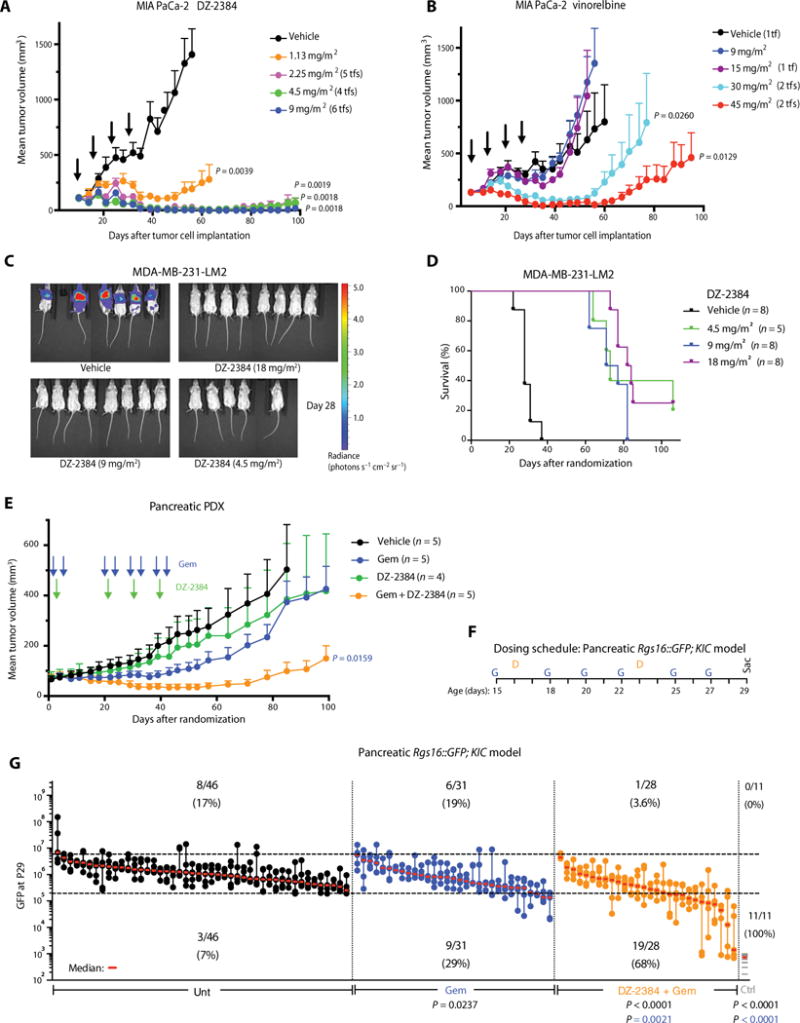
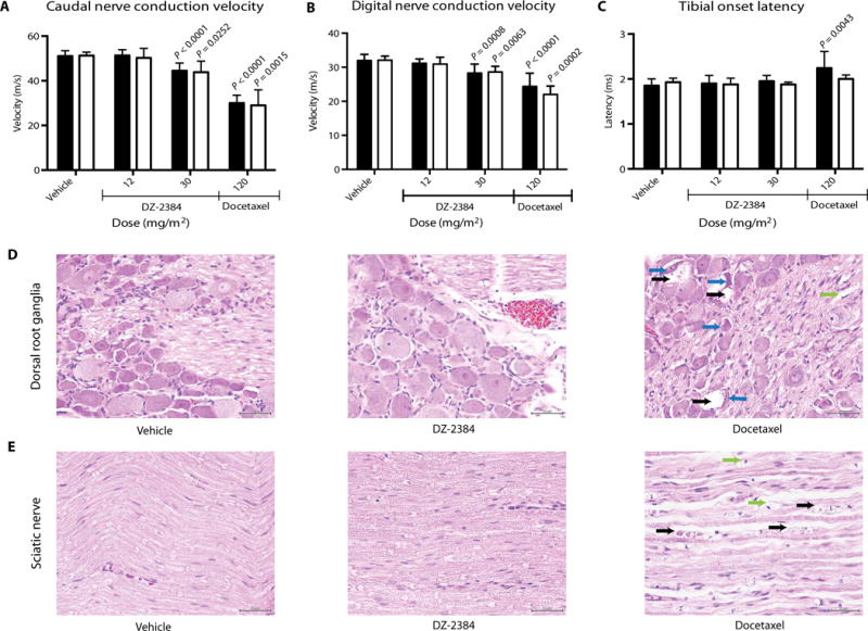
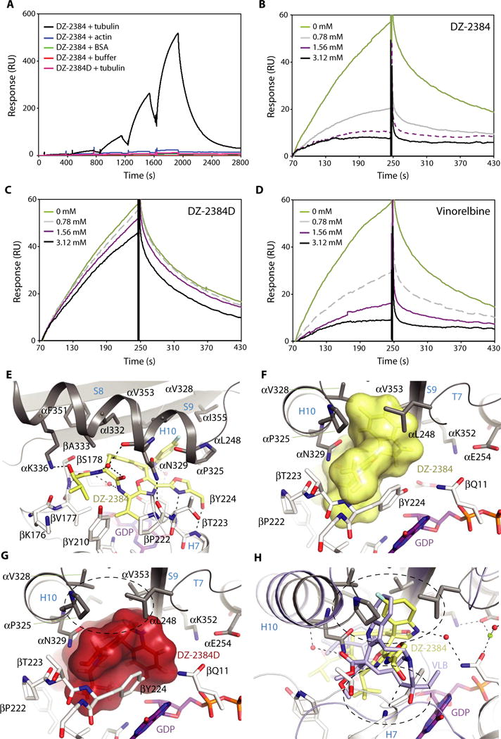
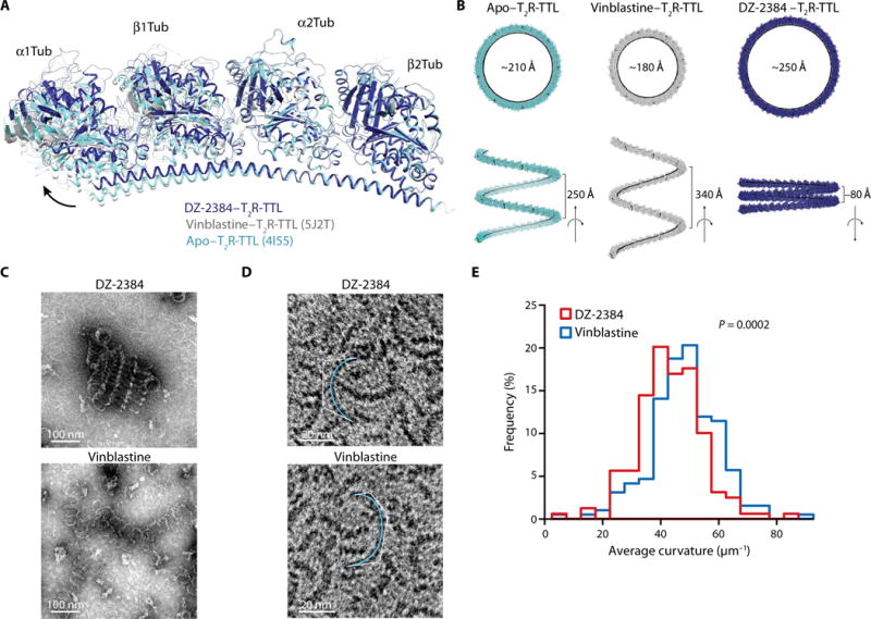
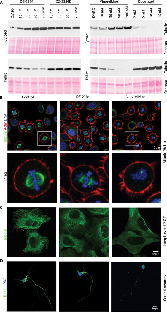
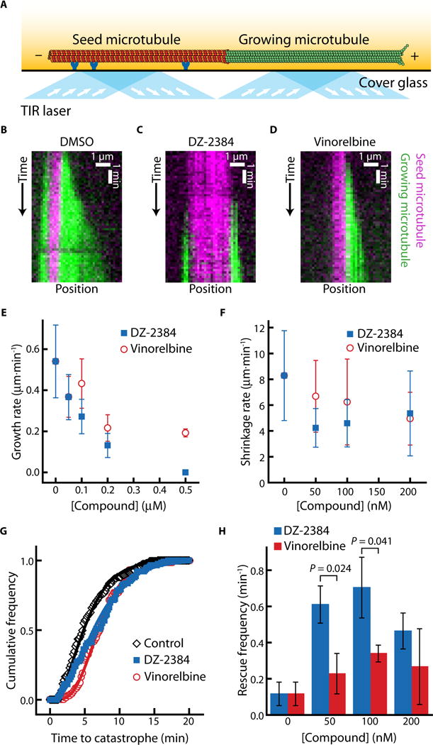
Microtubule-targeting agents (MTAs) are widely used anticancer agents, but toxicities such as neuropathy limit their clinical use. MTAs bind to and alter the stability of microtubules, causing cell death in mitosis. We describe DZ-2384, a preclinical compound that exhibits potent antitumor activity in models of multiple cancer types. It has an unusually high safety margin and lacks neurotoxicity in rats at effective plasma concentrations. DZ-2384 binds the vinca domain of tubulin in a distinct way, imparting structurally and functionally different effects on microtubule dynamics compared to other vinca-binding compounds. X-ray crystallography and electron microscopy studies demonstrate that DZ-2384 causes straightening of curved protofilaments, an effect proposed to favor polymerization of tubulin. Both DZ-2384 and the vinca alkaloid vinorelbine inhibit microtubule growth rate; however, DZ-2384 increases the rescue frequency and preserves the microtubule network in nonmitotic cells and in primary neurons. This differential modulation of tubulin results in a potent MTA therapeutic with enhanced safety.


Conflict of interest statement

Diazon Pharmaceuticals Inc. holds the rights to the Patent Cooperation Treaty publication no. WO2009/134938 that covers DZ-2384. None of the authors of this manuscript are named inventors on this patent. G.C.S., A.R., and P.H. are founders and shareholders in Diazon Pharmaceuticals Inc. R.A.B. and T.M.W. are founders of Tuevol Therapeutics Inc. and are inventors on patent applications (62/067,304; 62/067,276; 62/232,901; and 62/232,922) held or submitted by the UT Southwestern Medical Center that cover the use of the GEMM rapid in vivo assay (Rgs16∷GFP;KIC model) for screening therapeutics for pancreatic ductal adenocarcinoma.

Figures

Fig. 1
Fig. 1. DZ-2384 has potent antitumor activity as a single agent and in combination with gemcitabine
(A and B) Mean volume of MIA PaCa-2 tumors after 4 weeks of weekly bolus intravenous administrations (indicated by arrows) of vehicle, DZ-2384 (A), or vinorelbine (B). Error bars represent means ± SEM, n = 6. Tf, number of tumor-free animals on day 95. P values were determined on day 53. (C) In vivo bioluminescent imaging of luciferase-expressing MDA-MB-231-LM2 breast cancer metastases in the lungs is presented as a pseudocolor heat map of photon flux on day 28 after treatment initiation. (D) Kaplan-Meier curves representing survival of mice with MDA-MB-231-LM2 breast cancer metastases to the lung. P values <0.0001 for all treated mice were calculated using the log-rank (Mantel-Cox) test relative to vehicle-treated mice. (E) Mean tumor volume of a human pancreatic cancer PDX after treatment with gemcitabine (Gem) (100 mg/m2) or DZ-2384 (36 mg/m2) alone or in combination according to the schedule indicated by arrows. Error bars represent means ± SEM, n = 5. P values were determined on day 92. (F) Treatment schedule for the Rgs16∷GFP;KIC pancreatic model: DZ-2384 (7.5 and 30 mg/m2 for the first and second injection on P16 and P23, respectively) and gemcitabine (37.5 mg/m2 three times weekly as indicated). (G) Maximum tumor burden (GFP) signal of untreated (Unt; n = 46), gemcitabine-treated (n = 31), or gemcitabine + DZ-2384–treated (n = 28) groups of mice on P29 is represented quantitatively. Each aligned vertical dot column represents one pancreas, and dots are the first to fifth brightest nonoverlapping pancreas fields; the median is in red. GFP expression in Rgs16∷GFP; non-KIC control (Ctrl) mice is represented by one gray line per mouse. Top and bottom dashed horizontal lines mark the 95th and 1st percentile, respectively, of all untreated animal GFP values. Numbers below the 1st percentile represent the mildly or nontumorigenic pancreatic fields, and comparison with untreated animals represents the response rate (indicated below the bottom dashed line). All P values except in (D) were determined by Student’s unpaired two-tailed t test with Welch’s correction; the color matches the comparison group.
Fig. 2
Fig. 2. DZ-2384 has no effects on NCV, latency, and histopathology at effective antitumor concentrations
(A to C) Electrophysiological parameters were measured on caudal (A), digital (B), and tibial (C) nerves. Black bars represent measurements taken on day 24 after four weekly administrations of DZ-2384 or docetaxel at the doses indicated (n = 12). White bars represent measurements taken on day 44, allowing a 22-day recovery as indicated (n = 5). Error bars represent SD. P values were determined using an unpaired nonparametric Student’s t test. Each P value indicated is compared to vehicle bars of the corresponding color. (D and E) Histopathology of dorsal root ganglia (D) and sciatic nerve (E) tissue sections taken at necropsy on day 44. Arrows denote degenerating neurons with vacuolated cytoplasm (black) and condensed nuclei (blue). Black arrows in the sciatic nerve denote degenerating axons. Green arrows in the dorsal root ganglia and sciatic nerve denote digestion chambers. Scale bars, 50 μm.
Fig. 3
Fig. 3. DZ-2384 binds to the vinca domain of tubulin
(A) Specific, dose-dependent binding of tubulin dimers to DZ-2384 as assessed by SPR in single-cycle mode. Buffer (red), BSA (green), actin (blue), and tubulin (black) were titrated (0 to 10 mM; twofold dilution series) over biotinylated DZ-2384 [200 resonance units (RUs) immobilized]; under similar conditions, tubulin was titrated over biotinylated DZ-2384D surfaces (pink). (B to D) Multi-cycle SPR experiments in which the interaction between tubulin (3 μM) and DZ-2384 (biotin-immobilized) was tested in the presence of nonbiotinyl-ated DZ-2384 (B), DZ-2384D (C), and vinorelbine (D) competitors at the concentrations indicated. (E) Closeup view of the interaction between DZ-2384 (yellow sticks) and tubulin (gray and white ribbon). Interacting residues of α- and β-tubulin are shown in gray and white stick representation and are labeled. Hydrogen bonds are highlighted as dashed black lines. Secondary structural elements are labeled in blue. GDP, guanosine diphosphate. (F) View into the binding site turned by 90°. The ligand DZ-2384 is in yellow surface representation. The same display settings as in (E) are applied. (G) Surface representation of the inactive DZ-2384D diastereomer after modeling it into the DZ-2384 binding site. The hydrophobic pocket that interacts with the indoline moiety of DZ-2384 is devoid of any interaction in the case of the inactive compound (dashed black circle). (H) Superposition of the DZ-2384 (yellow) and vinblastine (VLB) [blue; PDB ID 4EB6 (60)] crystal structures. The top dashed circle indicates the hydrophobic pocket in α-tubulin that is occupied by the indoline moieties of both compounds in their complexes with tubulin. The bottom dashed circle highlights the bulky catharanthine moiety of vinblastine that is missing in DZ-2384.
Fig. 4
Fig. 4. DZ-2384 affects tubulin dimer curvature
(A) Ribbon diagram outlining the proposed difference in relative orientation between αβ-tubulin dimers in apo–T2R-TTL [aquamarine ribbon; PDB ID 4I55 (16)] compared to T2R-TTL complexed with vinblastine [gray ribbon; PDB ID 5J2T (61)] or DZ-2384 (blue ribbon). (B) Variation of the relative orientation of tubulin heterodimers in the different T2R-TTL complexes displayed in (A). Shown are models of helical oligomers obtained from the repetition of the indicated T2R-TTL complexes. The resulting helices are viewed along (bottom) and perpendicular (top) to their axes. Values for radii and pitches are given in black. (C and D) Electron micrographs of negatively stained MAP-rich (C) or purified (D) tubulin oligomers formed with 10 μM DZ-2384 (top) or 10 μM vinblastine (bottom). Curves used to quantitate oligomer curvature are shown in aquamarine (D). (E) Histogram of average curvature values for each condition [DZ-2384 (red) and vinblastine (blue)] with a bin of 5 μm−1 (n = 159 for DZ-2384 and n = 192 for vinblastine). P value was calculated using Welch’s t test.
Fig. 5
Fig. 5. DZ-2384 affects mitotic microtubules but minimally affects interphase and neuronal microtubules
(A) Western blot analysis of microtubule polymers (α-tubulin) isolated from H1299 cells treated with a dose range of DZ-2384, the inactive analog DZ-2384D, vinorelbine, or docetaxel for 4 hours. Ponceau staining shows equal protein loading. DMSO, dimethyl sulfoxide. (B) HeLa cells were synchronized with RO-3306 and then washed and released in DMSO (left) or at the IC80 of DZ-2384 (4 nM) (middle) and vinorel-bine (11 nM) (right) for 1 hour before fixation. Samples were immunostained with an antibody to α-tubulin (green) and phalloidin (red) and with 4′,6-diamidino-2-phenylindole (DAPI) (blue). Images were acquired by confocal microscopy with a 40× objective. Insets show an enlargement of single cells (bottom). (C) Non-synchronized U-2 osteosarcoma (OS) cells treated for 2 hours with the IC80 of each compound (300 nM each) and then immunostained with an α-tubulin antibody (green). (D) Treatment of E18 rat primary cortical neurons with 10 nM DZ-2384 or vinorelbine for 1 hour (IC50 values for DZ2384 and vinorelbine were 43 and 68 nM, respectively). Cells are immunostained with an α-tubulin antibody (green), and nuclei are stained with DAPI (blue).
Fig. 6
Fig. 6. DZ-2384 and vinorelbine affect microtubule dynamics differently in vitro
(A) Schematic of the in vitro assay for observing microtubule dynamics. The seed microtubule (red) is adhered to a cover glass surface by anti-rhodamine antibodies (dark blue). The dynamic microtubule (green) extends from the seed microtubule. Excitation by total internal reflection (TIR) lasers (light blue with arrows) allows for the observation of individual microtubules in the evanescent field (yellow fade). (B to D) Kymographs (distance versus time plot) depicting a dynamic microtubule (green) growing from a stabilized seed (magenta) in a standard polymerization buffer with DMSO [1% (v/v)] (B), DZ-2384 (200 nM) (C), or vinorelbine (200 nM) (D). (E) Microtubule growth rates plotted against increasing concentrations of DZ-2384 (blue squares) or vinorelbine (red circles). (F) Post-catastrophe shrinkage rates plotted against increasing concentrations of DZ-2384 (blue squares) or vinorelbine (red circles). (G) Cumulative frequency distribution of the time until catastrophe in the presence of DZ-2384 (200 nM) (blue squares), vinorelbine (200 nM) (red circles), or in 1% (v/v) DMSO (black diamonds). Solid lines are fits to the gamma distribution, as described in (62). (H) Column plot of rescue frequencies at increasing concentrations of DZ-2384 (blue bars) or vinorelbine (red bars). Error bars in (E) and (F) represent SD. Data in (E) and (G) contain 74 to 193 measurements from three different experiments. Data in (F) and (H) contain 75 to 192 measurements from three different experiments. P values were calculated using Welch’s t test.

Similar articles

Cited by

References

    1. Mandelkow EM, Mandelkow E, Milligan RA. Microtubule dynamics and microtubule caps: A time-resolved cryo-electron microscopy study. J Cell Biol. 1991;114:977–991. - PMC - PubMed
    1. Wang HW, Nogales E. Nucleotide-dependent bending flexibility of tubulin regulates microtubule assembly. Nature. 2005;435:911–915. - PMC - PubMed
    1. Jordan MA, Wilson L. Microtubules as a target for anticancer drugs. Nat Rev Cancer. 2004;4:253–265. - PubMed
    1. Castedo M, Perfettini JL, Roumier T, Andreau K, Medema R, Kroemer G. Cell death by mitotic catastrophe: A molecular definition. Oncogene. 2004;23:2825–2837. - PubMed
    1. Musacchio A, Hardwick KG. The spindle checkpoint: Structural insights into dynamic signalling. Nat Rev Mol Cell Biol. 2002;3:731–741. - PubMed

MeSH terms