. 2016 Nov 18;354(6314):893-897.
doi: 10.1126/science.aah5454.

The sacral autonomic outflow is sympathetic

Affiliations

The sacral autonomic outflow is sympathetic

I Espinosa-Medina et al. Science. .

Abstract

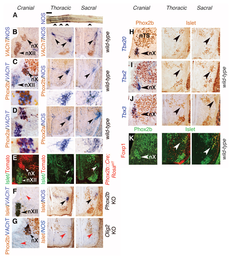
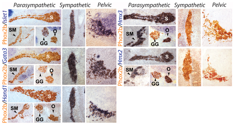
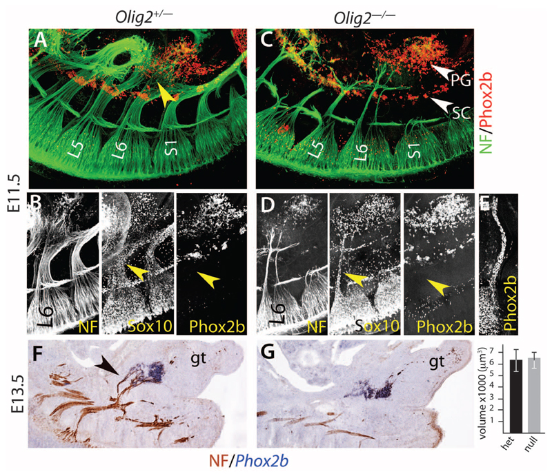
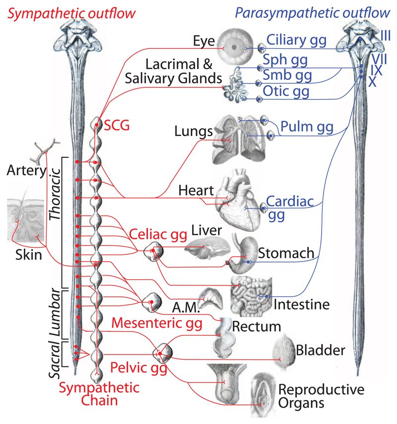
A kinship between cranial and pelvic visceral nerves of vertebrates has been accepted for a century. Accordingly, sacral preganglionic neurons are considered parasympathetic, as are their targets in the pelvic ganglia that prominently control rectal, bladder, and genital functions. Here, we uncover 15 phenotypic and ontogenetic features that distinguish pre- and postganglionic neurons of the cranial parasympathetic outflow from those of the thoracolumbar sympathetic outflow in mice. By every single one, the sacral outflow is indistinguishable from the thoracolumbar outflow. Thus, the parasympathetic nervous system receives input from cranial nerves exclusively and the sympathetic nervous system from spinal nerves, thoracic to sacral inclusively. This simplified, bipartite architecture offers a new framework to understand pelvic neurophysiology as well as development and evolution of the autonomic nervous system.


Figures

Fig. 1
Fig. 1. Sacral preganglionic neurons develop like sympathetic, not parasympathetic, ones.
(A) Longitudinal thick section of the spinal cord reacted for a reduced form of nicotinamide adenine dinucleotide phosphate (NADPH) diaphorase activity indicative of NOS expression, revealing the thoracolumbar and sacral visceromotor columns (arrowheads) separated by a gap. (B to K) Transverse sections at E13.5 through the right half of the medulla (left column in both panels), thoracolumbar spinal cord (middle), and sacral spinal cord (right), stained with the indicated antibodies and probes, or for NOS expression, in the genetic backgrounds indicated on the right. (B) The dorsal motor nucleus of the vagus nerve (nX) expresses VAChT but not NOS, whereas the thoracic and sacral preganglionic neurons (arrowheads) express NOS but not yet VAChT. The ventrally located somatic motoneurons, including the hypoglossal nucleus (nXII) in the hindbrain, express VAChT. [(C) and (D)] Phox2b (C) and Phox2a (D) are expressed in nX but in neither thoracic nor sacral preganglionic neurons (arrowheads). Lower panels in (C) and (D): higher magnifications of the preganglionic neurons. (E) Neurons of nX but neither thoracic nor sacral preganglionic ones (labeled by an antibody to Islet1/2, white arrowheads) derive from Phox2b+ precursors, permanently labeled in a Phox2b::Cre;RosatdT background. (F) nX is missing in Phox2b knockouts (red arrowhead), but thoracic and sacral preganglionic neurons are spared (black arrowheads). (G) nX is spared in Olig2 knockouts (black arrowhead), but thoracic and sacral preganglionic neurons are missing (red arrowheads). nXII is also missing, as expected of a somatic motor nucleus (red arrowhead). [(H) to (J)] Tbx20, Tbx2, and Tbx3 are expressed in all or a subset of nX neurons (arrowheads in panels of the left column) but in no thoracic or sacral preganglionic neuron (arrowheads in panels of the middle and right columns). (K) Foxp1 is not expressed in the nX (arrowhead in left column) but is a marker of both thoracic and sacral preganglionic neurons (arrowheads in middle and right columns). nTS, nucleus of the solitary tract. Scale bars: 1 mm (A), 100 μm [(B) to (K)].
Fig. 2
Fig. 2. All pelvic ganglionic cells have a sympathetic, not parasympathetic, transcriptional signature.
Sagittal sections through parasympathetic ganglia (columns headed “Parasympathetic”), the lumbar paravertebral sympathetic chain (columns headed “Sympathetic”), and the pelvic ganglion (columns headed “Pelvic”) at E13.5, stained by inmmunohistochemistry for Phox2b, a determinant of all autonomic ganglia (31), and in situ hybridization for the indicated probes. GG, geniculate ganglion (a cranial sensory ganglion); O, otic ganglion; S, sphenopalatine ganglion; SM, submandibular ganglion (all parasympathetic ganglia).
Fig. 3
Fig. 3. The pelvic ganglion forms independently of its nerve, like sympathetic and unlike parasympathetic ones.
(A and C). Whole-mount immunofluorescence with the indicated antibodies on E11.5 embryos either heterozygous (A) or homozygous (C) for an Olig2 null mutation. The nascent pelvic nerves [yellow arrowhead in (A)] seem to derive mostly from the L6 nerve at that stage. The Olig2 null mutation (C) spares two thin sensory pelvic projections. The pelvic ganglion (PG) lies ahead of most fibers in both heterozygous and mutant background. (B and D). View of the L6 nerve, covered with Sox10+ cells but no Phox2b+ cells (yellow arrowheads), unlike cranial nerves that give rise to parasympathetic ganglia at the same stage [Jacobson’s nerve in (E)]. (F and G) In situ hybridization for Phox2b and immunohistochemistry for neurofilament (NF) on heterozygous and homozygous Olig2 knockouts at E13.5, when parasympathetic ganglia have formed elsewhere in the body. Graph: the pelvic ganglion has the same volume whether its preganglionic nerve is present [black arrowhead in (F)] or not (6369 μm3 ± 1066 versus 6441 μm3 ± 919, P = 0.96, n = 5 embryos). gt, genital tubercle; L5 and L6, 5th and 6th lumbar roots; S1, 1st sacral root; SC, sympathetic chain.
Fig. 4
Fig. 4. Revised anatomy of the autonomic nervous system.
The efferent path of the autonomic nervous system is made up of a spinal sympathetic outflow (left, in red) and a cranial parasympathetic outflow (right, in blue). Sympathetic targets in the skin other than arteries are piloerector muscles and sweat glands. III, oculomotor nerve; VII, facial nerve; IX, glosso-pharyngeal nerve; X, vagus nerve; A.M., adrenal medulla; gg, ganglion; Pulm, pulmonary; SCG, superior cervical ganglion; Sph, sphenopalatine; Smb, submandibular. For a larger version, see fig. S11.

Comment in

References

    1. Gaskell WH. J Physiol. 1886;7:1–80.9. - PMC - PubMed
    1. Langley JN. The Autonomic Nervous System: Part I. W. Heffer; Cambridge: 1921.
    1. Kandel E, Schwartz J, Jessell T, Siegelbaum S, Hudspeth AJ. Principles of Neural Science. Fifth Edition. McGraw-Hill Professional; 2012.
    1. Langley JN, Anderson HK. J Physiol. 1895;19:71–139. - PMC - PubMed
    1. Briscoe J, et al. Nature. 1999;398:622–627. - PubMed

Publication types

MeSH terms

Substances