. 2016 Nov 18:7:13458.
doi: 10.1038/ncomms13458.

An elastic element in the protocadherin-15 tip link of the inner ear

Affiliations

An elastic element in the protocadherin-15 tip link of the inner ear

Raul Araya-Secchi et al. Nat Commun. .

Abstract

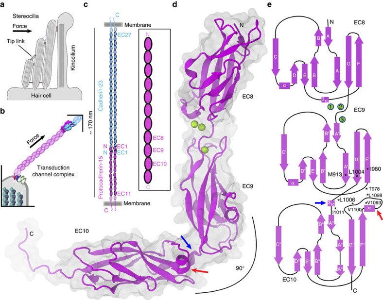
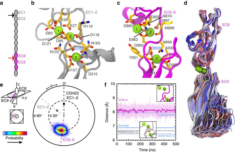
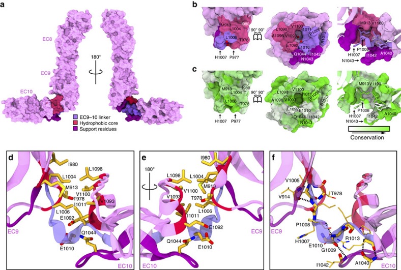
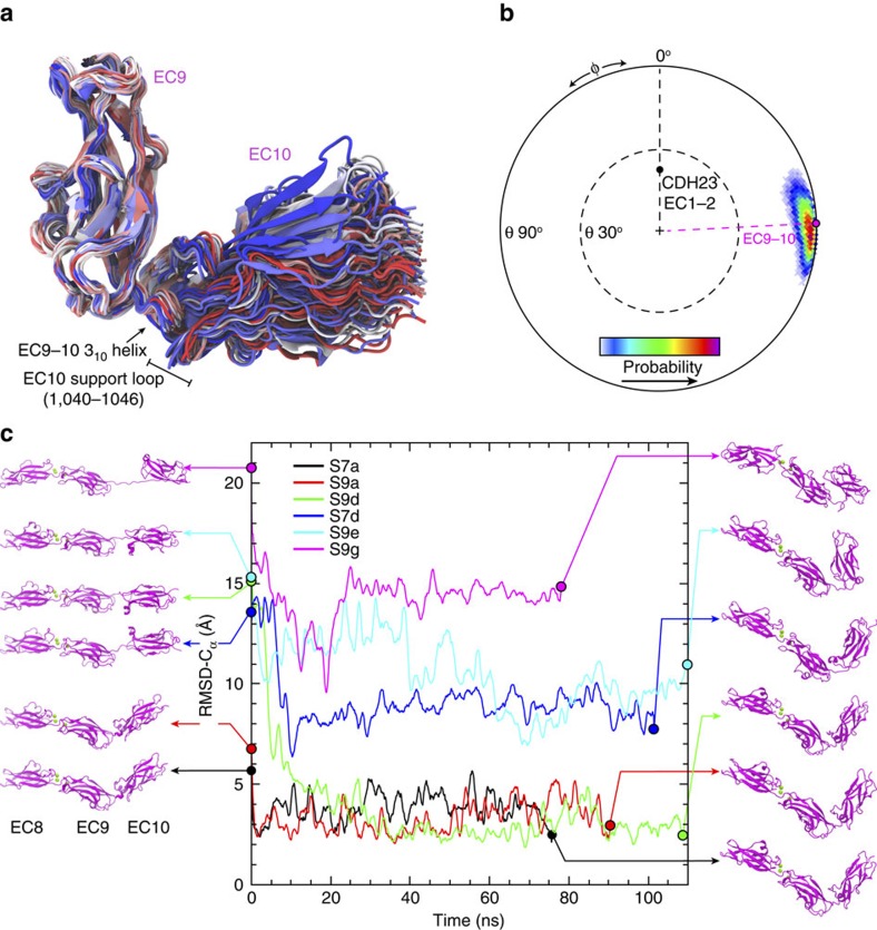
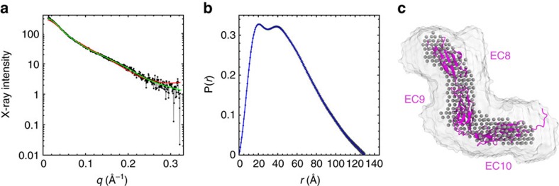
Tip link filaments convey force and gate inner-ear hair-cell transduction channels to mediate perception of sound and head movements. Cadherin-23 and protocadherin-15 form tip links through a calcium-dependent interaction of their extracellular domains made of multiple extracellular cadherin (EC) repeats. These repeats are structurally similar, but not identical in sequence, often featuring linkers with conserved calcium-binding sites that confer mechanical strength to them. Here we present the X-ray crystal structures of human protocadherin-15 EC8-EC10 and mouse EC9-EC10, which show an EC8-9 canonical-like calcium-binding linker, and an EC9-10 calcium-free linker that alters the linear arrangement of EC repeats. Molecular dynamics simulations and small-angle X-ray scattering experiments support this non-linear conformation. Simulations also suggest that unbending of EC9-10 confers some elasticity to otherwise rigid tip links. The new structure provides a first view of protocadherin-15's non-canonical EC linkers and suggests how they may function in inner-ear mechanotransduction, with implications for other cadherins.


Figures

Figure 1
Figure 1. Hair-cell mechanotransduction and structure of PCDH15.
(a) Schematic representation of a cochlear hair-cell stereocilia bundle highlighting the location of the tip link. (b) Mechanotransduction apparatus. PCDH15 directly conveys force to transduction channels. (c) The tip link is formed by the tip-to-tip interaction between CDH23 and PCDH15 parallel dimers. Inset shows the location of the repeats studied here. (d) Ribbon diagram of PCDH15 EC8–10. Calcium ions in the EC8–9 linker are shown as green spheres. The calcium-free EC9–10 linker is bent. (e) Topology diagram of PCDH15 EC8–10. A typical cadherin fold with seven β strands (labeled A to G) is observed for all EC repeats. The structure shows a novel EC9–10 310 helix (blue arrow) at the EC9–10 linker and an atypical EC10 FG-α loop (red arrow). Residues that form the EC9–10 interface are highlighted with an asterisk (*).
Figure 2
Figure 2. Structure and simulated dynamics of PCDH15 EC8–9.
(a) Schematics of PCDH15's extracellular domain and the location of linkers EC1–2 (black arrow) and EC8–9 (red arrow). (b,c) Detail of calcium-binding sites at the PCDH15 EC1–2 and EC8–9 linkers, respectively. Protein backbone shown as gray (EC1–2) and magenta (EC8–9) ribbon. Relevant residues are shown in stick representation. (d) Superposition of PCDH15 EC8–9 conformations taken every 5 ns from an equilibrium MD simulation (S1c; Table 2) using EC8 as a reference. Colour indicates time (blue–white–red). (e) To quantify the conformational freedom of EC9 with respect to EC8 the principal axis of EC8 was aligned to the z axis, and the projection in the xy plane of the principal axis of EC9 was plotted as a probability map for data taken every 20 ps of simulation S1c. The initial orientations for EC8–9, CDH23 EC1–2 (2WHV; φ≡0°), and PCDH15 EC1–2 (4APX) are shown as magenta, black and gray circles, respectively. (f) Distance between calcium ions at sites 2 and 3 measured every 20 ps during simulation S1c (pink). A 1 ns running average is shown in magenta; the average for the same distance measured for 41 cadherin linkers (Supplementary Table 1) is shown in blue (dashed lines show maximum and minimum). The same distance measured for PCDH15 EC1–2 is shown as a gray dotted line.
Figure 3
Figure 3. Structure of PCDH15 EC9–10 interface.
(a) Surface representation of PCDH15 EC8–10 (magenta). The EC9–10 interface is shown with its EC9–10 linker (violet), a hydrophobic core (dark pink) and supporting residues (purple). (b) PCDH15 EC9–10 interaction surfaces exposed (left and middle panels) and coloured as in a with interfacing residues labeled. Right panel shows side view as in a. Red spheres correspond to a pair of crystallographic water molecules. (c) Conservation of residues in the EC9–10 interface according to ConSurf and the alignment in Supplementary Fig. 4. (d,e) Detail of the EC9–10 interface. Protein backbone is shown as ribbons and relevant residues are shown as sticks. Some backbone atoms are omitted for clarity. (f) Detail of backbone-hydrogen bonds (dashed lines) found in the EC9–10 linker.
Figure 4
Figure 4. Simulated dynamics of PCDH15 EC9–10.
(a) Superposition of PCDH15 EC9–10 conformations taken every 5 ns from an equilibrium MD simulation and using EC9 as reference (S1c; Table 2). Colour indicates time (blue–white–red). The EC9–10 310 helix and EC10 support loop switch conformations throughout the trajectory. (b) Conformational freedom of EC10 with respect to EC9 as in Fig. 2e. The initial orientations for EC9–10 and CDH23 EC1–2 (2WHV, used as reference) are indicated. (c) r.m.s.d.-Cα from selected ‘relaxation' equilibrium MD simulations (Movie 1) started from stretched conformations (left). Final conformations of each relaxation run are shown on the right. r.m.s.d.-Cα is shown as 1 ns running average for all cases.
Figure 5
Figure 5. Low-resolution PCDH15 EC8–10 structure by SAXS.
(a) X-ray scattering intensity as a function of the scattering vector q (SAXS profile; black). Predicted scattering intensities from SAXS model (χ2=1.01; DAMMIF) and the PCDH15 EC8–10 structure (χ=1.59; FOXS) are shown as green and red lines, respectively. (b) Real-space pair distribution function (P(r)) from SAXS data. (c) Superposition of PCDH15 EC8–10 structure (magenta) with an averaged envelope (white surface) and a filtered solution (gray spheres) from ab initio models.
Figure 6
Figure 6. Constant-force SMD simulations of PCDH15 EC8–10.
(a) End-to-end distance measured for constant-force stretching of PCDH15 EC8–10 at 10 pN (simulation S3a, olive green), 25 pN (S3d, green), 50 pN (S3b, dark brown) and 100 pN (S3c, light brown). A 1 ns running average is shown in all cases. Interatomic distances for: (b) Val 1,005:Cα–Arg 1,013:Cα (light blue) His 1,007:O–Glu 1,010:N (red), Gly 1,009:O–Ile 1,042:N (orange), Arg 1,013:O–Ala 1,040:N (magenta) and Arg 1,013:N–Ala 1,040:O (purple), and (c) Leu 1,004:Cγ–Ala 1,096:Cβ (dark gray). (d) Snapshots of initial conformation and mechanically induced unbending of PCDH15 EC8–10 taken from the 10 pN simulation (S3a). Protein is shown as ribbons and coloured as in Fig. 3. Arrows indicate position and direction of the applied forces. (eh) Detail of the EC9–10 linker at time points indicated in ac.
Figure 7
Figure 7. Constant-velocity SMD simulations of PCDH15 EC8–10.
(a) Force applied to N terminus versus end-to-end distance for constant-velocity stretching of PCDH15 EC8–10 at 10 nm ns−1 (simulation S5d, black), 5 nm ns−1 (S5c, turquoise), 1 nm ns−1 (S5a1, light blue), 0.1 nm ns−1 (S5b, 1 ns running average, light green) and 0.02 nm ns−1 (S5e, 1 ns running average, dark green). (b) Snapshots of initial conformation and mechanically induced unbending and unfolding of PCDH15 EC8–10 taken from the 0.02 nm ns−1 simulation (S5e; Movie 2). Protein is shown in ribbon representation and coloured as in Fig. 3. Springs indicate position and direction of the applied forces. (cf) Force applied to PCDH15 EC8–10 N terminus (S5e, dark green) along with interatomic distances for (c,d) Arg 1,013:O–Ala 1,040:N (magenta) and Arg 1,013:N–Ala 1,040:O (purple); Tyr 1,019:N–Lys 1,108:O (light magenta); and Tyr 1,019:O–Tyr 1,110:N (pink). Rupture of these interactions correlates with unfolding force peaks. (e) Interatomic distances for His 1,007:O–Glu 1,010:N (red), Gly 1,009:O–Ile 1,042:N (orange), (f) Leu 1,004:Cγ–Leu 1,098:Cγ (maroon), and Leu 1,004:Cγ–Ala 1,096Cβ (dark gray). Rupture of these interactions correlates with unbending. A 1 ns running average is shown in all cases. (gj) Snapshots of the EC9–10 linker during S5e at time points indicated in e,f.
Figure 8
Figure 8. Mechanics of a chimeric tip link.
(a) Transduction apparatus diagram. The full-length PCDH15 (magenta), with its 11 EC repeats, forms a homophilic dimer that interacts with CDH23 (light blue). Known structures, including EC8–10, are shown. Inset shows a chimeric complex with CDH23 EC1–2 interacting with PCDH15 EC1–2–8–10. (b) Snapshots of initial conformation and mechanically induced unbending and unbinding of the chimeric complex taken from the 0.1 nm ns−1 simulation (S14a; Movie 3). Springs indicate position and direction of the applied forces. Unbinding with minimal unfolding follows unbending. (c) Force applied to one of the C termini versus end-to-end distance of the complex for constant velocity stretching at 1 (S14b1, teal) and 0.1 nm ns−1 (S14a, olive). (d) Models of tip-link function as a gating spring (left), stiff cable (middle) and a semi-elastic filament (right).

References

    1. Gillespie P. G. & Müller U. Mechanotransduction by hair cells: models, molecules, and mechanisms. Cell 139, 33–44 (2009). - PMC - PubMed
    1. Hudspeth A. J. Integrating the active process of hair cells with cochlear function. Nat. Rev. Neurosci. 15, 600–614 (2014). - PubMed
    1. Fettiplace R. & Kim K. X. The physiology of mechanoelectrical transduction channels in hearing. Physiol. Rev. 94, 951–986 (2014). - PMC - PubMed
    1. Hudspeth A. J. & Corey D. P. Sensitivity, polarity, and conductance change in the response of vertebrate hair cells to controlled mechanical stimuli. Proc. Natl Acad. Sci. USA 74, 2407–2411 (1977). - PMC - PubMed
    1. Pickles J. O., Comis S. D. & Osborne M. P. Cross-links between stereocilia in the guinea pig organ of Corti, and their possible relation to sensory transduction. Hear. Res. 15, 103–112 (1984). - PubMed

Publication types