. 2016 Nov 18:6:36478.
doi: 10.1038/srep36478.

Dormant cancer cells accumulate high protoporphyrin IX levels and are sensitive to 5-aminolevulinic acid-based photodynamic therapy

Affiliations

Dormant cancer cells accumulate high protoporphyrin IX levels and are sensitive to 5-aminolevulinic acid-based photodynamic therapy

Taku Nakayama et al. Sci Rep. .

Abstract

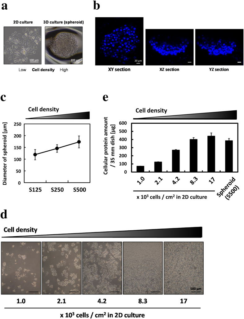
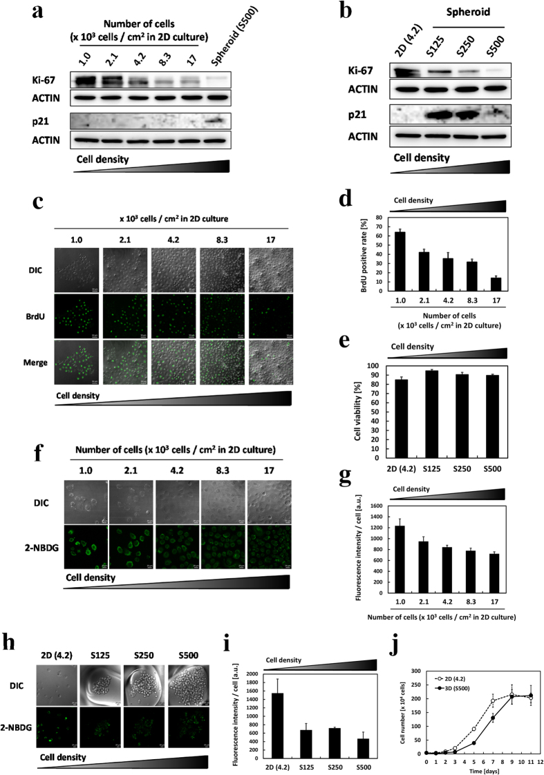
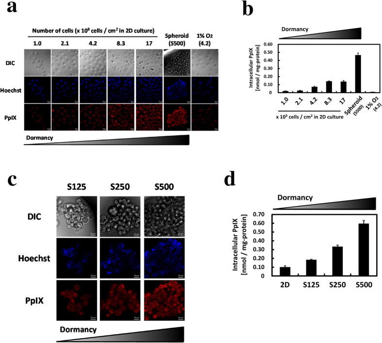
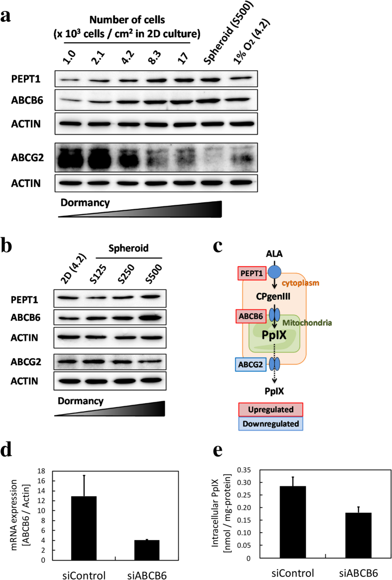
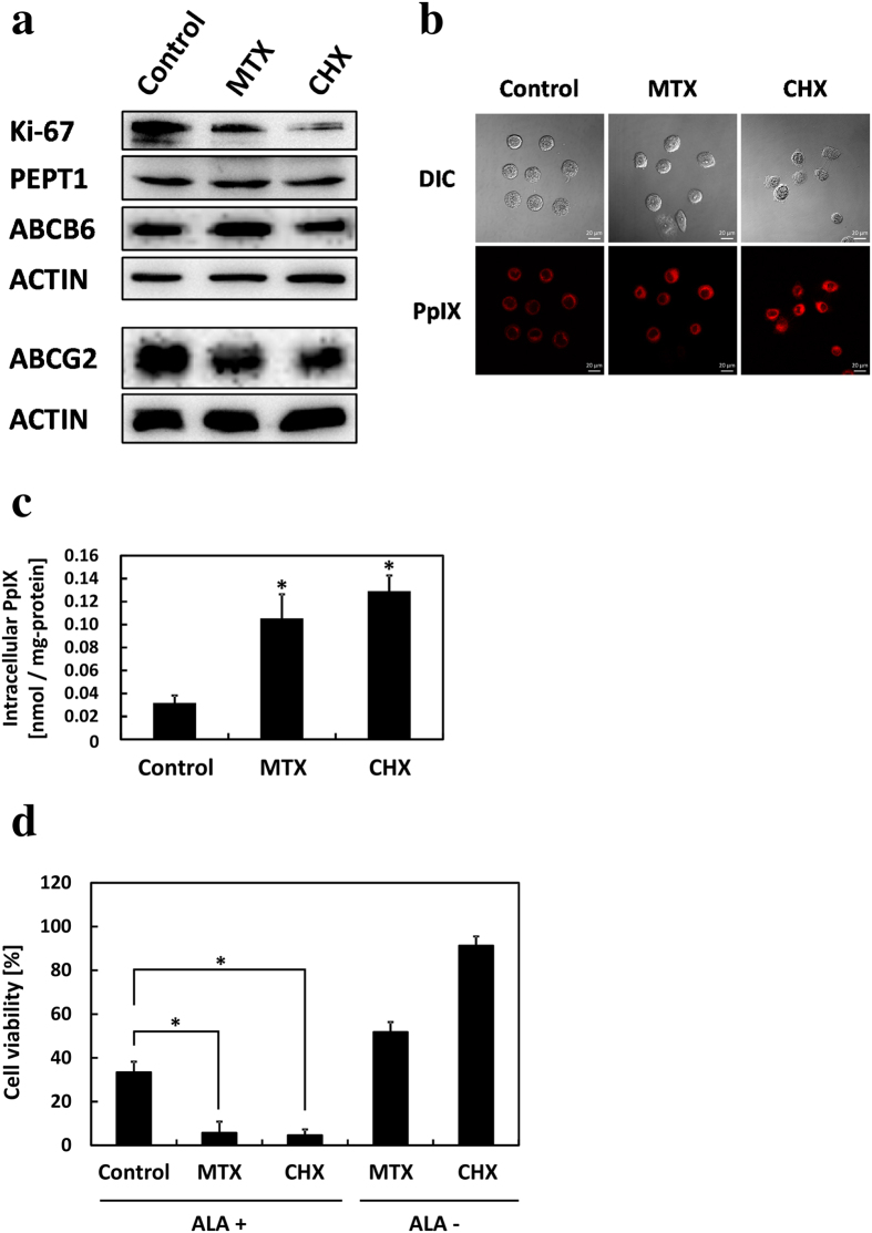
Photodynamic therapy (PDT) and diagnosis (PDD) using 5-aminolevulinic acid (ALA) to drive the production of an intracellular photosensitizer, protoporphyrin IX (PpIX), are in common clinical use. However, the tendency to accumulate PpIX is not well understood. Patients with cancer can develop recurrent metastatic disease with latency periods. This pause can be explained by cancer dormancy. Here we created uniformly sized PC-3 prostate cancer spheroids using a 3D culture plate (EZSPHERE). We demonstrated that cancer cells exhibited dormancy in a cell density-dependent manner not only in spheroids but also in 2D culture. Dormant cancer cells accumulated high PpIX levels and were sensitive to ALA-PDT. In dormant cancer cells, transporter expressions of PEPT1, ALA importer, and ABCB6, an intermediate porphyrin transporter, were upregulated and that of ABCG2, a PpIX exporter, was downregulated. PpIX accumulation and ALA-PDT cytotoxicity were enhanced by G0/G1-phase arrestors in non-dormant cancer cells. Our results demonstrate that ALA-PDT would be an effective approach for dormant cancer cells and can be enhanced by combining with a cell-growth inhibitor.


Conflict of interest statement

SBI Pharma CO., Ltd., provided support in the form of salaries for authors M.N. and T.T., but did not have any additional role in the study design, data collection and analysis, decision to publish, or preparation of the manuscript.

Figures

Figure 1
Figure 1. PC-3 spheroid formed in an EZSPHERE 3D culture plate and different cell density in the 2D culture.
All cells were incubated for 4 days. (a) Phase-contrast images of a spheroid. Scale bar, 100 μm. (b) Hoechst 33342 stains for nuclei. A z-stack section was obtained every 2 μm. Scale bar, 20 μm. (c) The diameter of formed spheroids. S125, 1.25; S250, 2.50; S500, 5.00 × 105 cells/35-mm dish. Over 40 spheroids were counted. (d) Phase-contrast images of cells in the 2D culture. Scale bar, 500 μm. (e) Cellular protein amount measured by Bradford assay; n = 3. All bars represent standard deviation (SD).
Figure 2
Figure 2. Investigation of cell proliferation, cell death, glucose uptake, and recovery of active status in the 2D culture and spheroids.
(a) Protein expression level of Ki-67 and p21 in the 2D culture was detected by Western blotting. (b) Protein expression levels of Ki-67 and p21 in the 3D culture were detected by Western blotting. (c) Immunochemistry images of BrdU-positive cells in the 2D culture. Scale bar, 50 μm. (d) Percent of BrdU-positive cells. n = 3. At least 400 cells were counted at each cell density. (e) Cell viability was measured by trypan blue staining. (f) 2-NBDG uptake in the 2D culture was analyzed by confocal fluorescence microscopy. Cells were incubated with glucose and FBS-free medium for 24 h, after which 100 μM 2-NBDG was added, followed by incubation for 25 min at 37 °C. (g) Fluorescence intensity of 2-NBDG in the 2D culture. (h) 2-NBDG uptake in the 3D culture was analyzed by confocal fluorescence microscopy. (i) Fluorescence intensity of 2-NBDG in the 3D culture. (j) Re-growth of degraded spheroids in the 3D culture compared with that in the 2D culture. Cells were incubated for 4 days to form S500 spheroids and for the same period for the 2D culture. After 4 days, cell numbers in the 2D culture and spheroids were confirmed to be similar. Then, cells were again placed in the 2D culture at 4.2 × 103 cells/cm2. Cells were counted by trypan blue staining. n = 3. Bars represent standard deviations (SD).
Figure 3
Figure 3. The effect of cell dormancy on PpIX accumulation.
After incubation for 3 days, cells were incubated for 24 h with 1 mM ALA-containing medium. (a) Confocal laser scanning microscopy images of differential interference contrast, Hoechst 33342 for nuclei and PpIX. Scale bar, 20 μm. (b) PpIX accumulations under different cell density conditions in the 2D culture were measured by HPLC. n = 3. (c) Confocal laser scanning microscopy images of different sizes of spheroids. Scale bar, 20 μm. (d) PpIX accumulation at different spheroid sizes was measured by HPLC. n = 3. Bars represent standard deviation (SD).
Figure 4
Figure 4. Effect of ALA-PDT on cell viability.
Cells were incubated with 1 mM ALA in a complete medium for 24 h and then exposed to 1080 mJ/cm2 light for 5 min. Cell viability was determined on the next day by the MTT assay after treatment. (a) ALA-PDT in the 2D culture. Cells were incubated for 1 day before ALA treatment. n = 6. (b) ALA-PDT in spheroids. Cells were incubated for 3 days before ALA treatment. The cell density of “2D” is the same as that of “S500”. n = 3. Bars represent standard deviation (SD) (Table 1) Percentage cell viability in the 2D culture. (Table 2) Percentage cell viability in spheroids.
Figure 5
Figure 5. The protein expressions of PEPT1, ABCB6, and ABCG2 were detected by Western blotting.
No cells were incubated with 1 mM ALA. (a) Expression at different cell density in the 2D culture. (b) Expression with different sizes of spheroids. (c) Regulation of transporters involved in porphyrin metabolism under high-cell-density 2D culture or in spheroids. (d) Expression of ABCB6 mRNA after incubation with siRNA in the high cell density-2D cultured cells. (e) PpIX accumulation after incubation with siRNA in the high cell density-2D cultured cells. CPgenIII: coproporphyrinogen III.
Figure 6
Figure 6. Porphyrin metabolism changed in cancer cells on addition of methotrexate (MTX) or cycloheximide (CHX).
MTX was cultured with 10 μM and CHX was cultured with 10 μg/ml for 48 h. (a) Protein expression was detected by Western blotting. (b) Confocal laser scanning microscopy images of PpIX accumulation. Scale bar, 20 μm. (c) PpIX accumulation was measured by HPLC. n = 3. *p < 0.005, compared with control. (d) Effect of ALA-PDT on cell viability. Cells were incubated with 1 mM ALA for 24 h and then exposed to 1080 mJ/cm2 light for 5 min. Cell viability was determined with trypan blue. Cell viability was normalized by untreated control samples. n = 3. *p < 0.003. All bars represent standard deviation (SD).

Similar articles

Cited by

References

    1. Krammer B. & Plaetzer K. ALA and its clinical impact, from bench to bedside. Photochem. Photobiol. Sci. 7, 283–289 (2008). - PubMed
    1. Kennedy J. C. & Pottier R. H. Endogenous protoporphyrin IX, a clinically useful photosensitizer for photodynamic therapy. J. Photochem. Photobiol. B 14, 275–292 (1992). - PubMed
    1. Tabata K., Ogura S. & Okura I. Photodynamic efficiency of protoporphyrin IX: comparison of endogenous protoporphyrin IXinduced by 5-aminolevulinic acid and exogenous porphyrin IX. Photochem. Photobiol. 66, 842–846 (1997).
    1. Stummer W. et al.. Fluorescence-guided surgery with 5-aminolevulinicacid for resection of malignant glioma: a randomised controlledmulticentre phase III trial. Lancet Oncol 7, 392–401 (2006). - PubMed
    1. Inoue K. et al.. Comparison between intravesical and oraladministration of 5-aminolevulinic acid in the clinical bene-fit of photodynamic diagnosis for non-muscle invasive bladdercancer. Cancer. 118, 1062–1074 (2012). - PubMed

Publication types

MeSH terms