. 2016 Jul-Sep;26(3):342-351.
doi: 10.4103/0971-3026.190426.

CT manifestations of small bowel ischemia due to impaired venous drainage-with a correlation of pathologic findings

Affiliations

CT manifestations of small bowel ischemia due to impaired venous drainage-with a correlation of pathologic findings

Chung Kuao Chou. Indian J Radiol Imaging. 2016 Jul-Sep.

Abstract

Acute abdominal pain may result from a wide variety of medical and surgical diseases. One of these diseases is small bowel ischemia, which may result in a catastrophic outcome if not recognized and treated promptly. Computed tomography (CT) by its faster image acquisition, thinner collimation, high resolution, and multiplanar reformatted images has become the most important imaging modality in evaluating the acute abdominal conditions. In this article, the author presents a description of the histology of the small bowel, pathophysiology of small bowel change, and a correlation of the pathologic and CT findings of the small bowel injuries due to impaired venous drainage. A convincing correlation of the microscopic mucosal condition with the enhancement pattern of the thickened small bowel wall on CT is useful in definitely describing the mucosal viability.

Keywords: Computed tomography; Intestines; Ischemia; Small bowel.


Figures
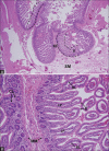
Figure 1 (A and B)
Figure 1 (A and B)
A 44-year-old woman with an adhesion-induced hernia in the pelvic cavity. (A) Photomicrograph (hematoxylin and eosin stain, original magnification ×40) shows the surgical margin of a resected small-bowel segment and a normal Kerckring fold (KF). Covering the surface of the fold is an aggregation of numerous villi (V), which forms the mucosa (bounded by dotted lines). These are visible on computed tomography. Although the underlying submucosa (SM) is slightly edematous, the core of the Kerckring fold remains uninvolved. (B) Photomicrograph (hematoxylin and eosin stain, original magnification ×200) shows that the villi are slender, covered by epithelium (E), and well separated from each other. They are approximately 0.5–1.0 mm tall. The mucosa at the surgical margin was judged viable, whereas the central portion of the resected bowel was necrotic (not shown). The basal lamina (BL) is barely visible, in contrast to the much thicker muscularismucosae (MM). CL = crypt of Lieberkühn; LP = lamina propria, the core of the villus
Figure 2 (A-D)
Figure 2 (A-D)
A 3-year-old boy with volvulus. (A) Photomicrograph (hematoxylin and eosin stain, original magnification ×40) shows that the columnar architecture of the villi (bounded by dotted lines) is still maintained, even though all of the epithelium has been sloughed off (necrotic mucosa). (B) Photomicrograph (hematoxylin and eosin stain, original magnification ×200) demonstrates the swollen lamina propria (LP) of the villi, which is predominantly filled with red blood cells and necrotic tissue. Inflammatory cells are relatively few. The covering epithelia are all disrupted, sloughed, and not visible. This kind of villus is called a ghost villus. These swollen ghost villi are compact together, in contrast to those normal, sufficiently separated slender villi shown in Figure 1. Most villi are still confined by a basal lamina (BL). (C) Non-enhanced computed tomography (CT) scan depicts the target-like, thickened wall with a high-attenuating inner layer (arrow), which is nearly isodense to the adjacent muscles (arrowhead). This finding is compatible with a necrotic mucosa composed of closely contact, RBC-filled ghost villi, as shown in A. The submucosa is edematous or hemorrhagic. (D) On the contrast-enhanced CT scan, the inner layer (arrow) is not enhancing and has become hypoattenuating compared with the abdominal wall muscles (arrowhead), indicating poor blood supply to the mucosa. The edematous submucosa is enhanced and becomesisodense to hemorrhagic mucosa, even though still hypodense to muscles
Figure 3 (A and B)
Figure 3 (A and B)
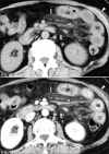
A 58-year-old woman with mesenteric panniculitis, which is a preceding process of retractile mesenteritis. Varying degrees of mesenteric fibrosis may constrict venous branches and result in impaired venous drainage. (A) Non-enhanced scan. The small bowel wall (SB) is symmetrically thickened and hypoattenuating relative to the muscles (arrowheads). The mesenteric fat (M) is diffusely hyperattenuated to the other fatty tissues and partly covered anteriorly by the thickened visceral peritoneum (arrow) of the mesentery. (B) On this contrast-enhanced scan, the congested wall has become hyperdense to the muscles
Figure 4 (A-D)
Figure 4 (A-D)
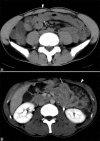
A 70-year-old man with adhesion-induced strangulation. (A) Photomicrograph (hematoxylin and eosin stain, original magnification ×40) shows an edematous and hemorrhagic submucosa (SM). (B) Photomicrograph (hematoxylin and eosin stain, original magnification ×100) shows some villi with ulceration of their tips (arrow), evidence of earliest ischemic necrosis. However, most villi (arrowheads) remain slender and architecturally intact. MM = muscularis mucosae. (C) Non-enhanced scan shows a thickened wall (arrowhead) that is somewhat homogeneous in attenuation and darker than the muscles (arrow). (D) On contrast-enhanced computed tomography, attenuation of the normally enhancing mucosa (arrowhead) is higher than that of the muscles (arrow). The swollen Kerckring folds closely contact together, forming a black-white interlacing appearance composed of the enhancing mucosa and submucosal edema. The patient received surgical enterolysis of the adhesion with segmental small-bowel resection. The pathologic specimen was not judged as necrotic
Figure 5 (A-D)
Figure 5 (A-D)
(A–C) An 83-year-old woman with adhesion-induced strangulation. (A) Photomicrograph (hematoxylin and eosin stain, original magnification ×40) shows a predominantly edematous submucosa (SM) and a severely swollen Kerckring fold (KF). (B) On this photomicrograph (hematoxylin and eosin stain, original magnification ×200), ghost villi (V) are swollen and compacted together. Their mean height is approximately 0.5 mm, similar to that of normal villi. Boundaries of some villi are intact; others are lost (arrowheads). The villi are filled with red blood cells, inflammatory cells, and necrotic tissue. BL = basal lamina. (C) Contrast-enhanced computed tomography (CT) scan shows a thickened wall (arrow) of homogeneous attenuation without a recognizable, enhancing inner layer. The thickened wall is hypodense to the adjacent muscles. This appearance is consistent with mucosal necrosis. The patient recovered after surgery. (D) Contrast-enhanced CT scan in an 81-year-old woman with episodes, pathologic results, and an outcome similar to those of the patient in A–C. The thickened ileum (arrow), approximately 2 cm in outer diameter, showed no inner-layer enhancement
Figure 6 (A-D)
Figure 6 (A-D)
A 42-year-old man with superior mesenteric venous thrombosis. (A) Photomicrograph (hematoxylin and eosin stain, original magnification ×40) shows the submucosa (SM) stuffed with red blood cells (RBCs). (B) Photomicrograph (hematoxylin and eosin stain, original magnification ×200) shows ghost villi that are swollen and compacted. Some are still confined by a basal lamina (BL), whereas others (arrowhead) are disrupted and merged together. The lamina propria (LP) is filled with RBCs and necrotic tissue. MM = muscularis mucosa. (C) On non-enhanced computed tomography (CT), attenuation is higher on the left half (arrow) of the thickened wall than on the right (arrowhead), and it is similar to the adjacent muscles. This finding suggests an increased hemorrhagic component in the left half. (D) On contrast-enhanced CT scan, the right half (arrowhead) has normally enhancing mucosa, including Kerckring folds, and is now clearly seen. The left half (arrow) does not show similar inner-layer and fold enhancement (an appearance consistent with mucosal necrosis), and it is hypoattenuating relative to the muscles
Figure 7
Figure 7
A 73-year-old man with an incarcerated left inguinal hernia. The covering mucosa of upper segment (arrow) is poorly enhancing on contrast-enhanced computed tomography. The patient recovered after surgical reduction without resection of the small bowel. This outcome indicates that transmural necrosis did not occur, though the mucosa might have been damaged or necrotic. The normally enhancing mucosa of the lower segment (arrowhead) was about 1 mm thick. The heaved Kerckring fold, approximately 4–6 mm tall, is composed of a dark edematous submucosa (like a mountain) and covered by a bright mucosa (like a forest)
Figure 8 (A and B)
Figure 8 (A and B)
A 54-year-old man with superior mesenteric venous thrombosis. (A). Early phase of computed tomographic arterioportography. The mucosa of the target-like, thickened wall (arrowhead) is hypodense to that of the uninvolved segment (arrow). (B). Delayed phase shows a contrary appearance. The mucosa of the thickened wall (arrowhead) becomes hyperdense to that of the uninvolved segment (arrow). These appearances reflect a slow blood inflow and delayed extravasation of molecules of the contrast material into the extravascular space, resulting in a prolonged mucosal enhancement in contrast to that of the normally circulated segment
Figure 9
Figure 9
A 30-year-old woman who received total colectomy. Contrast-enhanced scan shows a nonoccluding thrombus (arrowhead) in the superior mesenteric vein and an adjacent abscess (arrow)
Figure 10 (A-F)
Figure 10 (A-F)
A 32-year-old woman with an adhesion-induced internal hernia. (A–F) Contrast-enhanced skipped, nonadjacent images. The circumscribed segment is totally filled with fluid and can be successfully traced from one end (1) to the other (18). Both ends are in close contact. The wall is approximately 2.75–3.75 mm thick. It is poorly enhancing and darker than the muscles. The patient received small-bowel resection and recovered. The specimen showed hemorrhagic necrosis of the full thickness of the wall
Figure 11
Figure 11
A 34-year-old man with an adhesion-induced closed-loop obstruction. Coronal reformatted image clearly shows a beak sign and the constriction point (arrows), and submucosal edema (arrowheads) of the wall
Figure 12
Figure 12
A 23-year-old woman with polyposis-induced intussusception. Mesenteric vessels and fatty tissue are clearly seen in the bowel lumen. The thickened submucosa is either covered by normally enhancing, viable mucosa (arrow) or poorly enhancing, necrotic mucosa (arrowhead) which is invisible. The pathologic specimen showed transmural necrosis
Figure 13 (A and B)
Figure 13 (A and B)
A 30-year-old man with nonspecific abdominal pain. (A) On this non-enhanced scan, the collapsed small bowel (arrow) has a wall thickness larger than 1cm and an outer diameter of approximately 25mm. Its attenuation is lower than that of the adjacent muscle (arrowhead). (B) Strongly enhancing bowel (arrow) is brighter than the muscles (arrowhead) on this contrast-enhanced scan. The patient recovered uneventfully
Figure 14
Figure 14
Same patient as in Figure 13. Kerckring folds (arrows) of the longitudinally sectioned, distended bowel isapproximately 4–6 mm high and 2–3 mm thick, with a 2–3 mm interfold distance. The section thickness is 7.5mm. A transverse scan of the wall (rectangles) would involve two folds at most like that of the adjacent, continuous, transversely sectioned bowel (arrowheads); this can mimic a target sign or a poorly enhancing thickened wall. This appearance should not be interpreted as wall thickening

References

    1. Wiesner W, Khurana B, Ji H, Ros PR. CT of acute bowel ischemia. Radiology. 2003;226:635–50. - PubMed
    1. Federle MP, Chun G, Jeffrey RB, Rayor R. Computed tomographic findings in bowel infarction. AJR Am J Roentgenol. 1984;142:91–5. - PubMed
    1. Alpern MB, Glazer GM, Francis IR. Ischemic or infracted bowel: CT findings. Radiology. 1988;166:149–52. - PubMed
    1. Bartnicke BJ, Balfe DM. CT appearance of intestinal ischemia and intramural hemorrhage. RadiolClin North Am. 1994;32:845–60. - PubMed
    1. Klein HM, Lensing R, Klosterhalfen B, Tons C, RW Gunther. Diagnostic imaging of mesenteric infarction. Radiology. 1995;197:79–82. - PubMed