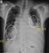
Young patient with generalized lymphangiomatosis: Differentiating the differential
- PMID: 27857472
- PMCID: PMC5036344
- DOI: 10.4103/0971-3026.190416
Young patient with generalized lymphangiomatosis: Differentiating the differential
Abstract
We present the case of a 19-year-old man who was extensively evaluated in multiple centres for long-standing cough, dyspnea, and hemoptysis without a definitive diagnosis. His chest radiograph at presentation showed mediastinal widening, bilateral pleural effusions, and Kerley B lines. Computed tomography of the thorax showed a confluent, fluid-density mediastinal lesion enveloping the mediastinal viscera without any mass effect. There were bilateral pleural effusions, prominent peribronchovascular interstitial thickening, interlobular septal thickening and lobular areas of ground glass density with relative sparing of apices. There were a few dilated retroperitoneal lymphatics and well-defined lytic lesions in the bones. In this case report, we aim to systematically discuss the relevant differentials and arrive at a diagnosis. We also briefly discuss the treatment options and prognosis along with our patient's course in the hospital and final outcome.
Keywords: Cystic mediastinal mass; generalized lymphangiomatosis; pulmonary interstitial thickening; pulmonary lymphangiectasia.
Figures


References
-
- Azouz EM, Saigal G, Rodriguez MM, Podda A. Langerhans' cell histiocytosis: Pathology, imaging and treatment of skeletal involvement. Pediatr Radiol. 2005;35:103–15. - PubMed
-
- Faul JL, Berry GJ, Colby TV, Ruoss SJ, Walter MB, Rosen GD, et al. Thoracic lymphangiomas, lymphangiectasis, lymphangiomatosis, and lymphatic dysplasia syndrome. Am J Respir Crit Care Med. 2000;161(Pt 1):1037–46. - PubMed
-
- de Lima AS, Martynychen MG, Florêncio RT, Rabello LM, de Barros JA, Escuissato DL. Pulmonary lymphangiomatosis: A report of two cases. J Bras Pneumol. 2007;33:229–33. - PubMed
-
- Wunderbaldinger P, Paya K, Partik B, Turetschek K, Hörmann M, Horcher E, et al. CT and MR imaging of generalized cystic lymphangiomatosis in pediatric patients. AJR Am J Roentgenol. 2000;174:827–32. - PubMed
LinkOut - more resources
Full Text Sources
Other Literature Sources

