. 2010 May 19:6:37-51.
doi: 10.2142/biophysics.6.37. eCollection 2010.

Modeling of the gap junction of pancreatic β-cells and the robustness of insulin secretion

Affiliations

Modeling of the gap junction of pancreatic β-cells and the robustness of insulin secretion

Tomoki Kitagawa et al. Biophysics (Nagoya-shi). .

Abstract

Pancreatic β-cells are interconnected by gap junctions, which allow small molecules to pass from cell to cell. In spite of the importance of the gap junctions in cellular communication, modeling studies have been limited by the complexity of the system. Here, we propose a mathematical gap junction model that properly takes into account biological functions, and apply this model to the study of the β-cell cluster. We consider both electrical and metabolic features of the system. Then, we find that when a fraction of the ATP-sensitive K+ channels are damaged, robust insulin secretion can only be achieved by gap junctions. Our finding is consistent with recent experiments conducted by Rocheleau et al. Our study also suggests that the free passage of potassium ions through gap junctions plays an important role in achieving metabolic synchronization between β-cells.

Keywords: gap junctions; pancreatic β-cells; robustness; synchronization.


Figures
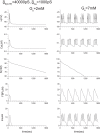
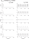
Figure 13.
Figure 13.
Single β-cell model calculations of Bedersen et al. together with Eq. (A3). The original model used the approximation of constant potassium ion concentration. Where v is the cell membrane potential, Ca is Ca2+ ion concentration, K is K+ ion concentration, and FBP is the concentration of fructose 1,6-bisphosphate.
Figure 14.
Figure 14.
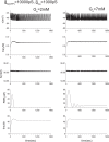
Modified single β-cell model calculations, where Eq. (A4) was used instead of Eq. (A3).
Figure 15.
Figure 15.
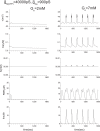
Effect of K(ATP) variation. Original K(ATP) value is 40000 pS. Note that K value is slightly higher than 95 mM here.
Figure 16.
Figure 16.
Effect of the Ca variation. Original Ca value is 1000 pS.
Figure 17.
Figure 17.
Effect of the Ca variation. Original Ca value is 1000 pS.
Figure 1.
Figure 1.
Essential processes of the insulin secretion. Glucose is taken into the β-cell by GLUT-2 transporters, and broken down during glycolysis. Glycolytic product pyruvate is taken into the mitochondria in order to produce ATP. The ATP-sensitive K+ channel regulates membrane potential, and Ca2+ flow into the cell, and the insulin release into the blood is prompted by the elevated cytosolic Ca2+ concentration.
Figure 2.
Figure 2.
A scheme for insulin secretion adopted by Pedersen et al. Ge is the extracellular glucose concentration; Gi is the intracellular glucose concentration; G6P is glucose 6-phosphate concentration; FBP is fructose 1-6-bisphosphate concentration; and SERCA means the SERCA pumps.
Figure 3.
Figure 3.
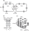
The general structure of gap junctions. Within a gap junction, there are a few to many thousands channels of diameter 1.5 nm. The length of the cell gap is about 2∼4 nm. The thickness of the cell membrane is about 5 nm. Thus, the length of the gap junctions d is nearly 13 nm. The channels allow inorganic ions and molecules with a mass of less than 1 kDa to pass into the other cell.
Figure 4.
Figure 4.
Synchronization is achieved immediately when both potassium ions and calcium ions are allowed to pass through gap junctions between two β-cells (a), but is not achieved when only calcium ions are allowed to pass through gap junctions (b). Here, Ca =1000 pS, and K(ATP) =40000 pS, and the extracellular glucose concentration is 7 mM. The solid line and dotted line indicate different β-cells.
Figure 5.
Figure 5.
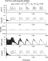
Synchronization of 10×10 β-cell square lattice with periodic boundary conditions. Here, Ca =1000 pS, and K(ATP) = 40000 pS.
Figure 6.
Figure 6.
Two β-cells connected with gap junctions. (a) Wild type cells connected with gap junctions. (b) The K(ATP) channel in one of two β-cells is partially blocked. (c) The K(ATP) channel in one β-cell is blocked, and additionally, the gap junction is non-functional.
Figure 7.
Figure 7.
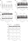
Partial blocking of the ATP-sensitive K+ channels of the two β-cell system when Ge= 2 mM. (a) K(ATP) =40000 pS (Cell 2), 40000 pS (Cell 1), (b) K(ATP) =40000 pS (Cell 2), 10000 pS (Cell 1). The above figures correspond to the cases of Figs. 6(a) and 6(b), respectively. In the case of Fig. 6(c), the result becomes the simple sum of Fig. 14 and Fig. 15 since the gap junction is closed. This means that the insulin secretion cannot be stopped, even though the glucose level is low.
Figure 8.
Figure 8.
ATP-sensitive K+ current and gap currents of the cases in Fig. 7, where Ge= 2 mM. (a) IK(ATP) of the case of Fig. 7(a), (b) IK(ATP) of the case of Fig. 7(b), (c) gap currents IG(K) and IG(Ca) of the cases of Figs. 7(a) and 7(b). When the ATP-sensitive potassium channels of both cells are fully functional, the gap currents are negligibly small. However, when one of them is blocked, the outward gap currents from the blocked cell and IK(ATP) of the normal cell are significantly increased, (d) a comparison of IK(ATP)blocked+IG(K) and IK(ATP)normalIG(K), where IK(ATP)blocked and IK(ATP)normal are ATP-sensitive potassium current of the blocked cell and that of the normal cell, respectively. Note that IK(ATP)blocked+IG(K) and IK(ATP)normalIG(K) in Fig. 8(d) are roughly equal to IK(ATP) in Fig. 8(a). An increase of IG(K) brings the intracellular potassium density of the blocked cell back to the normal level, and ultimately the insulin secretion is ceased. The intercellular collaboration of ATP-sensitive potassium channels via gap junctions is a key of this robustness. Here the positive direction of IK(ATP) is outward from the cell.
Figure 9.
Figure 9.
ATP-sensitive K+ current and gap currents, where Ge = 7 mM. Similar to Fig. 8, the blocked or damaged cell significantly increases the outward gap currents IG(K) and IG(Ca) from the cell. (a) IK(ATP) of the case of Fig. 4(a), (b) IK(ATP) of the case of Fig. 9(e), (c) gap currents IG(K) and IG(Ca) of the cases of Figs. 4(a) and 9(e). (d) a comparison of IK(ATP)blocked+IG(K) and IK(ATP)normalIG(K). They are nearly identical and in the same range of value of IK(ATP) in equilibrium state (2000 fA∼10000 fA) in Fig. 9(a).
Figure 10.
Figure 10.
Two β-cells with different Ca values, where Ca =1100 pS (Cell 2), 900 pS (Cell 1), and Ge = 7 mM.
Figure 11.
Figure 11.
Results of the linear coupling scheme. Here, two β-cells with the same and different Ca values when Ge = 7 mM. (a) Ca =1000 pS (Cell 2), 1000 pS (Cell 1), and (b) Ca =1100 pS (Cell 2), 900 pS (Cell 1).
Figure 12.
Figure 12.
Results of the linear coupling scheme. Here, two β-cells with the same and different K(ATP) values when Ge = 2 mM. (a) K(ATP) =40000 pS (Cell 2), 40000 pS (Cell 1), (b) K(ATP) =40000 pS (Cell 2), 10000 pS (Cell 1).

Similar articles

Cited by

References

    1. Nagano S. Modeling the model organism Dictyostelium discoideum. Develop. Growth Differ. 2000;42:541–550. - PubMed
    1. Nagano S. Diffusion-assisted aggregation and synchronization in Dictyostelium discoideum. Phys. Rev. Lett. 1998;80:4826–4829.
    1. Nagano S. Biological receptor scheme for the robust synchronization of limit cycle oscillators. Prog. Theor. Phys. 2002;107:861–877.
    1. Nagano S. Receptors as a master key for synchronization of rhythms. Phys. Rev. E. 2003;67:0562151–0562154. - PubMed
    1. Yokoyama A, Nagano S. Biological receptor scheme for the external synchronization of mutually coupled oscillator systems. J. Phys. Soc. Jpn. 2008;77:0240021–0240029.