. 2016 Dec;77(6):510-520.
doi: 10.1055/s-0036-1584198. Epub 2016 May 31.

STAT3 Inhibition as a Therapeutic Strategy for Chordoma

Affiliations

STAT3 Inhibition as a Therapeutic Strategy for Chordoma

Anthony C Wang et al. J Neurol Surg B Skull Base. 2016 Dec.

Abstract

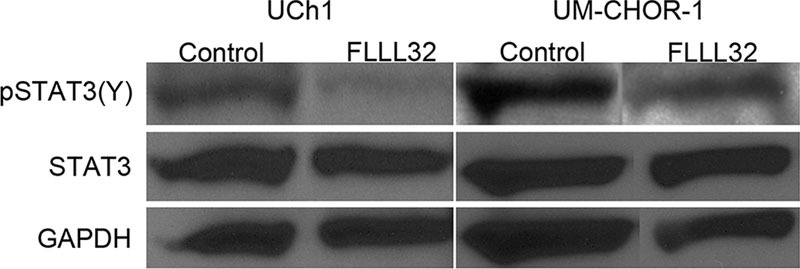
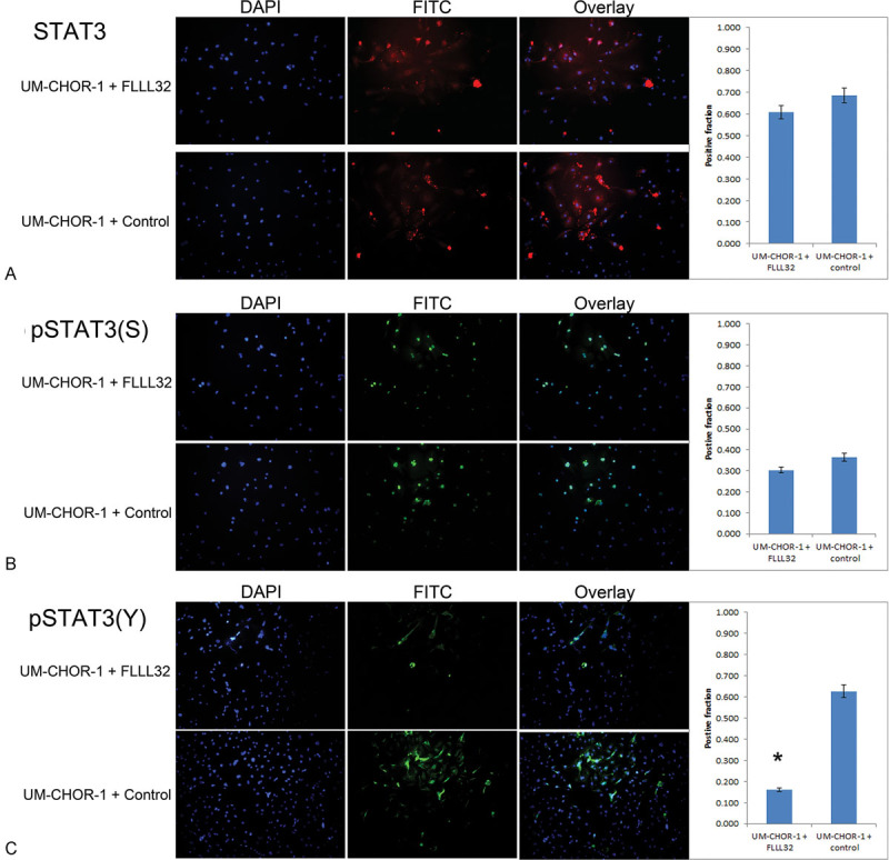
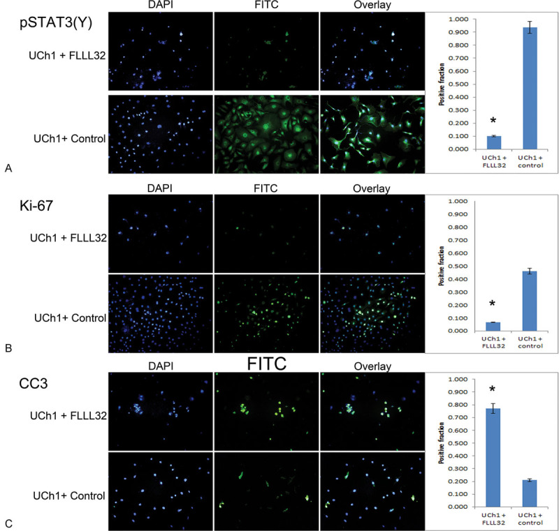
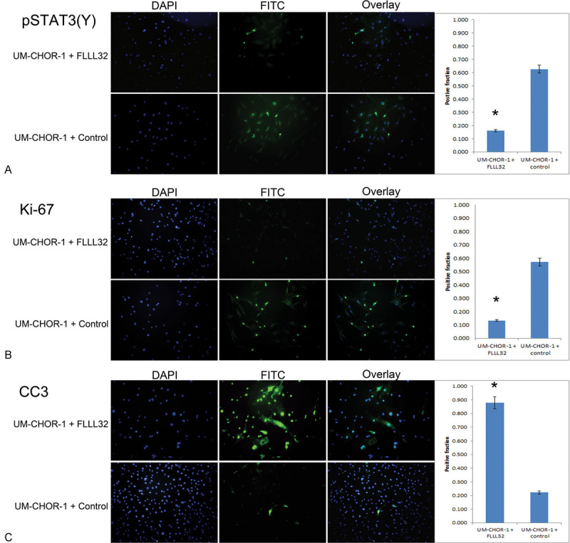
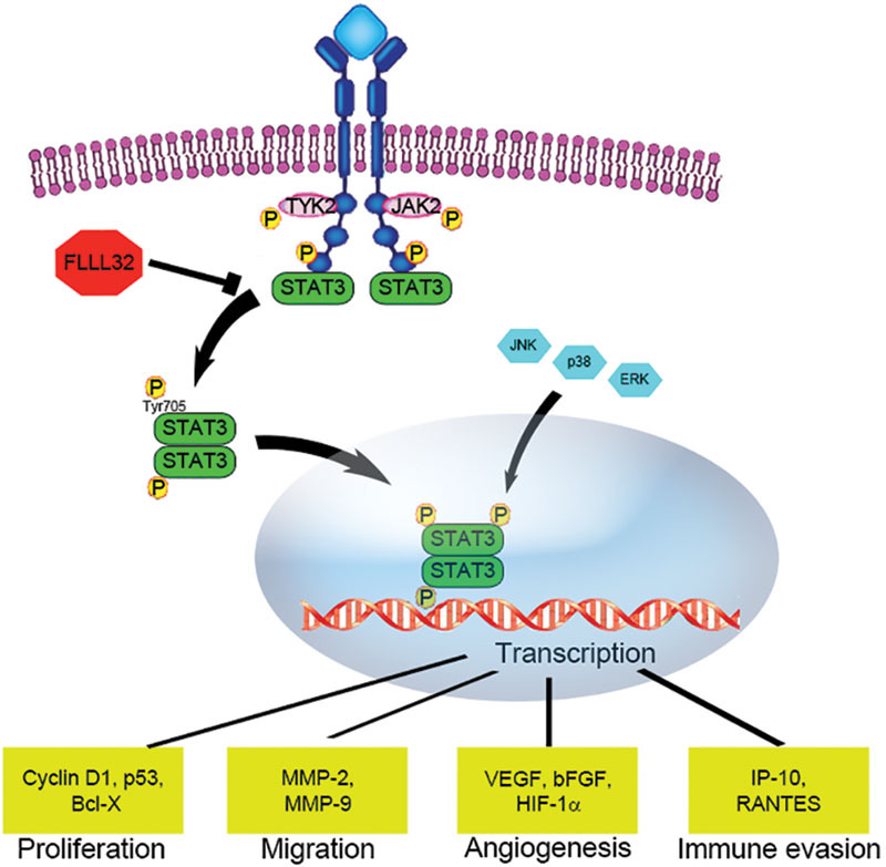
Objective Signal transducer and activator of transcription (STAT) proteins regulate key cellular fate decisions including proliferation and apoptosis. STAT3 overexpression induces tumor growth in multiple neoplasms. STAT3 is constitutively activated in chordoma, a tumor with a high recurrence rate despite maximal surgical and radiation treatment. We hypothesized that a novel small molecule inhibitor of STAT3 (FLLL32) would induce significant cytotoxicity in sacral and clival chordoma cells. Methods Sacral (UCh1) and clival (UM-CHOR-1) chordoma cell lines were grown in culture (the latter derived from primary tumor explants). FLLL32 dosing parameters were optimized using cell viability assays. Antitumor potential of FLLL32 was assessed using clonal proliferation assays. Potential mechanisms underlying observed cytotoxicity were examined using immunofluorescence assays. Results FLLL32 induced significant cytotoxicity in UCh1 and UM-CHOR-1 chordoma cells, essentially eliminating all viable cells, correlating with observed downregulation in activated, phosphorylated STAT3 upon administration of FLLL32. Mechanisms underlying the observed cytotoxicity included increased apoptosis and reduced cellular proliferation through inhibition of mitosis. Conclusion As a monotherapy, FLLL32 induces potent tumor kill in vitro in chordoma cell lines derived from skull base and sacrum. This effect is mediated through inhibition of STAT3 phosphorylation, increased susceptibility to apoptosis, and suppression of cell proliferation.

Keywords: FLLL32; STAT3; chordoma; sacrum; skull base.


Conflict of interest statement

Conflict of Interest The authors have no conflict of interest to report pertaining to the materials or methods used in this study or the findings specified in this article.

Figures

Fig. 1
Fig. 1
FLLL32 inhibits cellular proliferation and exhibits cytotoxicity. (A) Resazurin cell viability assays were performed on the UCh1 cell line in vitro. Cells were plated at a concentration of 2,500 cells per well, then treated with 5 µM FLLL32 or equal volume of 0.1% DMSO vehicle as a control. Cell viability in FLLL32-treated cells was dramatically reduced, relative to the normal growth pattern exhibited by control cells. Growth arrest was witnessed after approximately 2 days, with a significantly diminished population of viable cells following treatment with FLLL32 (p < 0.01). (B) Resazurin cell viability assays were performed on the UM-CHOR-1 cell line in vitro. Cell viability in FLLL32-treated cells was significantly reduced, relative to the normal growth pattern exhibited by control cells. Growth arrest was witnessed after approximately 2 days, with a significantly diminished population of viable cells following treatment with FLLL32 (p < 0.05). (C) Clonal proliferation assays were performed to evaluate the cytotoxicity of FLLL32 on the UCh1 cell line in vitro. Cells were plated at a concentration of 10,000 cells per well and allowed to grow for 72 days. Cells were then treated with 5 µM FLLL32 or equal volume 0.1% DMSO vehicle as a control. Cytotoxicity was demonstrated after 3 days in FLLL32-treated cells. Complete arrest of cell proliferation was witnessed in UCh1 cells with a decline of cell numbers to 2.6% in the treated cells by day 9 (p < 0.01). (D) Clonal proliferation assays were performed to evaluate the cytotoxicity of FLLL32 on the UM-CHOR-1 cell line in vitro. Though not immediate, cytotoxicity was demonstrated after 2 days in FLLL32-treated cells. Within 24 hours of initiating treatment with FLLL32 (day 4), no viable cells could be detected. This effect was sustained for the remainder of the assay period and was statistically significant when compared with untreated controls (p < 0.01). DMSO, dimethyl sulfoxide.
Fig. 2
Fig. 2
FLLL32 inhibits pSTAT3(Y) but not STAT3 expression. Western blot analysis demonstrates a relative paucity of pSTAT3(Y) expression in FLLL32-treated cells, using both the UCh1 and UM-CHOR-1 cell lines, when compared with control cells. Total STAT3 expression, however, did not change with FLLL32 treatment. GAPDH was used as loading control. GAPDH, glyceraldehyde 3-phosphate dehydrogenase; pSTAT, phosphorylated signal transducer and activator of transcription; STAT, signal transducer and activator of transcription.
Fig. 3
Fig. 3
FLLL32 inhibits STAT3 phosphorylation at the Y705 residue. (A) STAT3 immunofluorescence staining was performed on UM-CHOR-1 cells treated with FLLL32 or with equal volume 0.1% DMSO vehicle as a control. Of cells treated with FLLL32, 60.8% exhibited positive staining, compared with 68.7% of control-treated cells, showing no significant difference in total STAT3 expression. (B) Staining for pSTAT3(S) residue was not significantly different between FLLL32-treated (30.4%) and control-treated (36.6%) UM-CHOR-1 cells. (C) Staining for pSTAT3(Y), however, showed a significant reduction with FLLL32 treatment (16.1%), relative to control-treated (62.7%) UM-CHOR-1 cells (p < 0.05). (*Statistically significant.) DMSO, dimethyl sulfoxide; pSTAT, phosphorylated signal transducer and activator of transcription; STAT, signal transducer and activator of transcription.
Fig. 4
Fig. 4
FLLL32 inhibition of pSTAT3 (Y705) results in reduced cellular proliferation and increased apoptosis in UCh1 cells. (A) Immunofluorescence staining was performed on UCh1 cells treated with FLLL32 or with equal volume 0.1% DMSO vehicle as a control. Of cells treated with FLLL32, 10.0% exhibited positive staining, compared with 93.8% of control-treated cells; as such, a significant reduction in pSTAT3(Y) expression was seen in the FLLL32-treated cells (p < 0.05). (*Statistically significant.) (B) Ki-67 staining was significantly higher in control-treated UCh1 cells (46.0%) relative to FLLL32-treated cells (6.7%), indicating decreased proliferative activity with FLLL32 treatment (p < 0.05). (C) CC3 staining was significantly higher in FLLL32-treated UCh1 cells (77.1%) relative to control-treated cells (21.1%), indicating increased apoptosis induced by FLLL32 treatment (p < 0.05). DMSO, dimethyl sulfoxide; pSTAT, phosphorylated signal transducer and activator of transcription.
Fig. 5
Fig. 5
FLLL32 inhibition of pSTAT3 (Y705) results in reduced cellular proliferation and increased apoptosis in UM-CHOR-1 cells. (A) Immunofluorescence staining was performed on UM-CHOR-1 cells treated with FLLL32 or with equal volume 0.1% DMSO vehicle as a control. Of cells treated with FLLL32, 16.1% exhibited positive staining compared with 62.7% in control-treated cells, showing a significant reduction in pSTAT3(Y) expression in the FLLL32-treated cells (p < 0.05). (*Statistically significant.) (B) Ki-67 staining was significantly higher in control-treated UM-CHOR-1 cells (57.1%) relative to FLLL32-treated cells (13.3%), indicating decreased proliferative activity with FLLL32 treatment (p < 0.05). (C) CC3 was significantly higher in FLLL32-treated UM-CHOR-1 cells (87.9%) relative to control-treated cells (22.2%), indicating increased apoptosis induced by FLLL32 treatment (p < 0.05). CC3, cleaved caspase 3; DMSO, dimethyl sulfoxide; pSTAT, phosphorylated signal transducer and activator of transcription.
Fig. 6
Fig. 6
STAT3 activation is associated with cellular proliferation, inhibition of apoptosis, cell migration, and immune modulation. Cytoplasmic STAT3 monomers dimerize when phosphorylated at the tyrosine 705 (Y705) residue by Janus kinases associated with cytokine receptors, and then translocate to the nucleus, where the homodimers activate target gene transcription. STAT, signal transducer and activator of transcription.

References

    1. Walcott B P, Nahed B V, Mohyeldin A, Coumans J V, Kahle K T, Ferreira M J. Chordoma: current concepts, management, and future directions. Lancet Oncol. 2012;13(2):e69–e76. - PubMed
    1. McMaster M L, Goldstein A M, Bromley C M, Ishibe N, Parry D M. Chordoma: incidence and survival patterns in the United States, 1973-1995. Cancer Causes Control. 2001;12(1):1–11. - PubMed
    1. Williams B J, Raper D M, Godbout E. et al.Diagnosis and treatment of chordoma. J Natl Compr Canc Netw. 2013;11(6):726–731. - PubMed
    1. Stacchiotti S, Casali P G. Systemic therapy options for unresectable and metastatic chordomas. Curr Oncol Rep. 2011;13(4):323–330. - PubMed
    1. Di Maio S, Temkin N, Ramanathan D, Sekhar L N. Current comprehensive management of cranial base chordomas: 10-year meta-analysis of observational studies. J Neurosurg. 2011;115(6):1094–1105. - PubMed