Review
. 2016 Nov 3:4:124.
doi: 10.3389/fcell.2016.00124. eCollection 2016.

Septins As Modulators of Endo-Lysosomal Membrane Traffic

Affiliations
Review

Septins As Modulators of Endo-Lysosomal Membrane Traffic

Kyungyeun Song et al. Front Cell Dev Biol. .

Abstract

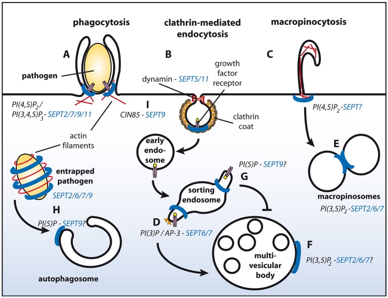
Septins constitute a family of GTP-binding proteins, which assemble into non-polar filaments in a nucleotide-dependent manner. These filaments can be recruited to negatively charged membrane surfaces. When associated with membranes septin filaments can act as diffusion barriers, which confine subdomains of distinct biological functions. In addition, they serve scaffolding roles by recruiting cytosolic proteins and other cytoskeletal elements. Septins have been implicated in a large variety of membrane-dependent processes, including cytokinesis, signaling, cell migration, and membrane traffic, and several family members have been implicated in disease. However, surprisingly little is known about the molecular mechanisms underlying their biological functions. This review summarizes evidence in support of regulatory roles of septins during endo-lysosomal sorting, with a particular focus on phosphoinositides, which serve as spatial landmarks guiding septin recruitment to distinct subcellular localizations.

Keywords: endocytosis; endosome; membrane; phosphoinositides; septins; sorting.


Figures

Figure 1
Figure 1
Schematic representation of endosomal sorting events with proven or putative involvement of septins. Septin filaments are indicated in blue, actin filaments in red. SEPT2/7/9/11 promote phagocytosis (A). Through associating with dynamin SEPT5/11 might function during clathrin-mediated endocytosis (B). Interactions of septins with actin filaments might modulate macropinocytosis (C). Complex formation between AP-3 and SEPT6/7 facilitates degradative sorting (D). SEPT2/6/7 promote fusion of macropinosomes (E) and potentially of MVBs with lysosomes (F). PI(5)P at endosomes protects cargo from degradative sorting, and might recruit SEPT9 to sorting endosomes (G). SEPT2/6/7/9 form cages around intracellular pathogens and promote their sorting to autophagosomes (H). SEPT9 inhibits ubiquitylation of EGF receptors at the plasma membrane by associating with CIN85, and thereby attenuates degradative sorting at endosomes (I). See text for details.

References

    1. Akil A., Peng J., Omrane M., Gondeau C., Desterke C., Marin M., et al. . (2016). Septin 9 induces lipid droplets growth by a phosphatidylinositol-5-phosphate and microtubule-dependent mechanism hijacked by HCV. Nat. Commun. 7:12203. 10.1038/ncomms12203 - DOI - PMC - PubMed
    1. Baust T., Anitei M., Czupalla C., Parshyna I., Bourel L., Thiele C., et al. . (2008). Protein networks supporting AP-3 function in targeting lysosomal membrane proteins. Mol. Biol. Cell 19, 1942–1951. 10.1091/mbc.E08-02-0110 - DOI - PMC - PubMed
    1. Boal F., Mansour R., Gayral M., Saland E., Chicanne G., Xuereb J. M., et al. . (2015). TOM1 is a PI5P effector involved in the regulation of endosomal maturation. J. Cell Sci. 128, 815–827. 10.1242/jcs.166314 - DOI - PubMed
    1. Bridges A. A., Jentzsch M. S., Oakes P. W., Occhipinti P., Gladfelter A. S. (2016). Micron-scale plasma membrane curvature is recognized by the septin cytoskeleton. J. Cell Biol. 213, 23–32. 10.1083/jcb.201512029 - DOI - PMC - PubMed
    1. Bridges A. A., Zhang H., Mehta S. B., Occhipinti P., Tani T., Gladfelter A. S. (2014). Septin assemblies form by diffusion-driven annealing on membranes. Proc. Natl. Acad. Sci. U.S.A. 111, 2146–2151. 10.1073/pnas.1314138111 - DOI - PMC - PubMed