Review
. 2017 Aug;126(4):443-455.
doi: 10.1007/s00412-016-0620-7. Epub 2016 Nov 17.

Structure of centromere chromatin: from nucleosome to chromosomal architecture

Affiliations
Review

Structure of centromere chromatin: from nucleosome to chromosomal architecture

Thomas Schalch et al. Chromosoma. 2017 Aug.

Abstract

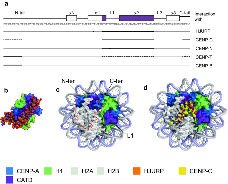
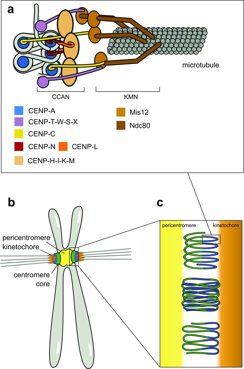
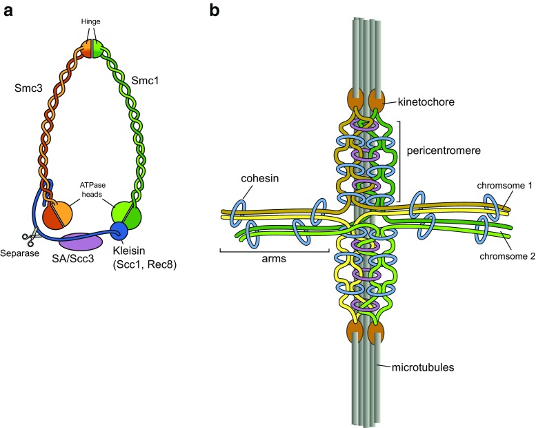
The centromere is essential for the segregation of chromosomes, as it serves as attachment site for microtubules to mediate chromosome segregation during mitosis and meiosis. In most organisms, the centromere is restricted to one chromosomal region that appears as primary constriction on the condensed chromosome and is partitioned into two chromatin domains: The centromere core is characterized by the centromere-specific histone H3 variant CENP-A (also called cenH3) and is required for specifying the centromere and for building the kinetochore complex during mitosis. This core region is generally flanked by pericentric heterochromatin, characterized by nucleosomes containing H3 methylated on lysine 9 (H3K9me) that are bound by heterochromatin proteins. During mitosis, these two domains together form a three-dimensional structure that exposes CENP-A-containing chromatin to the surface for interaction with the kinetochore and microtubules. At the same time, this structure supports the tension generated during the segregation of sister chromatids to opposite poles. In this review, we discuss recent insight into the characteristics of the centromere, from the specialized chromatin structures at the centromere core and the pericentromere to the three-dimensional organization of these regions that make up the functional centromere.

Keywords: CENP-A; Centromere; Chromatin; Cohesin; Pericentromere.


Conflict of interest statement

Conflict of interest

The authors declare that they have no conflict of interest.

Figures

Fig. 1
Fig. 1
CENP-A residues that specify the centromere. a Primary sequence and schematic representation of the secondary structure of human CENP-A (top). The CATD is highlighted in purple. Regions of the CENP-A protein that interact with HJURP and different CCAN components (bottom). b Structural model of the CENP-A/H4 heterodimer in complex with HJURP (PDB ID 3R45) (Hu et al. 2011). c Structural model of the CENP-A nucleosome (PDB ID 3AN2) (Tachiwana et al. 2011). The CENP-A/H4 heterodimer on the left is shown in the same orientation as in b (framed by red dotted line), illustrating how HJURP binding prevents formation of a (CENP-A/H4)2 tetramer and association with DNA. d Structural model of CENP-C bound to the CENP-A nucleosome (CENP-C from PDB ID 4X23 modeled on CENP-A nucleosome from PDB ID 3AN2) (Tachiwana et al. ; Kato et al. 2013)
Fig. 2
Fig. 2
Three-dimensional arrangement of the centromere during mitosis. a Cartoon of kinetochore complex linking CENP-A chromatin to the microtubules. CENP-A nucleosomes coordinate the protein network of the CCAN, which recruits the outer kinetochore (KMN network) that attaches to the microtubules. b The pericentromere provides cohesion between sister chromatids and acts as a foundation for the centromere core, which assembles the kinetochore complexes for the attachment of microtubules. c Chromatin of the centromere core is folded to expose the CENP-A nucleosomes to the surface of the primary constriction. Several models for this assembly have been proposed: solenoid model (top) (Blower et al. 2002), layered boustrophedon (middle) (Ribeiro et al. 2010), and looping model (bottom) (Blower et al. 2002)
Fig. 3
Fig. 3
Cohesin shapes the centromere. a Model of the S. cerevisiae cohesin molecule. b Schematic representation of two S. cerevisiae sister chromosomes attached to the mitotic spindle showing how cohesin shapes the centromeres in yeast mitosis. Cohesin connects sister chromatids at chromosome arms but also links together centromeric chromatin, both by intrachromosome (blue) and interchromosome (purple) strand trapping, thereby forming a barrel-shaped structure underneath the kinetochore

References

    1. Aldrup-Macdonald ME, Sullivan BA. The past, present, and future of human centromere genomics. Genes. 2014;5:33–50. doi: 10.3390/genes5010033. - DOI - PMC - PubMed
    1. Alper BJ, Lowe BR, Partridge JF. Centromeric heterochromatin assembly in fission yeast—balancing transcription, RNA interference and chromatin modification. Chromosom Res. 2012;20:521–534. doi: 10.1007/s10577-012-9288-x. - DOI - PMC - PubMed
    1. Amano M, Suzuki A, Hori T, et al. The CENP-S complex is essential for the stable assembly of outer kinetochore structure. J Cell Biol. 2009;186:173–182. doi: 10.1083/jcb.200903100. - DOI - PMC - PubMed
    1. Bailis JM, Bernard P, Antonelli R, et al. Hsk1-Dfp1 is required for heterochromatin-mediated cohesion at centromeres. Nat Cell Biol. 2003;5:1111–1116. doi: 10.1038/ncb1069. - DOI - PubMed
    1. Bassett EA, DeNizio J, Barnhart-Dailey MC, et al. HJURP uses distinct CENP-A surfaces to recognize and to stabilize CENP-A/histone H4 for centromere assembly. Dev Cell. 2012;22:749–762. doi: 10.1016/j.devcel.2012.02.001. - DOI - PMC - PubMed

Publication types

LinkOut - more resources