

AGEs induce ectopic endochondral ossification in intervertebral discs
- PMID: 27858401
- PMCID: PMC5482230
- DOI: 10.22203/eCM.v032a17
AGEs induce ectopic endochondral ossification in intervertebral discs
Abstract
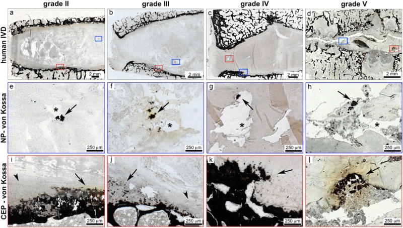
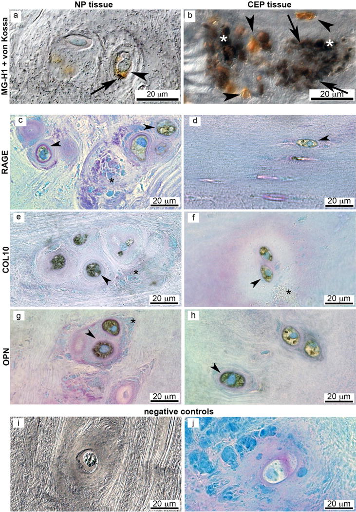
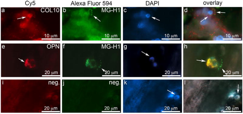
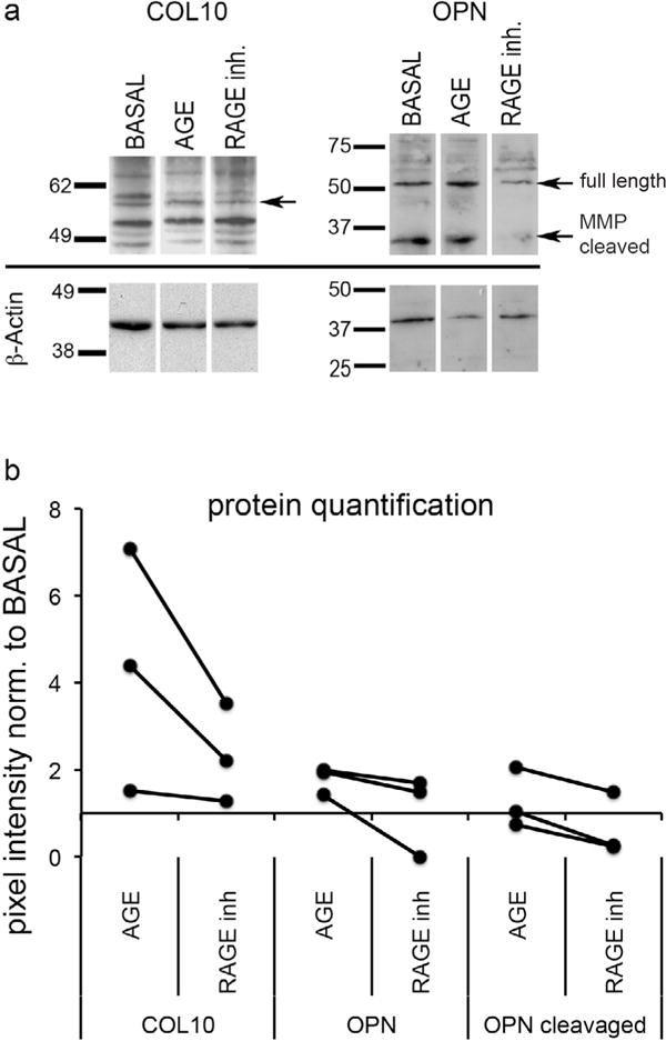
Ectopic calcifications in intervertebral discs (IVDs) are known characteristics of IVD degeneration that are not commonly reported but may be implicated in structural failure and dysfunctional IVD cell metabolic responses. This study investigated the novel hypothesis that ectopic calcifications in the IVD are associated with advanced glycation end products (AGEs) via hypertrophy and osteogenic differentiation. Histological analyses of human IVDs from several degeneration stages revealed areas of ectopic calcification within the nucleus pulposus and at the cartilage endplate. These ectopic calcifications were associated with cells positive for the AGE methylglyoxal-hydroimidazolone-1 (MG-H1). MG-H1 was also co-localised with Collagen 10 (COL10) and Osteopontin (OPN) suggesting osteogenic differentiation. Bovine nucleus pulposus and cartilaginous endplate cells in cell culture demonstrated that 200 mg/mL AGEs in low-glucose media increased ectopic calcifications after 4 d in culture and significantly increased COL10 and OPN expression. The receptor for AGE (RAGE) was involved in this differentiation process since its inhibition reduced COL10 and OPN expression. We conclude that AGE accumulation is associated with endochondral ossification in IVDs and likely acts via the AGE/RAGE axis to induce hypertrophy and osteogenic differentiation in IVD cells. We postulate that this ectopic calcification may play an important role in accelerated IVD degeneration including the initiation of structural defects. Since orally administered AGE and RAGE inhibitors are available, future investigations on AGE/RAGE and endochondral ossification may be a promising direction for developing non-invasive treatment against progression of IVD degeneration.
Figures

References
-
- Adams MA, Roughley PJ. What is intervertebral disc degeneration, and what causes it? Spine (Phila Pa 1976) 2006;31:2151–2161. - PubMed
-
- Ahmed N. Advanced glycation endproducts – role in pathology of diabetic complications. Diabetes Res Clin Pract. 2005;67:3–21. - PubMed
-
- Benneker LM, Heini PF, Alini M, Anderson SE, Ito K. 2004 Young Investigator Award Winner: vertebral endplate marrow contact channel occlusions and intervertebral disc degeneration. Spine (Phila Pa 1976) 2005;30:167–173. - PubMed
-
- Berezin AE, Kremzer AA. Circulating osteopontin as a marker of early coronary vascular calcification in type two diabetes mellitus patients with known asymptomatic coronary artery disease. Atherosclerosis. 2013;229:475–481. - PubMed
Web references
-
- Andersson G. United States bone and joint initiative: The Burden of Musculoskeletal Diseases in the United States (BMUS) Amer Acad Orthop Surg. 2014 http://www.boneandjointburden.org/2014-report/iia0/definition.
Additional References
-
- Tsuru M, Nagata K, Jimi A, Irie K, Yamada A, Nagai R, Horiuchi S, Sata M. Effect of AGEs on human disc herniation: intervertebral disc hernia is also effected by AGEs. Kurume Med J. 2002;49:7–13. - PubMed
-
- Wagner DR, Reiser KM, Lotz JC. Glycation increases human annulus fibrosus stiffness in both experimental measurements and theoretical predictions. J Biomech. 2006;39:1021–1029. - PubMed
-
- Watson AM, Soro-Paavonen A, Sheehy K, Li J, Calkin AC, Koitka A, Rajan SN, Brasacchio D, Allen TJ, Cooper ME, Thomas MC, Jandeleit-Dahm KJ. Delayed intervention with AGE inhibitors attenuates the progression of diabetes-accelerated atherosclerosis in diabetic apolipoprotein E knockout mice. Diabetologia. 2011;54:681–689. - PubMed
Publication types
MeSH terms
Substances
Grants and funding
LinkOut - more resources
Full Text Sources
Other Literature Sources
Research Materials