. 2017 Feb;241(3):362-374.
doi: 10.1002/path.4845. Epub 2016 Dec 29.

Vessel co-option is common in human lung metastases and mediates resistance to anti-angiogenic therapy in preclinical lung metastasis models

Affiliations

Vessel co-option is common in human lung metastases and mediates resistance to anti-angiogenic therapy in preclinical lung metastasis models

Victoria L Bridgeman et al. J Pathol. 2017 Feb.

Abstract

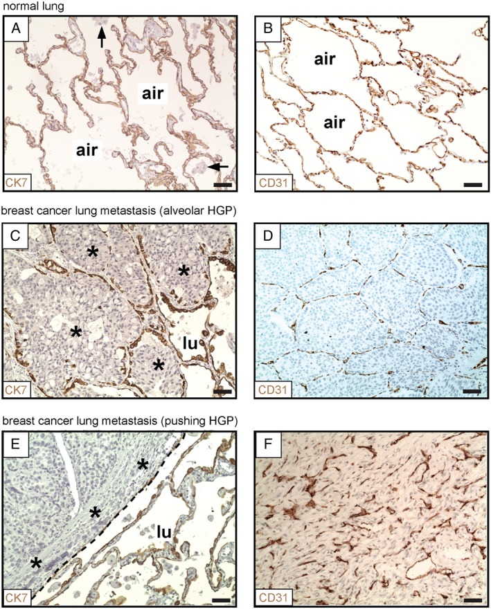
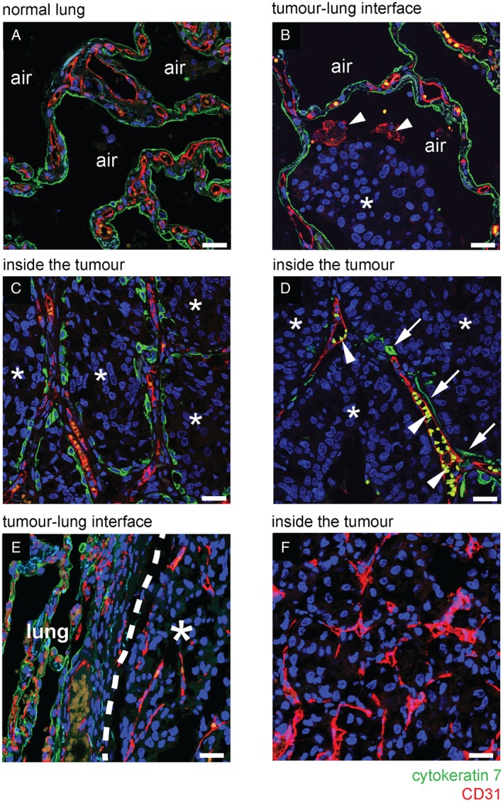
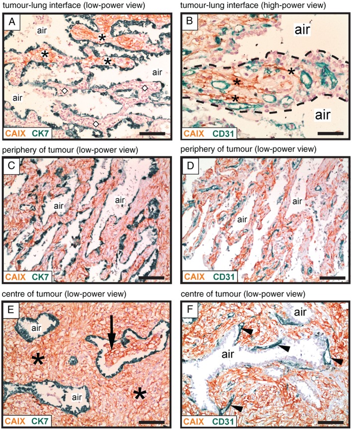
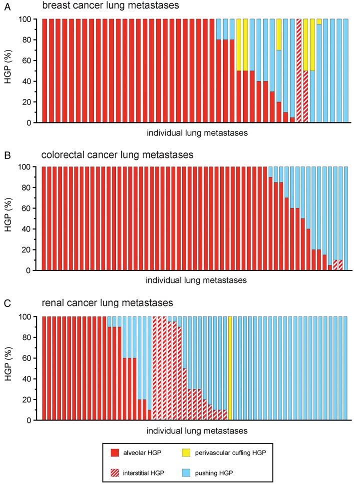
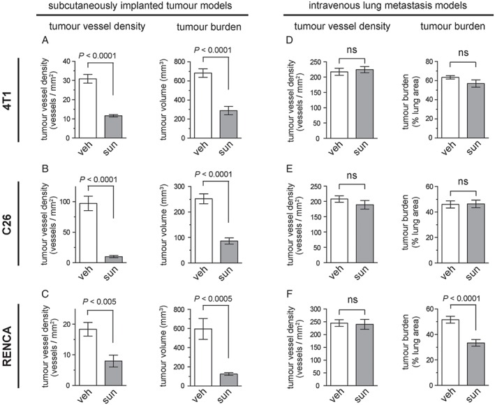
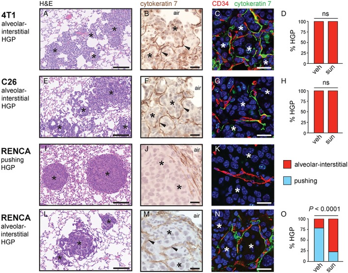
Anti-angiogenic therapies have shown limited efficacy in the clinical management of metastatic disease, including lung metastases. Moreover, the mechanisms via which tumours resist anti-angiogenic therapies are poorly understood. Importantly, rather than utilizing angiogenesis, some metastases may instead incorporate pre-existing vessels from surrounding tissue (vessel co-option). As anti-angiogenic therapies were designed to target only new blood vessel growth, vessel co-option has been proposed as a mechanism that could drive resistance to anti-angiogenic therapy. However, vessel co-option has not been extensively studied in lung metastases, and its potential to mediate resistance to anti-angiogenic therapy in lung metastases is not established. Here, we examined the mechanism of tumour vascularization in 164 human lung metastasis specimens (composed of breast, colorectal and renal cancer lung metastasis cases). We identified four distinct histopathological growth patterns (HGPs) of lung metastasis (alveolar, interstitial, perivascular cuffing, and pushing), each of which vascularized via a different mechanism. In the alveolar HGP, cancer cells invaded the alveolar air spaces, facilitating the co-option of alveolar capillaries. In the interstitial HGP, cancer cells invaded the alveolar walls to co-opt alveolar capillaries. In the perivascular cuffing HGP, cancer cells grew by co-opting larger vessels of the lung. Only in the pushing HGP did the tumours vascularize by angiogenesis. Importantly, vessel co-option occurred with high frequency, being present in >80% of the cases examined. Moreover, we provide evidence that vessel co-option mediates resistance to the anti-angiogenic drug sunitinib in preclinical lung metastasis models. Assuming that our interpretation of the data is correct, we conclude that vessel co-option in lung metastases occurs through at least three distinct mechanisms, that vessel co-option occurs frequently in lung metastases, and that vessel co-option could mediate resistance to anti-angiogenic therapy in lung metastases. Novel therapies designed to target both angiogenesis and vessel co-option are therefore warranted. © 2016 The Authors. The Journal of Pathology published by John Wiley & Sons Ltd on behalf of Pathological Society of Great Britain and Ireland.

Keywords: angiogenesis; anti-angiogenic therapy; drug resistance; lung metastasis; sunitinib; vessel co-option.


Figures

Figure 1
Figure 1
Alveolar and pushing growth patterns of human lung metastases. (A, B) Normal human lung parenchyma, stained for CK7 (A) or CD31 (B). (C, D) Alveolar HGP of human breast cancer lung metastasis. Staining for CK7 (at the tumour–lung interface) is shown in (C). Staining for CD31 (within the tumour) is shown in (D). (E, F) Pushing HGP of human breast cancer lung metastasis. Staining for CK7 (at the tumour–lung interface) is shown in (E). Staining for CD31 (within the tumour) is shown in (F). Cancer cells: asterisks. Alveolar macrophages: arrows. Alveolar air space: air. Tumour–lung interface: dashed line. Normal lung: lu. Scale bar: 50 µm.
Figure 2
Figure 2
Vessel co‐option occurs in the alveolar growth pattern of human lung metastases. (A–D) Immunofluorescence co‐staining for CD31 (red) and CK7 (green) in a case of human breast cancer lung metastasis that presented with an alveolar HGP. (A) In areas of tumour‐free normal lung parenchyma, the alveolar walls are composed of CD31‐positive alveolar capillaries (red) that are sheathed by CK7‐positive pneumocytes (green). (B) At the tumour–lung interface, cancer cells (asterisks) invade an alveolar air space. Arrowheads indicate two CD31‐positive alveolar macrophages in the alveolar air space that also reacted with the CD31 antibody. (C) Behind the tumour–lung interface, cancer cells (asterisks) completely fill the alveolar air spaces, preserving the alveolar walls and the associated alveolar capillaries. (D) Towards the centre of the metastatic lesion, co‐opted alveolar capillaries can be found that are only partially coated by pneumocytes. Arrows indicate pneumocytes that are still associated with co‐opted alveolar capillaries. Arrowheads indicate autofluorescent erythrocytes in the lumen of co‐opted alveolar capillaries. (E, F) Immunofluorescence co‐staining for CD31 (red) and CK7 (green) in a sample of human renal cancer lung metastasis with a pushing HGP. At the tumour–lung interface, cancer cells push the alveolar walls away (E). No incorporation of alveolar walls was observed either at the tumour–lung interface (E) or deeper into the metastasis (F). Alveolar air space: air. Normal lung: lung. Scale bar: 25 µm.
Figure 3
Figure 3
Vessel co‐option occurs in the interstitial growth pattern of human lung metastases. Immunohistochemical analysis of a renal cancer lung metastasis with an interstitial HGP, illustrating growth of cancer cells within the alveolar walls. Staining for CAIX (brown) was used to detect cancer cells, in combination with either CK7 staining (green) to detect pneumocytes (A, C, E) or CD31 staining (green) to detect blood vessels (B, D, F). (A) Tumour–lung interface: alveolar walls filled with cancer cells are present at the top of the image (asterisks), whereas tumour‐free alveolar walls of the normal lung are present below (diamond symbols). (B) High‐power view of an alveolar wall (delineated with a dashed line). Asterisks indicate cancer cells that are infiltrating around pre‐existing alveolar capillaries. (C, D) The area just behind the tumour–lung interface: the alveolar walls are now completely filled with cancer cells. The intervening alveolar air spaces remain intact. (E, F) The centre of the metastasis. In (E), asterisks indicate cancer cells that are filling the expanded alveolar walls, while the intervening alveolar air spaces remain intact. The arrow indicates an alveolar air space that has become partially filled with cancer cells. In (F), arrowheads indicate blood vessels that are closely associated with the abluminal side of an alveolar air space. Alveolar air space: air. Scale bars: 100 µm (A, C, D, E, F) and 50 µm (B).
Figure 4
Figure 4
Frequency of the different HGPs in lung metastases of human breast, colorectal and renal cancer. Lung metastases of human breast cancer (A), human colorectal cancer (B) and human renal cancer (C) were scored for their growth pattern. Each bar represents an individual case of metastasis showing the percentage of the tumour–lung interface scored as alveolar, interstitial, perivascular cuffing or pushing HGP. n = 46 breast cancer lung metastases (A), n = 57 colorectal cancer lung metastases (B), and n = 61 renal cancer lung metastases (C).
Figure 5
Figure 5
Limited efficacy of sunitinib in lung metastasis models as compared with subcutaneously implanted tumours. (A–C) The efficacy of sunitinib was tested in mice injected subcutaneously with 4 T1 (A), C26 (B) or RENCA (C) cells. The graphs show tumour vessel density ± standard error of the mean (SEM) (left) or tumour burden ± SEM (right) in subcutaneous 4 T1 (A), C26 (B) or RENCA (C) tumours after 10 days of treatment with either 40 mg/kg per day sunitinib or vehicle alone. n = 10 mice per experimental group for tumour burden graphs. n = 6 mice per experimental group for tumour vessel density graphs. (D–F) Mice were injected via the tail vein with 4 T1 (D), C26 (E) or RENCA (F) cells. The graphs show tumour vessel density ± SEM (left) or tumour burden ± SEM (right) in the lungs after 10 days of treatment with either 40 mg/kg per day sunitinib or vehicle alone. n = 9 or 10 mice per experimental group for tumour burden graphs. n = 5 mice per experimental group for tumour vessel density graphs. ns, no significant difference; sun, sunitinib; veh, vehicle.
Figure 6
Figure 6
Evidence for vessel co‐option in lung metastasis models. Histopathological characterization was performed on lung metastases formed by 4 T1 cells (A–D), C26 cells (E–H) and RENCA cells (I–O) after tail vein injection. (A, E, I, L) Low‐power views of lung metastasis morphology by H&E staining. (B, C, E, F, I, J, L, M) Higher‐power views of CK7‐stained lung metastases (B, F, J, M) or lung metastases co‐stained for CD34 (red) and CK7 (green) (C, G, K, N). The graphs show percentage alveolar/interstitial HGP and percentage pushing HGP scored in 4 T1 (D), C26 (H) and RENCA (O) lung metastases from vehicle (veh)‐treated or sunitinib (sun)‐treated mice (n = 9 or 10 mice per experimental group). Cancer cells: asterisks. Alveolar air spaces: air. ns, no significant difference. Scale bars: 125 µm (A, E, I, L), 20 µm (B, F, J, M) and 20 µm (C, G, K, N).

References

    1. Bergers G, Hanahan D. Modes of resistance to anti‐angiogenic therapy. Nat Rev 2008; 8 : 592–603. - PMC - PubMed
    1. Ebos JM, Kerbel RS. Antiangiogenic therapy: impact on invasion, disease progression, and metastasis. Nat Rev Clin Oncol 2011; 8 : 210–221. - PMC - PubMed
    1. Carmeliet P, Jain RK. Molecular mechanisms and clinical applications of angiogenesis. Nature 2011; 473 : 298–307. - PMC - PubMed
    1. Sennino B, McDonald DM. Controlling escape from angiogenesis inhibitors. Nat Rev 2012; 12 : 699–709. - PMC - PubMed
    1. Vasudev NS, Reynolds AR. Anti‐angiogenic therapy for cancer: current progress, unresolved questions and future directions. Angiogenesis 2014; 17 : 471–494. - PMC - PubMed

MeSH terms