. 2017 Mar 1;595(5):1533-1546.
doi: 10.1113/JP273189. Epub 2017 Jan 15.

Post-translational cleavage of Hv1 in human sperm tunes pH- and voltage-dependent gating

Affiliations

Post-translational cleavage of Hv1 in human sperm tunes pH- and voltage-dependent gating

Thomas K Berger et al. J Physiol. .

Abstract

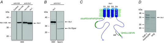
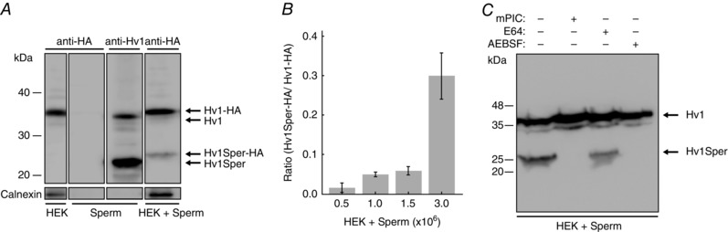
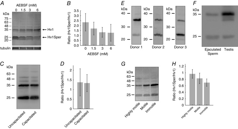
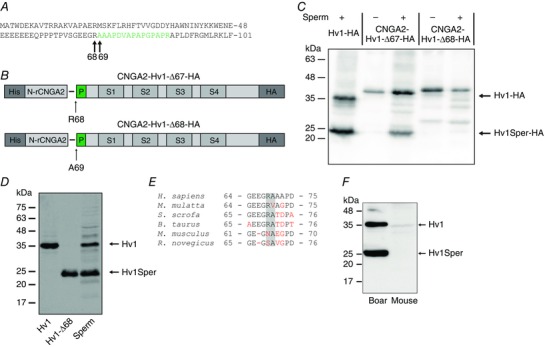
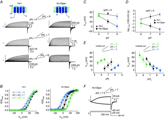
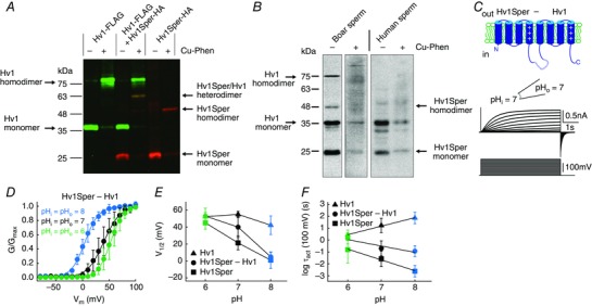
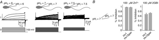
Key points: In human sperm, proton flux across the membrane is controlled by the voltage-gated proton channel Hv1. We show that sperm harbour both Hv1 and an N-terminally cleaved isoform termed Hv1Sper. The pH-control of Hv1Sper and Hv1 is distinctively different. Hv1Sper and Hv1 can form heterodimers that combine features of both constituents. Cleavage and heterodimerization of Hv1 might represent an adaptation to the specific requirements of pH control in sperm.

Abstract: In human sperm, the voltage-gated proton channel Hv1 controls the flux of protons across the flagellar membrane. Here, we show that sperm harbour Hv1 and a shorter isoform, termed Hv1Sper. Hv1Sper is generated from Hv1 by removal of 68 amino acids from the N-terminus by post-translational proteolytic cleavage. The pH-dependent gating of the channel isoforms is distinctly different. In both Hv1 and Hv1Sper, the conductance-voltage relationship is determined by the pH difference across the membrane (∆pH). However, simultaneous changes in intracellular and extracellular pH that leave ΔpH constant strongly shift the activation curve of Hv1Sper but not that of Hv1, demonstrating that cleavage of the N-terminus tunes pH sensing in Hv1. Moreover, we show that Hv1 and Hv1Sper assemble as heterodimers that combine features of both constituents. We suggest that cleavage and heterodimerization of Hv1 represents an adaptation to the specific requirements of pH control in sperm.

Keywords: electrophysiology; ion channels; sperm; voltage-gated channels.


Figures

Figure 1
Figure 1. Human sperm harbour full‐length Hv1 and a short Hv1 variant
A, western blot of HEK cells heterologously expressing Hv1‐HA or Hv1, probed with a C‐terminal anti‐Hv1 or an anti‐HA antibody. B, western blot of HEK cells expressing Hv1 and of human sperm, probed with a C‐terminal anti‐Hv1 antibody; in sperm, the antibody recognizes an additional protein of ∼25 kDa (termed Hv1Sper). C, cartoon of Hv1 showing the proteotypic peptides detected by MS (green) and the epitope of the anti‐Hv1 antibody (AB). D, western blot of human monocyte cell line THP‐1 and human lung epithelial cell lysate, probed with the C‐terminal anti‐Hv1 antibody.
Figure 2
Figure 2. Hv1 is post‐translationally cleaved by a protease
A, western blot of HEK cells expressing Hv1‐HA, of human sperm, and of a mix of sperm and Hv1‐HA‐expressing HEK cells, probed with a C‐terminal anti‐Hv1 or an anti‐HA antibody. B, Hv1Sper‐HA/Hv1‐HA band intensity is dependent on the ratio of the number of sperm in the sperm/HEK cell mix. C, western blot of a mix of sperm and Hv1‐HA‐expressing HEK cells in the presence or absence of protease inhibitors mPIC, E64, and AEBSF, as probed with an anti‐HA antibody. Error bars indicate the SD.
Figure 3
Figure 3. The Hv1Sper/Hv1 ratio varies among donors and sperm subpopulations, but is independent of capacitation
A, western blot of sperm lysed in the presence of various AEBSF concentrations, probed with a C‐terminal anti‐Hv1 antibody. B, mean Hv1Sper/Hv1 ratio at various AEBSF concentrations. C, western blot of uncapacitated and capacitated sperm, probed with a C‐terminal anti‐Hv1 antibody. D, mean Hv1Sper/Hv1 ratio of uncapacitated and capacitated sperm (uncapacitated, 1.4 ± 0.7, n = 4; capacitated, 1.3 ± 0.5, n = 4). E, western blot of sperm from different donors, probed with a C‐terminal anti‐Hv1 antibody. F, western blot of sperm and testis lysate, probed with a C‐terminal anti‐Hv1 antibody. G, western blot of highly motile, motile, and immotile sperm subpopulations, probed with a C‐terminal anti‐Hv1 antibody. H, mean Hv1/Hv1Sper ratio of the three sperm subpopulations (highly motile, 0.94 ± 0.13; motile, 0.81 ± 0.14; immotile, 0.67 ± 0.11; n = 4). Error bars indicate the SD.
Figure 4
Figure 4. Hv1 is N‐terminally cleaved at position R68
A, amino acid sequence of the N‐terminal part of Hv1. The peptide shown in green was identified by MS in the protein band corresponding to Hv1Sper (Fig. 1B). B, Hv1‐HA constructs in which the first 67 (Hv1‐Δ67) or 68 amino acids (Hv1‐Δ68) were replaced by an N‐terminal segment of a CNGA2 channel (His‐N‐rCNGA2). C, western blot of HEK cells, alone (–) or mixed with sperm (+), expressing Hv1‐HA or the CNGA2‐Hv1‐HA construct, probed with the anti‐HA antibody. D, western blot of oocytes expressing Hv1 or Hv1‐Δ68 and of human sperm, probed with the C‐terminal anti‐Hv1 antibody. E, sequence alignment of the N‐terminal cleavage site (highlighted in grey) among six mammalian species (human, macaque, boar, bull, mouse, rat; non‐conserved amino acids in red). F, western blot of boar and mouse sperm, probed with the C‐terminal anti‐Hv1 antibody.
Figure 5
Figure 5. Hv1Sper has altered pH‐voltage dependence and activation kinetics
A, inside‐out patch clamp recording from X. laevis oocytes expressing Hv1 (left) or Hv1Sper (right) at pHi = pHo = 6 (top), 7 (middle) and 8 (bottom). Voltage steps of increasing amplitudes (–80 to +100 mV in 10 mV increments) elicited voltage‐dependent outward currents. B, GV relationships derived from tail currents at the three different pH conditions (leaving ΔpH = 0), fitted with a single Boltzmann function. C, half‐maximal activation voltage (V 1/2) derived from the Boltzmann fits of the data in (B). D, activation‐time constants of Hv1Sper and Hv1 obtained from single (Hv1Sper) and weighted double (Hv1) exponential fits. E, voltage of half‐maximal activation (V 1/2) as a function of changes in pHi and pHo in the inside‐out (left) and outside‐out (right) configuration. The shift of V 1/2 in the inside‐out configuration for pHo of 6, 7 and 8 was –15.6, –30.9 and –20.4 mV/pH unit, respectively. The shift of V 1/2 in the outside‐out configuration for pHi of 6, 7 and 8 was –44, –46 and –19.4 mV/pH unit, respectively. F, inward and outward currents of Hv1Sper, depending on the pH conditions. Error bars indicate the SD.
Figure 6
Figure 6. Hv1Sper and Hv1 form heterodimers with unique biophysical properties
A, western blot of X. laevis oocytes expressing Hv1‐FLAG or Hv1Sper‐HA, or both, in the presence or absence of the oxidant Cu‐phenanthroline, probed with an anti‐FLAG (green) and anti‐HA (red) antibody. B, western blot of boar and human sperm incubated in the presence or absence of the cysteine cross‐linker Cu‐phenanthroline (Cu‐Phen), probed with a C‐terminal anti‐Hv1 antibody. C, inside‐out patch clamp recording from Xenopus oocytes expressing a tandem heterodimer consisting of Hv1Sper linked to Hv1 with a flexible linker, recorded at pHi = pHo = 7. Voltage steps of increasing amplitudes (–80 to +100 mV) elicited voltage‐dependent outward currents. D, GV relationships derived from tail currents at three different pH conditions (leaving ΔpH = 0), fitted with a single Boltzmann function. E, half‐maximal activation voltage (V 1/2) derived from the Boltzmann fits shown in (C) (Hv1 and Hv1Sper taken from Fig. 5 C). F, activation time constants of the Hv1Sper–Hv1 tandem heterodimer, as well as Hv1 and Hv1Sper (taken from Fig. 5 D). Error bars indicate the SD.
Figure 7
Figure 7. Control of sperm proton currents by pH and block of Hv1Sper by Zn2+ and 2GBI
A, whole‐cell patch clamp recording from human sperm at pHi = pHo = 6, 7, and 7.5. Voltage‐dependent outward currents (top traces) evoked by voltage steps (bottom traces) of increasing amplitude (–80 to +100 mV, pH 6 and 7; –20 to +100 mV, pH 7.5). B, inhibition of outward currents in excised patches of X. laevis oocytes heterologously expressing Hv1 or Hv1Sper by extracellular Zn2+ and intracellular 2GBI at pHi = pHo = 7. Currents were evoked by a voltage step from –80 mV to +80 mV (Zn2+: Hv1 99.7 ± 0.3% inhibition, n = 3; Hv1Sper 98.9 ± 1%, n = 4; 2GBI: Hv1 69.4 ± 3.7% inhibition, n = 3; Hv1Sper 74.2 ± 1.4%, n = 3). Error bars indicate the SD.

References

    1. Armstrong CM & Bezanilla F (1977). Inactivation of the sodium channel. II. Gating current experiments. J Gen Physiol 70, 567–590. - PMC - PubMed
    1. Austin CR (1951). Observations on the penetration of the sperm in the mammalian egg. Aust J Sci Res B 4, 581–596. - PubMed
    1. Batra‐Safferling R, Abarca‐Heidemann K, Körschen HG, Tziatzios C, Stoldt M, Budyak I, Willbold D, Schwalbe H, Klein‐Seetharaman J & Kaupp UB (2006). Glutamic acid‐rich proteins of rod photoreceptors are natively unfolded. J Biol Chem 281, 1449–1460. - PubMed
    1. Berger TK & Isacoff EY (2011). The pore of the voltage‐gated proton channel. Neuron 72, 991–1000. - PMC - PubMed
    1. Bönigk W, Loogen A, Seifert R, Kashikar N, Klemm C, Krause E, Hagen V, Kremmer E, Strünker T & Kaupp UB (2009). An atypical CNG channel activated by a single cGMP molecule controls sperm chemotaxis. Sci Signal 2, ra68. - PubMed

MeSH terms

LinkOut - more resources