Review
. 2017 Apr 26:35:31-52.
doi: 10.1146/annurev-immunol-041015-055354. Epub 2016 Nov 14.

The Lymphatic System: Integral Roles in Immunity

Affiliations
Review

The Lymphatic System: Integral Roles in Immunity

Gwendalyn J Randolph et al. Annu Rev Immunol. .

Abstract

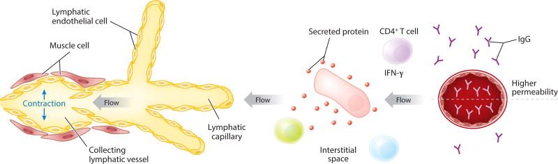
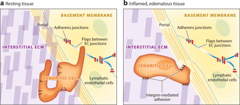
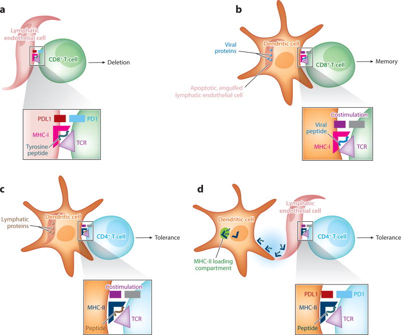
The lymphatic vasculature is not considered a formal part of the immune system, but it is critical to immunity. One of its major roles is in the coordination of the trafficking of antigen and immune cells. However, other roles in immunity are emerging. Lymphatic endothelial cells, for example, directly present antigen or express factors that greatly influence the local environment. We cover these topics herein and discuss how other properties of the lymphatic vasculature, such as mechanisms of lymphatic contraction (which immunologists traditionally do not take into account), are nonetheless integral in the immune system. Much is yet unknown, and this nascent subject is ripe for exploration. We argue that to consider the impact of lymphatic biology in any given immunological interaction is a key step toward integrating immunology with organ physiology and ultimately many complex pathologies.

Keywords: adhesion; endothelium; lymph; lymph node; migration.


Figures

Figure 1
Figure 1
Schema depicting the two major parts of the lymphatic vasculature: lymphatic capillaries and lymphatic collecting vessels. With contraction of lymphatic collecting vessels, flow through the lymphatic vessel occurs, and this dictates the direction of interstitial fluid flow within the adjacent tissue. The larger cell in the tissue (pink) is depicted as secreting a protein. Because of the distribution of the protein in the flow environment, the green cell would be subjected to more interaction with the protein than the nearby cell in light blue. The image also shows the distribution of IgG in the bloodstream and in the vasculature, with the concentration of IgG being lower in the interstitial space than in plasma under all conditions except when the permeability of the vessel has been increased, for instance, because of secretion of IFN-γ by T helper cells.
Figure 2
Figure 2
Entry of immune cells into lymphatic capillaries. (a) Immune cells (dendritic cells are the most thoroughly studied) migrate through the extracellular matrix of a tissue (purple) with amoeboid movement by using matrix fibers (purple blocks) as structural supports, pushing against them with force generated in the cytoskeleton (arrows) to make their way to the lymphatic vessel. No specific adhesion is necessary between the cell and the extracellular matrix. Immune cells continue to utilize adhesion molecule–independent amoeboid motion to enter the lymphatic vessel, finding areas around the vessels with the least dense basement membrane (yellow) and flap-like areas (blue) between lymphatic endothelial cells that are not sealed by adherens junctions (gray and red). (b) In the context of an inflamed and edematous tissue, extracellular matrix fibers may become less dense as fluid accumulates in the tissue and increases the space between such fibers, making conditions unfavorable for amoeboid movement without use of adhesion molecules. Under these circumstances, immune cells employ integrins to anchor themselves to extracellular matrix fibers and lymphatic endothelial cells to make their way into the lymphatic lumen. Abbreviations: EC, endothelial cell; ECM, extracellular matrix.
Figure 3
Figure 3
Morphological features of lymphatic collecting vessels and lymph node lymphatic endothelium using lineage tracer mice and immunostaining. (a) Human mesenteric lymphatic collecting vessel stained for podoplanin (red) and smooth muscle actin (green) to reveal the veiled-like pattern of muscle around the collecting vessels and the bulge often seen around the valves. (b,c) Two single z-stacks of a branched collecting lymphatic vessel outside of the mouse popliteal lymph node, acquired from two-photon imaging in the Prox-1 ERCre × Tomatofl/fl mouse crossed with the CD11cYFP mouse. The images show the extremes at diastole and systole for the lymphangions in view. Lymphatics are red, dendritic cells are green, and collagen is blue.
Figure 4
Figure 4
Lymphatic endothelial cells in antigen presentation require cooperation with dendritic cells. The schema depict four scenarios described in the literature and covered in this review where lymphatic endothelium in the lymph node participates in presenting antigen, most generally to promote peripheral tolerance but also to serve as a long-term reservoir for antigen presentation late in a response for the promotion of CD8+ T cell memory. The scenario in panel a is the only one that does not involve dendritic cells as critical intermediates. (a) A subset of lymphatic endothelial cells express peripheral antigens, or acquire them through uptake of dying cells, for subsequent MHC-I-mediated presentation to CD8+ T cells, leading to immunological tolerance. (b) Lymphatic endothelial cells capable of long-term retention of antigens, such as viral proteins or particles, in lymph nodes undergo apoptosis during lymph node contraction in the late phases of an immune response. The dying lymphatic endothelial cells are engulfed by DCs that cross-present foreign antigens originally present in lymphatic endothelial cells. This mechanism fosters the generation of CD8+ T cell memory against viral antigens. (c) Lymphatic endothelial cells can express MHC-II but lack HLA-DM for appropriate peptide loading of the MHC. CD4+ T cell–associated immunological tolerance can be fostered when proteins from lymphatic endothelial cells are taken up by DCs, allowing for peptides derived from lymphatic endothelial cells to be loaded onto MHC-II molecules of the DCs. (d) Another mechanism that allows lymphatic endothelial cells to support CD4+ T cell immunity despite the lack of HLA-DM occurs when lymphatic endothelial cells acquire intact MHC class II–peptide complexes from DCs, resulting in their presentation of antigen to CD4+ T cells through a mechanism referred to as cross-dressing.

References

    1. Jiang X, Clark RA, Liu L, Wagers AJ, Fuhlbrigge RC, Kupper TS. Skin infection generates non-migratory memory CD8+ TRM cells providing global skin immunity. Nature. 2012;483:227–31. - PMC - PubMed
    1. Steinert EM, Schenkel JM, Fraser KA, Beura LK, Manlove LS, et al. Quantifying Memory CD8 T cells reveals regionalization of immunosurveillance. Cell. 2015;161:737–49. - PMC - PubMed
    1. Masopust D, Vezys V, Marzo AL, Lefrancois L. Preferential localization of effector memory cells in nonlymphoid tissue. Science. 2001;291:2413–17. - PubMed
    1. Reinhardt RL, Khoruts A, Merica R, Zell T, Jenkins MK. Visualizing the generation of memory CD4 T cells in the whole body. Nature. 2001;410:101–5. - PubMed
    1. Roozendaal R, Mebius RE. Stromal cell–immune cell interactions. Annu. Rev. Immunol. 2011;29:23–43. - PubMed

Publication types