Review
. 2017 Jan 6:57:535-565.
doi: 10.1146/annurev-pharmtox-010715-103335.

Mitochondrial Dysfunction and Myocardial Ischemia-Reperfusion: Implications for Novel Therapies

Affiliations
Review

Mitochondrial Dysfunction and Myocardial Ischemia-Reperfusion: Implications for Novel Therapies

Edward J Lesnefsky et al. Annu Rev Pharmacol Toxicol. .

Abstract

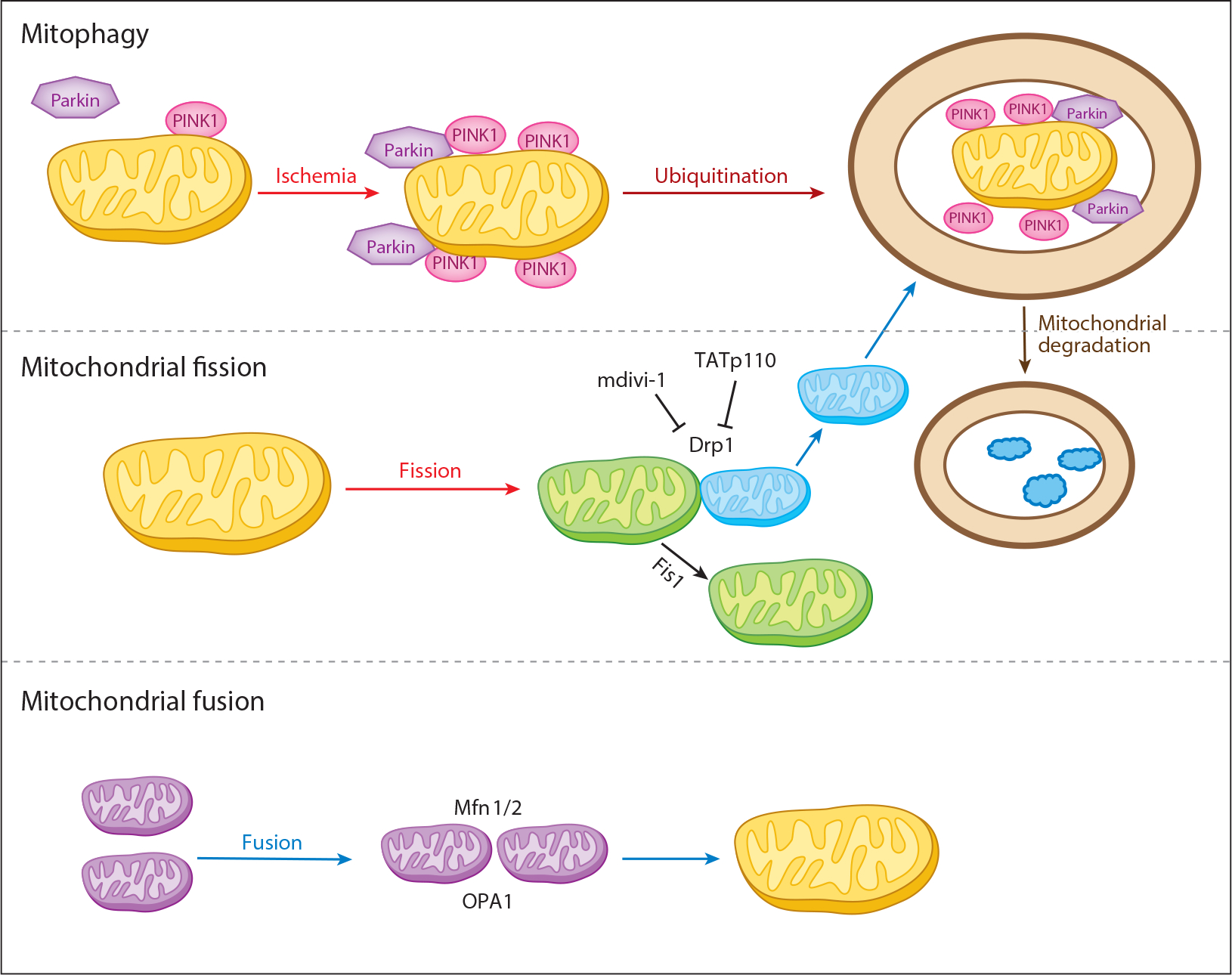
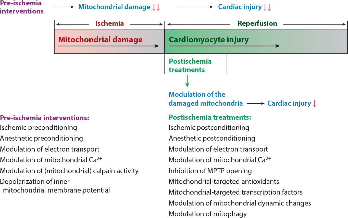
Mitochondria have emerged as key participants in and regulators of myocardial injury during ischemia and reperfusion. This review examines the sites of damage to cardiac mitochondria during ischemia and focuses on the impact of these defects. The concept that mitochondrial damage during ischemia leads to cardiac injury during reperfusion is addressed. The mechanisms that translate ischemic mitochondrial injury into cellular damage, during both ischemia and early reperfusion, are examined. Next, we discuss strategies that modulate and counteract these mechanisms of mitochondrial-driven injury. The new concept that mitochondria are not merely stochastic sites of oxidative and calcium-mediated injury but that they activate cellular responses of mitochondrial remodeling and cellular reactions that modulate the balance between cell death and recovery is reviewed, and the therapeutic implications of this concept are discussed.

Keywords: cardiolipin; electron transport chain; fatty acid oxidation; oxidative phosphorylation; reactive oxygen species; ubiquinol:cytochrome c oxidoreductase.


Figures

Figure 1
Figure 1
Structure and function of normal mitochondria. (a) The locations of subsarcolemmal and interfibrillar mitochondria in the cardiomyocyte are depicted schematically. The various cellular components are not drawn to scale. (b) Shown are the locations of the voltage-dependent anion channel (VDAC) in the outer mitochondrial membrane (OMM); the electron transport chain (ETC) in the cristae of the inner mitochondrial membrane (IMM) with the adenine nucleotide translocase (ANT), monocarboxylate transporter (MCT), and dicarboxylate translocase (DCT) in the IMM; the intermembrane space (IMS); and contact sites as fusion points of the outer and inner boundary membrane. (c) The ETC. Reduced nicotinamide adenine dinucleotide (NADH) donates an electron to complex I (CI) with flow through to complex IV (CIV) coupled with H+ pumping into the IMS at CI, CIII, and CIV. Reduced flavin adenine dinucleotide (FADH2) feeds electrons into CII with flow to CIV. However, no H+ is pumped into the IMS at complex II. H+ moves into the matrix coupled with the phosphorylation of ADP to ATP by CV. Other abbreviation: Q, coenzyme Q.
Figure 2
Figure 2
Sites of ischemia-mediated damage to electron transport. (a) Sites of ischemia damage to the electron transport chain (ETC) complexes. (b) Sites within these complexes where electrons leak to form superoxides and subsequently other reactive oxygen species, including hydrogen peroxide (H2O2). (c) Sites of action of classical chemical inhibitors (red) and potential therapeutic agents that modulate the ETC (blue). Potential therapeutic modulators of complex I include amobarbital acting at the quinol binding site (39), SNO donors (99), and nitric oxide (NO) (100) which likely interact with iron-sulfur (Fe-S) centers, and STAT3 (121). Other compounds that inhibit complex I partially and that have been reported to result in cardioprotection include metformin (31), phenformin (31), and ranolazine (97) (specific sites not depicted). Malonate competes for oxidation of succinate by complex II (29), whereas AP5A (60), diazoxide (200), and sevoflurane (201) alter other mechanisms. The peptide SS-31 binds to cardiolipin and also inhibits cytochrome c peroxidase (, , –204). Other abbreviations: AP5A, diadenosine pentaphosphate; C, cytochrome c; FMN, flavin mononucleotide; I, II, III, and IV, complexes I, II, III, and IV; ISP, iron-sulfur protein; NADH, nicotinamide adenine dinucleotide; Q, coenzyme Q; STAT3, signal transducer and activator of transcription 3; TTFA, thenoyltrifluoroacetone.
Figure 3
Figure 3
Mitochondrial morphology is preserved following ischemia alone. Electron micrographs of in situ subsarcolemmal mitochondria (SSM) and interfibrillar mitochondria (IFM) in nonischemic controls (a) and myocytes following 45 min of stop-flow ischemia (b) in the rabbit heart. Mitochondria are intact and, following ischemia, exhibit minimal change, confirming their morphological integrity at the end of the ischemic period. Micrographs of isolated SSM from nonischemic control (c) and ischemic hearts (d) confirm purity of the isolated mitochondrial population and reinforce the paucity of ischemia-induced damage to these organelles. Figure courtesy of American Physiological Society, copyright 1997 (13).
Figure 4
Figure 4
Mechanisms of mitochondrial-driven injury that result from ischemic damage to mitochondria, especially the mitochondrial electron transport chain (ETC). These include activation of the mitochondrial permeability transition pore (MPTP) and permeabilization of the outer mitochondrial membrane, leading to release of cytochrome c (Cyt c) and activation of mitochondrial calpains (mit-CPN1), which leads to the cleavage and release of truncated apoptosis-inducing factor (t-AIF) and the export of reactive oxygen species generated by the ETC by the voltage-dependent anion channel (VDAC), including mitochondrial contact sites. Antioxidant systems include superoxide dismutase 2 (SOD2) in the mitochondrial matrix and superoxide dismutase 1 (SOD1) in the intermembrane space. Superoxide (•O2) in the mitochondrial matrix is converted to hydrogen peroxide (H2O2) by SOD2, whereas superoxide in the intermembrane space is converted by SOD1 to H2O2.
Figure 5
Figure 5
Mitochondrial dynamics. Mitophagy (mitochondrial autophagy) is mediated by Parkin and PINK1. Mitochondrial fission with Drp1 and Fis1 is a key component of the division of mitochondria. Drp1 can be inhibited by mdivi-1 and TATp110 peptides. Mitochondrial fusion utilizes the outer mitochondrial membrane Mfn1/2 and the inner membrane OPA1. Fission of damaged mitochondria can lead to destruction of the daughter organelles by mitophagy. Abbreviations: Drp1, dynamin-related protein 1; Fis1, mitochondrial fission protein 1; Mfn1/2, mitofusin 1 and 2; OPA1, optic atrophy 1; Parkin, component of a multiprotein E3 ubiquitin ligase complex; PINK1, PTEN-induced putative kinase 1.
Figure 6
Figure 6
Strategies to decrease mitochondria and myocardial injury during ischemia-reperfusion. Mitochondria, which are damaged progressively during ischemia, set the stage for progressive myocardial injury during reperfusion. Therefore, pre-ischemic interventions, which include ischemic preconditioning (205), anesthetic preconditioning (111), modulation of electron transport (1), modulation of mitochondrial Ca2+ (108, 110, 111), modulation of mitochondrial calpain activity (206), and depolarization of inner mitochondrial membrane potential (66, 67), are more effective at reducing cardiac injury during reperfusion in that these interventions protect mitochondria from ischemic damage. Modulation of the ischemic-damaged mitochondria during early reperfusion, which include ischemic postconditioning (–209), anesthetic postconditioning (134, 135), modulation of electron transport (93, 114), modulation of mitochondrial Ca2+ (110, 111), inhibition of mitochondrial permeability transition pore (MPTP) opening (112), mitochondrial-targeted antioxidants (86, 210), mitochondrial-targeted transcription factors (126, 127), modulation of mitochondrial dynamic changes (174), and modulation of mitophagy (187), can attenuate myocardial injury during reperfusion. Treatments during early reperfusion are less effective than interventions before ischemia but have more clinical relevance in that the imminent onset of a myocardial infarction is rarely known.

References

    1. Chen Q, Camara AK, Stowe DF, Hoppel CL, Lesnefsky EJ. 2007. Modulation of electron transport protects cardiac mitochondria and decreases myocardial injury during ischemia and reperfusion. Am. J. Physiol. Cell Physiol. 292:C137–47 - PubMed
    1. Murphy E, Steenbergen C. 2008. Mechanisms underlying acute protection from cardiac ischemia-reperfusion injury. Physiol. Rev. 88:581–609 - PMC - PubMed
    1. Yellon DM, Hausenloy DJ. 2007. Myocardial reperfusion injury. N. Engl. J. Med. 357:1121–35 - PubMed
    1. Reimer KA, Lowe JE, Rasmussen MM, Jennings RB. 1977. The wavefront phenomenon of ischemic cell death. 1. Myocardial infarct size versus duration of coronary occlusion in dogs. Circulation 56:786–94 - PubMed
    1. Lesnefsky EJ, Chen Q, Hoppel CL. 2016. Mitochondrial metabolism in aging heart. Circ. Res. 118:1593–611 - PMC - PubMed

Publication types

MeSH terms

Substances