Review
. 2017 Feb 10:79:93-117.
doi: 10.1146/annurev-physiol-022516-034055. Epub 2016 Nov 16.

A Critical and Comparative Review of Fluorescent Tools for Live-Cell Imaging

Affiliations
Review

A Critical and Comparative Review of Fluorescent Tools for Live-Cell Imaging

Elizabeth A Specht et al. Annu Rev Physiol. .

Abstract

Fluorescent tools have revolutionized our ability to probe biological dynamics, particularly at the cellular level. Fluorescent sensors have been developed on several platforms, utilizing either small-molecule dyes or fluorescent proteins, to monitor proteins, RNA, DNA, small molecules, and even cellular properties, such as pH and membrane potential. We briefly summarize the impressive history of tool development for these various applications and then discuss the most recent noteworthy developments in more detail. Particular emphasis is placed on tools suitable for single-cell analysis and especially live-cell imaging applications. Finally, we discuss prominent areas of need in future fluorescent tool development-specifically, advancing our capability to analyze and integrate the plethora of high-content data generated by fluorescence imaging.

Keywords: cellular dynamics; fluorescent dyes; fluorescent proteins; live-cell imaging; probes.


Figures
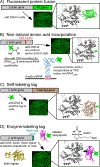
Figure 1
Figure 1
Methods of fluorescently tagging proteins. As an example, tagging of β-actin by various tools is presented where the genetically encoded parts are drawn as bars (to scale). (a) FPs can be fused at the N- or C-termini of a protein of interest. The structures of β-actin (PDB ID 2BTF) and GFP (PDB ID 1EMA) are shown at the same relative scale. (b) A modified tRNA (loaded by an evolved aminoacyl-tRNA synthetase) can incorporate an unnatural amino acid at the amber (TAG) codon. A representative tRNA structure (ID 1EHZ) is shown to illustrate the incorporation of an unnatural amino acid in response to the amber codon (UAG) in the mRNA. (c) The HaloTag can be genetically fused to a protein of interest (HaloTag, PDB ID 4KAA). The Halo Tag consists of a modified dehalogenase that covalently binds to a membrane-permeant synthetic ligand. (d) A short biotin tag sequence is genetically fused to a protein of interest. Biotin is then bound to the tag by biotin ligase, which in turn tightly binds labeled streptavidin. The structures of β-actin and streptavidin (PDB ID 4JNJ) in the fusion construct are shown at the same relative scale (biotin ligase: PDB ID 1BIA). Abbreviation: FP, fluorescent protein.
Figure 2
Figure 2
Methods of fluorescently labeling RNA in fixed or live samples. (a) Endogenous RNA is labeled by sequence-specific fluorescent oligo probes that bind the RNA of interest in fixed permeabilized cells. (b) A small aptamer (called Spinach/Broccoli) is genetically incorporated as a fusion of the RNA of interest, and fluorescence activation is achieved after binding of the cell-permeable small molecule DFHBI. (c) Short aptamers are incorporated at the 3′ end of the RNA of interest in series. Each aptamer binds an aptamer-binding protein fused to one or several FPs. (d) An FP is fused to a genetically modified version of Cas9 that remains nuclear, unless it is retained in the cytosol upon binding to an endogenous RNA of interest via a sequence-specific PAMmer. Abbreviation: FP, fluorescent protein; PAMmer, protospacer adjacent motif-presenting oligomer.

References

    1. Spence MTZ, Johnson ID. 2010. Molecular Probes Handbook: A Guide to Fluorescent Probes and Labeling Technologies. Carlsbad, CA: Life Tech. Corp. 11th ed.
    1. Lavis LD, Raines RT. 2008. Bright ideas for chemical biology. ACS Chem. Biol. 3(3):142–55 - PMC - PubMed
    1. Lavis LD, Raines RT. 2014. Bright building blocks for chemical biology. ACS Chem. Biol. 9(4):855–66 - PMC - PubMed
    1. Wysocki LM, Lavis LD. 2011. Advances in the chemistry of small molecule fluorescent probes. Curr. Opin. Chem. Biol. 15(6):752–59 - PubMed
    1. Grimm JB, English BP, Chen J, Slaughter JP, Zhang Z, et al. 2015. A general method to improve fluorophores for live-cell and single-molecule microscopy. Nat. Methods 12(3):244–50 - PMC - PubMed

Publication types

Substances

LinkOut - more resources