Review
. 2017 Feb 10:79:237-260.
doi: 10.1146/annurev-physiol-022516-034102. Epub 2016 Nov 14.

Regulation of Mammalian Oocyte Meiosis by Intercellular Communication Within the Ovarian Follicle

Affiliations
Review

Regulation of Mammalian Oocyte Meiosis by Intercellular Communication Within the Ovarian Follicle

Laurinda A Jaffe et al. Annu Rev Physiol. .

Abstract

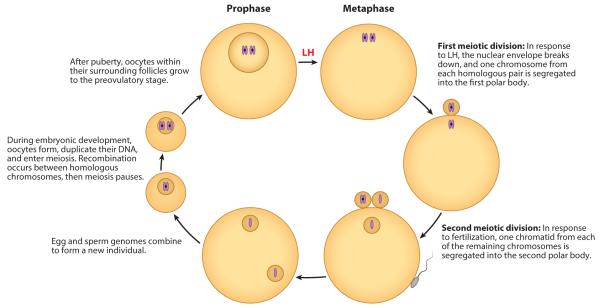
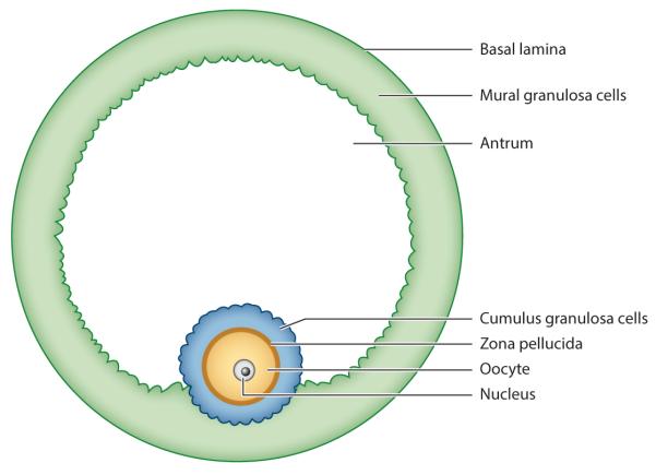
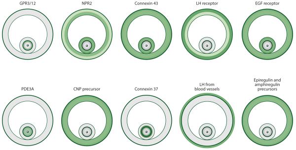
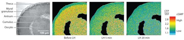
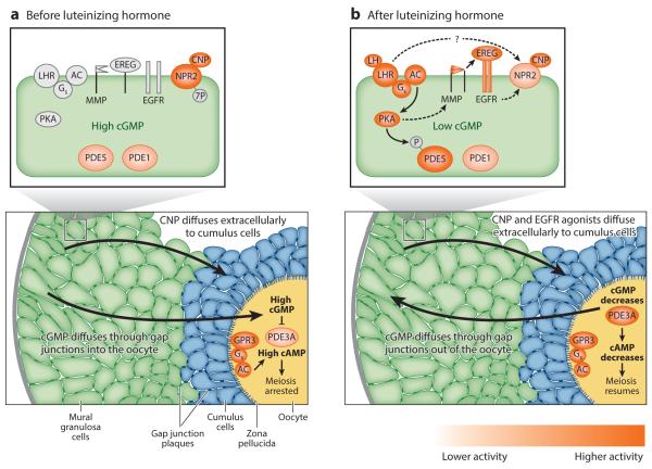
Meiotic progression in mammalian preovulatory follicles is controlled by the granulosa cells around the oocyte. Cyclic GMP (cGMP) generated in the granulosa cells diffuses through gap junctions into the oocyte, maintaining meiotic prophase arrest. Luteinizing hormone then acts on receptors in outer granulosa cells to rapidly decrease cGMP. This occurs by two complementary pathways: cGMP production is decreased by dephosphorylation and inactivation of the NPR2 guanylyl cyclase, and cGMP hydrolysis is increased by activation of the PDE5 phosphodiesterase. The cGMP decrease in the granulosa cells results in rapid cGMP diffusion out of the oocyte, initiating meiotic resumption. Additional, more slowly developing mechanisms involving paracrine signaling by extracellular peptides (C-type natriuretic peptide and EGF receptor ligands) maintain the low level of cGMP in the oocyte. These coordinated signaling pathways ensure a fail-safe system to prepare the oocyte for fertilization and reproductive success.

Keywords: cyclic GMP; gap junctions; intercellular communication; luteinizing hormone; oocyte meiosis; ovarian follicle.


Figures

Figure 1
Figure 1
Life cycle of a mammalian oocyte. The diagram places the prophase-to-metaphase transition in response to luteinizing hormone (LH) into the context of the overall meiotic cell cycle of the oocyte. One set of homologous chromosomes, each composed of two chromatids (purple), is shown.
Figure 2
Figure 2
Tissue layers of a mammalian preovulatory follicle. The oocyte with its prophase-arrested nucleus is surrounded by 2–3 layers of cumulus granulosa cells, which are attached in one region to the 5–10 layers of mural granulosa cells. Elsewhere, a fluid-filled antrum separates the two types of granulosa cells.
Figure 3
Figure 3
Localization of some of the signaling proteins that regulate meiotic arrest and resumption in preovulatory follicles. Protein distribution is either determined directly, by ligand binding, immunofluorescence, or Western blotting; or it is inferred from mRNA distribution, by in situ hybridization, or from RT-qPCR. Green indicates the presence of the protein, and lighter green indicates a lesser amount of the protein. White indicates that the protein (or mRNA) was either not detected or detected at a level ≤10% of that elsewhere. Figure is based on data from the following references: CNP precursor (66); connexin 43 (9), connexin 37 (9), EGF receptor (; LA Jaffe and JR Egbert, unpublished data); epiregulin and amphiregulin precursors (99); GPR3/12 (40, 46); LH from blood vessels (14); LH receptor (84); NPR2 (66, 70); and PDE3A (, which used a previous nomenclature, PDE3B). Abbreviations: CNP, C-type natriuretic peptide; EGF, epidermal growth factor; LH, luteinizing hormone; NPR2, natriuretic peptide receptor 2; PDE3A, phosphodiesterase 3A.
Figure 4
Figure 4
LH-induced cGMP decreases in a mouse ovarian follicle from a transgenic mouse expressing the cGMP FRET sensor cGi500. A larger CFP/YFP emission ratio indicates higher cGMP. Ratios are color-coded: Yellow indicates high cGMP, and turquoise indicates low cGMP. Before LH perfusion, cGMP is high throughout the follicle but low in the theca cells. At 5 min after LH perfusion, cGMP has decreased in the mural cells but not in the cumulus cells or oocyte. At 20 min, cGMP has decreased throughout the follicle. Figure modified from Reference 71. Abbreviations: CFP, cyan fluorescent protein; cGMP, cyclic GMP; FRET, Förster resonance energy transfer; LH, Luteinizing hormone; YFP, yellow fluorescent protein.
Figure 5
Figure 5
Working model of signaling pathways that regulate meiotic arrest and resumption in preovulatory follicles. Panel a shows a follicle and an expanded view of a mural granulosa cell before LH exposure. Panel b depicts events occurring in response to LH. Some of the slower events are not shown, such as the decrease in CNP and the increase in mRNA encoding epiregulin and amphiregulin. Higher levels of enzymatic activity are depicted by darker shades of orange. Abbreviations: AC, adenylyl cyclase; CNP, C-type natriuretic peptide; EGFR, epidermal growth factor receptor; EREG, epiregulin (and amphiregulin); GPR3, G-protein receptor 3; Gs, Gs G protein; LH, luteinizing hormone; LHR, luteinizing hormone receptor; MMP, matrix metalloprotease; NPR2, natriuretic peptide receptor 2; PDE1, 3A, 5, phosphodiesterases; PKA, protein kinase A. Figure modified from Reference 71, with additional details about the phosphodiesterases from Reference 109. The cellular structures were drawn based on electron microscopic images of a mouse follicle from Valentina Baena and Mark Terasaki.

References

    1. Eppig JJ, Viveiros MM, Marin-Bivens C, De La Fuente R. Regulation of mammalian oocyte maturation. In: Leung PCK, Adashi EY, editors. The Ovary. 2nd ed Elsevier/Academic; San Diego, CA: 2004. pp. 113–29.
    1. Conti M, Hsieh M, Zamah AM, Oh JS. Novel signaling mechanisms during oocyte maturation and ovulation. Mol. Cell. Endocrinol. 2012;356:65–73. - PMC - PubMed
    1. Clift D, Schuh M. Restarting life: fertilization and the transition from meiosis to mitosis. Nat. Rev. Mol. Cell Biol. 2013;14:549–62. - PMC - PubMed
    1. Holt JE, Lane SIR, Jones KT. The control of meiotic maturation in mammalian oocytes. Curr. Top. Dev. Biol. 2013;102:207–26. - PubMed
    1. Adhikari D, Liu K. The regulation of maturation promoting factor during prophase I arrest and meiotic entry in mammalian oocytes. Mol. Cell. Endocrinol. 2014;382:480–87. - PubMed

Publication types

LinkOut - more resources