. 2016 Dec 27;7(52):86359-86373.
doi: 10.18632/oncotarget.13411.

An NK cell line (haNK) expressing high levels of granzyme and engineered to express the high affinity CD16 allele

Affiliations

An NK cell line (haNK) expressing high levels of granzyme and engineered to express the high affinity CD16 allele

Caroline Jochems et al. Oncotarget. .

Abstract

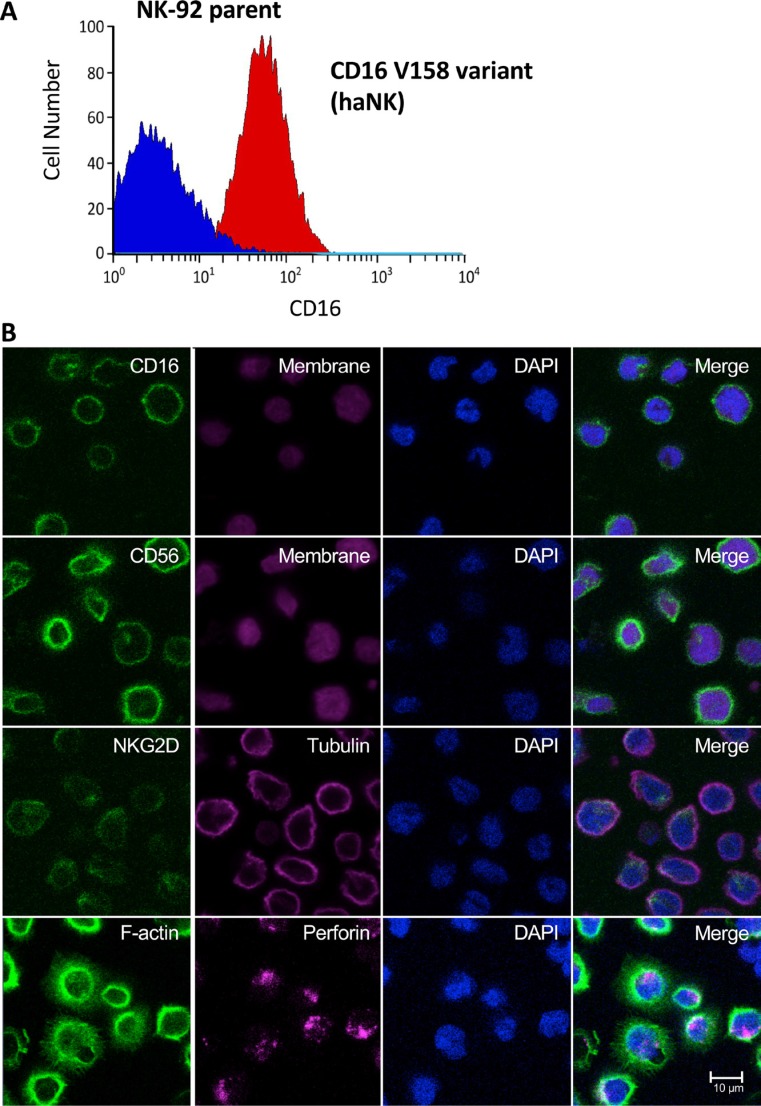
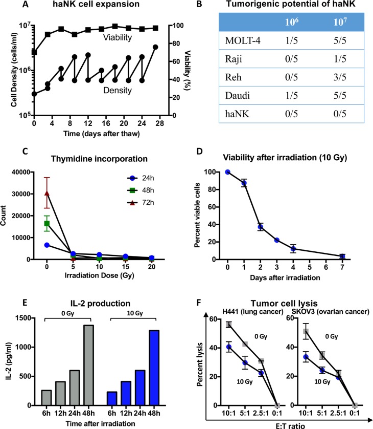
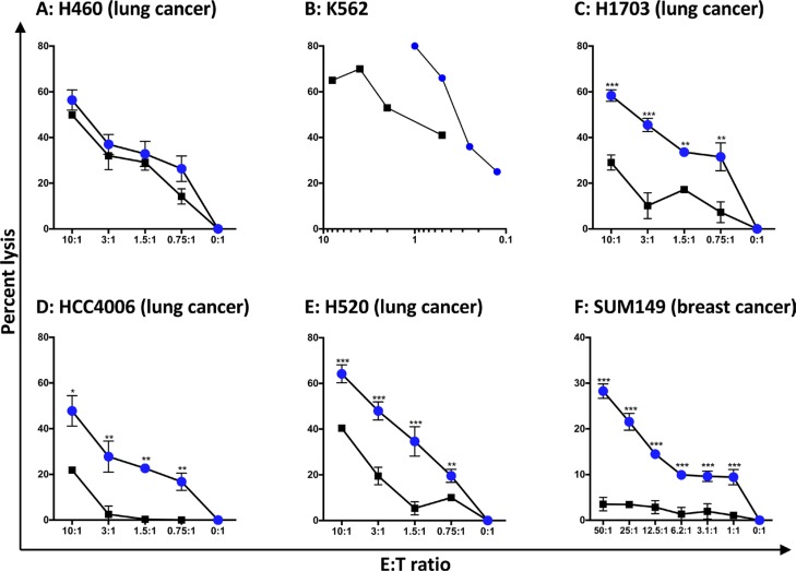
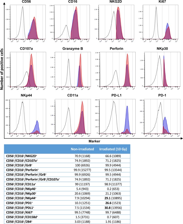
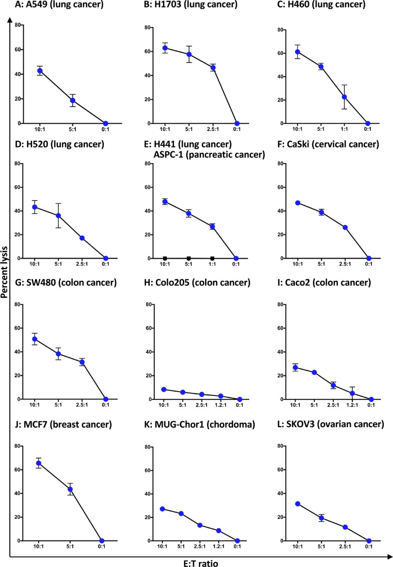
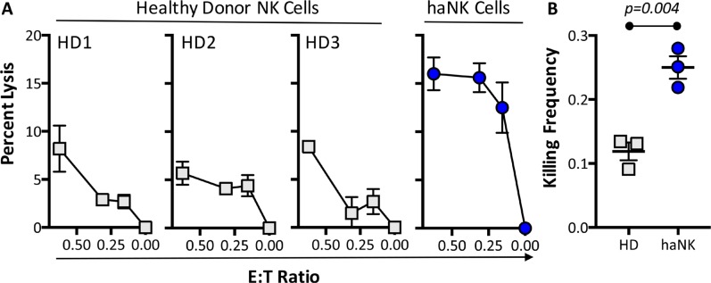
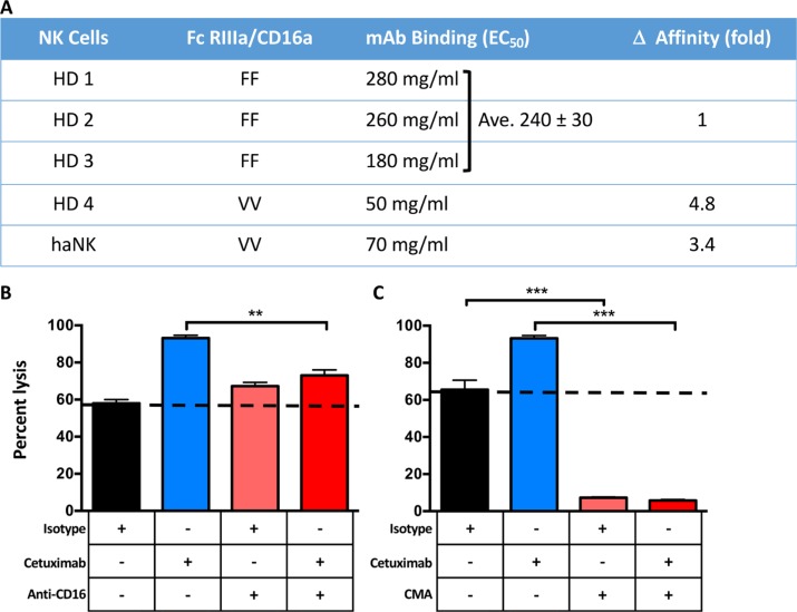
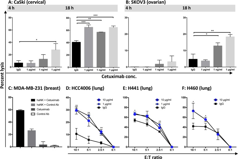
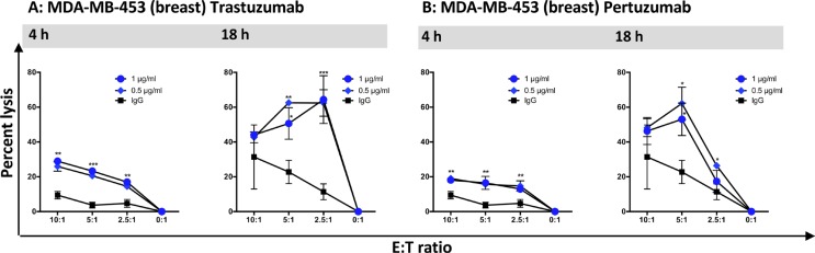
Natural killer (NK) cells are known to play a role in mediating innate immunity, in enhancing adaptive immune responses, and have been implicated in mediating anti-tumor responses via antibody-dependent cell-mediated cytotoxicity (ADCC) by reactivity of CD16 with the Fc region of human IgG1 antibodies. The NK-92 cell line, derived from a lymphoma patient, has previously been well characterized and adoptive transfer of irradiated NK-92 cells has demonstrated safety and shown preliminary evidence of clinical benefit in cancer patients. The NK-92 cell line, devoid of CD16, has now been engineered to express the high affinity (ha) CD16 V158 FcγRIIIa receptor, as well as engineered to express IL-2; IL-2 has been shown to replenish the granular stock of NK cells, leading to enhanced perforin- and granzyme-mediated lysis of tumor cells. The studies reported here show high levels of granzyme in haNK cells, and demonstrate the effects of irradiation of haNK cells on multiple phenotypic markers, viability, IL-2 production, and lysis of a spectrum of human tumor cells. Studies also compare endogenous irradiated haNK lysis of tumor cells with that of irradiated haNK-mediated ADCC using cetuximab, trastuzumab and pertuzumab monoclonal antibodies. These studies thus provide the rationale for the potential use of irradiated haNK cells in adoptive transfer studies for a range of human tumor types. Moreover, since only approximately 10% of humans are homozygous for the high affinity V CD16 allele, these studies also provide the rationale for the use of irradiated haNK cells in combination with IgG1 anti-tumor monoclonal antibodies.

Keywords: ADCC; NK lysis; cetuximab; high affinity CD16; immunotherapy.


Conflict of interest statement

CONFLICTS OF INTEREST

Laboratory of Tumor Immunology And Biology, National Cancer Institute

The authors have no potential conflicts of interest to disclose. The Laboratory of Tumor Immunology and Biology, National Cancer Institute, has a Cooperative Research and Development Agreement (CRADA) with NantBioScience.

Immune Cell Development And Host Defense Program, Institute For Cancer Research, Fox Chase Cancer Center

No potential conflicts of interest to disclose.

NantBioScience, Inc., Affiliates

Hans Klingemann and Laurent Boissel are employees of NantKwest. Patrick Soon-Shiong is a founder and an executive at NantKwest and NantCell, Shahrooz Rabizadeh is a contributor to NantCell.

Figures

Figure 1
Figure 1. Analyses of CD16 high affinity variant (V158) in haNK cells
(A) The NK-92 parent cell line was modified to express a high affinity CD16 variant. (B) Immunofluorescence imaging of haNK cells. haNK cells were stained for expression of common NK markers as described in Materials and Methods. The expression of CD16 (green), CD56 (green), NKG2D (green), F-actin (green), CellMask plasma membrane stain (magenta), tubulin (magenta), perforin (magenta), and DAPI nuclear stain (blue) were visualized by confocal microscopy. Scale bar = 10 μm.
Figure 2
Figure 2. Characteristics of haNK cells before and after irradiation
(A) haNK cell expansion (subcultured every 3 days) with cell density (cells/ml) and viability (%). haNK cells were cultured in X-VIVO-10 medium without phenol red, and followed for 28 days. (B) Evaluation of the tumorigenic potential of non-irradiated haNK cells. Male athymic nude mice were injected s.c. with 106 or 107 of different tumor cells (see Methods), and monitored daily for the presence of tumors at injection sites. At study conclusion (day 63) the number of resultant viable tumors (≥ 50 mm3) was quantified for each tumor cell line and cell number injected. (C) 3H-thymidine incorporation was measured to evaluate proliferation of haNK cells irradiated at different doses. Irradiation at ≥ 5 Gy was adequate to render haNK cells replication incompetent. (D) haNK cell viability was evaluated daily after irradiation (10 Gy) using AO/PI staining. (E) Evaluation of haNK cell cumulative cytokine secretion after irradiation (10 Gy). Results are in pg/ml for 5 × 10⁵ haNK cells/ml. (F) haNK cell lysis of the human carcinoma cell lines H441 (lung) and SKOV3 (ovarian) before irradiation (gray squares), and 24 h after irradiation (10 Gy, blue circles) using an 18-h 111In release assay.
Figure 3
Figure 3. Comparison of tumor cell lysis by haNK cells 1 h vs. 24 h post-irradiation
(AF) haNK cells were either irradiated (10 Gy) and assayed immediately (squares) or after 24 h in culture (circles). The 18-h haNK lysis assay was performed with different tumor cell targets at the E:T ratios shown. K562 was analyzed by flow cytometry. The human lung cancer cell lines (H460, H1703, HCC4006, and H520) were analyzed by CeligoS, and the human breast cancer cell line SUM149 was analyzed by 111In-release. Results shown are the means (SD) of triplicate measurements from 1 of 3 comparable repeat experiments. T-tests were employed to compare lysis at 0 h and 24 h post-irradiation at all E:T ratios. ***P < 0.001, **P < 0.01, *P < 0.05.
Figure 4
Figure 4. Phenotyping of haNK cells with and without irradiation
haNK cells were stained for flow cytometry either non-irradiated, or irradiated (10 Gy) and rested for 24 h before staining. Figures show the expression of different markers for FMO controls (open), non-irradiated haNK cells (red), and irradiated haNK cells (blue). Table: Values shown are percent of CD56+CD16+ haNK cells (MFI). Bolded figures denote a significant increase after irradiation.
Figure 5
Figure 5. haNK lysis of human tumor cell lines
haNK cells were irradiated (10 Gy) and kept in culture for 24 h prior to an 18-h lysis assay at different E:T ratios. Four lung cancer cell lines A549, H1703, H460, and H520 were analyzed by CeligoS (AD). In a separate series of studies, human tumor cell line lysis was analyzed by 111In-release (EL). Results shown are the means (SD) of triplicate measurements from one of at least three comparable repeat experiments.
Figure 6
Figure 6. haNK cells have a higher killing frequency than healthy donor NK cells
(A) Primary human NK cells purified from three independent healthy donors (HD) or haNK (10 Gy irradiated) cells were co-incubated with 111In-labeled MDA-MB-231 (breast cancer) target cells at low E:T ratios for 18 h. (B) % specific lysis was used to calculate killing frequency by dividing the number of target cells killed by the number of effector cells used for the 0.625:1 ratio.
Figure 7
Figure 7. Comparative affinities of CD16 allotypes and factors influencing haNK-mediated lysis
(A) Influence of CD16 FCGR3A-158V/F polymorphism on the concentration-effect relationship of cetuximab-dependent NK cell-mediated cytotoxicity. Purified NK cells from homozygous F/F, V/V healthy donors (HD) or haNK cells were incubated with varying concentrations of cetuximab for 30 min at 4°C followed by FITC-conjugated anti-CD16 3G8 MAb and then analyzed by flow cytometry. Percentages of inhibition of 3G8 binding were calculated as described in Methods, and the results are expressed as effective MAb concentration to achieve 50% inhibition of 3G8 MAb binding to NK cells (EC50). (B and C) The H460 human lung carcinoma cell line was used as a target in an 18-h 111In-release assay to evaluate if haNK ADCC mediated by cetuximab (10 μg/ml) could be blocked using anti-CD16 antibody (50 μg/ml) (B), or concanamycin A (CMA) (C). haNK cells were irradiated (10 Gy) and used at a 20:1 E:T ratio. Results shown are the mean (SEM) lysis of triplicate measurements in one of 3 repeat experiments. The dotted lines indicate endogenous haNK-mediated lysis. T-tests were employed to compare the treatments. ***P < 0.001, **P < 0.01.
Figure 8
Figure 8. haNK ADCC mediated by cetuximab was evaluated with 4 h and 18 h 111In-release assays
haNK cells (irradiated 10 Gy) were used as effector cells at different E:T ratios. Cetuximab was used at the specified concentrations. (A) CaSki: human cervical carcinoma (5:1 ratio); (B) SKOV3: human ovarian carcinoma (5:1 ratio). (C) MDA-MB-231 (human breast carcinoma) cells were used as a target at an E:T ratio of 7.5:1. Target cells were also incubated with cetuximab (1 μg/ml) or isotype control IgG1 antibody (1 μg/ml) alone, without effector cells. haNK ADCC mediated by cetuximab (black bar) and haNK killing (gray bar), cetuximab alone (dark gray bar) and control antibody alone (white bar) are shown. (DF) 18-h lytic assays employing irradiated haNK cells with three different human lung carcinoma lines (HCC4006, H441, H460) at different E:T ratios. Results shown are the averages (SD) of triplicate measurements from one of at least three comparable repeat experiments. Multiple t-tests were used to compare each dose with IgG control at all E:T ratios. ***P < 0.001, **P < 0.01, *P < 0.05.
Figure 9
Figure 9. haNK cell ADCC mediated by trastuzumab and pertuzumab was evaluated with 4 h and 18 h 111In-release assays
haNK cells (irradiated 10 Gy) were used as effector cells at different E:T ratios, and MAbs were used at the specified concentrations. MDA-MB-453: human breast carcinoma. Results shown are the averages (SD) of triplicate measurements from one of at least three comparable repeat experiments. Multiple t-tests were used to compare each dose with IgG control at all E:T ratios. ***P < 0.001, **P < 0.01, *P < 0.05.

References

    1. Gras Navarro A, Bjorklund AT, Chekenya M. Therapeutic potential and challenges of natural killer cells in treatment of solid tumors. Front Immunol. 2015;6:202. - PMC - PubMed
    1. Paust S, Senman B, von Andrian UH. Adaptive immune responses mediated by natural killer cells. Immunol Rev. 2010;235:286–296. - PMC - PubMed
    1. Weiner LM, Dhodapkar MV, Ferrone S. Monoclonal antibodies for cancer immunotherapy. Lancet. 2009;373:1033–1040. - PMC - PubMed
    1. Klingemann H, Boissel L, Toneguzzo F. Natural killer cells for immunotherapy – advantages of the NK-92 cell line over blood NK cells. Front Immunol. 2016:7. - PMC - PubMed
    1. Boissel L, Betancur-Boissel M, Lu W, Krause DS, Van Etten RA, Wels WS, Klingemann H. Retargeting NK-92 cells by means of CD19- and CD20-specific chimeric antigen receptors compares favorably with antibody-dependent cellular cytotoxicity. OncoImmunology. 2013;2:e26527. - PMC - PubMed

MeSH terms