. 2016 Nov 18;11(11):e0167063.
doi: 10.1371/journal.pone.0167063. eCollection 2016.

Cellular Basis of Pineal Gland Development: Emerging Role of Microglia as Phenotype Regulator

Affiliations

Cellular Basis of Pineal Gland Development: Emerging Role of Microglia as Phenotype Regulator

María P Ibañez Rodriguez et al. PLoS One. .

Abstract

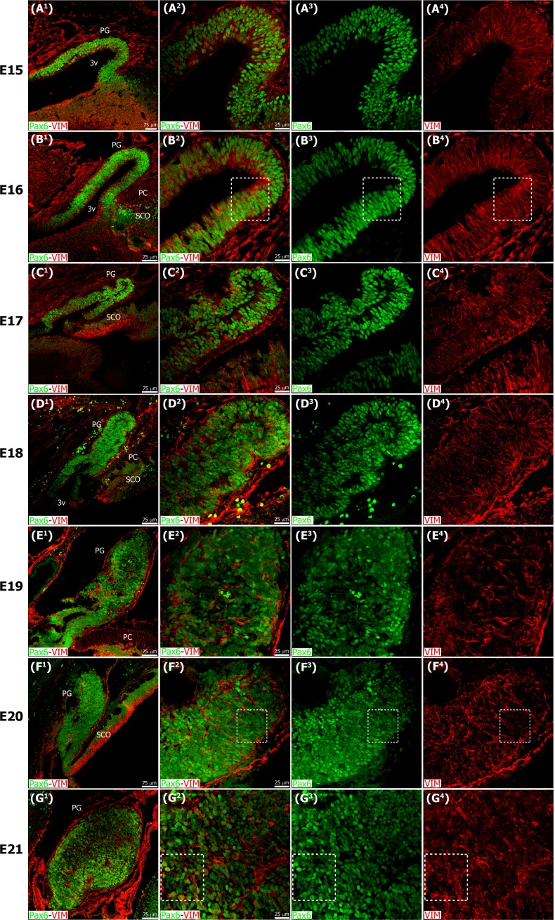
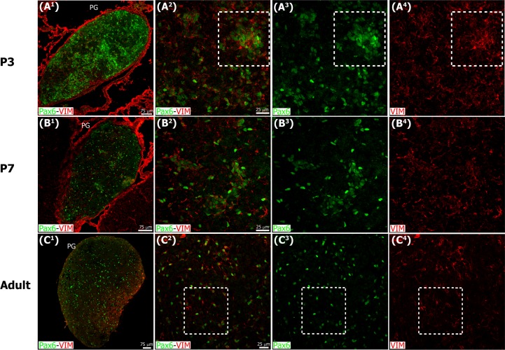
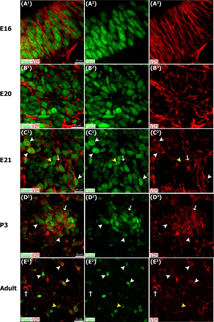
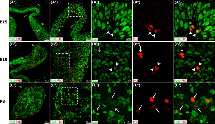
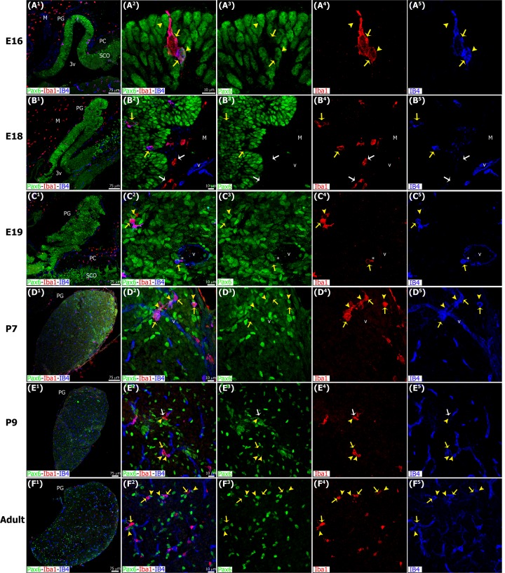
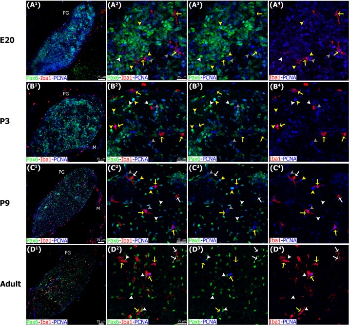
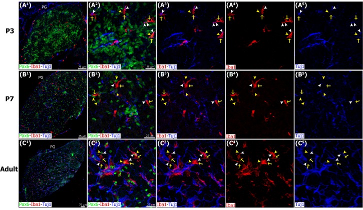
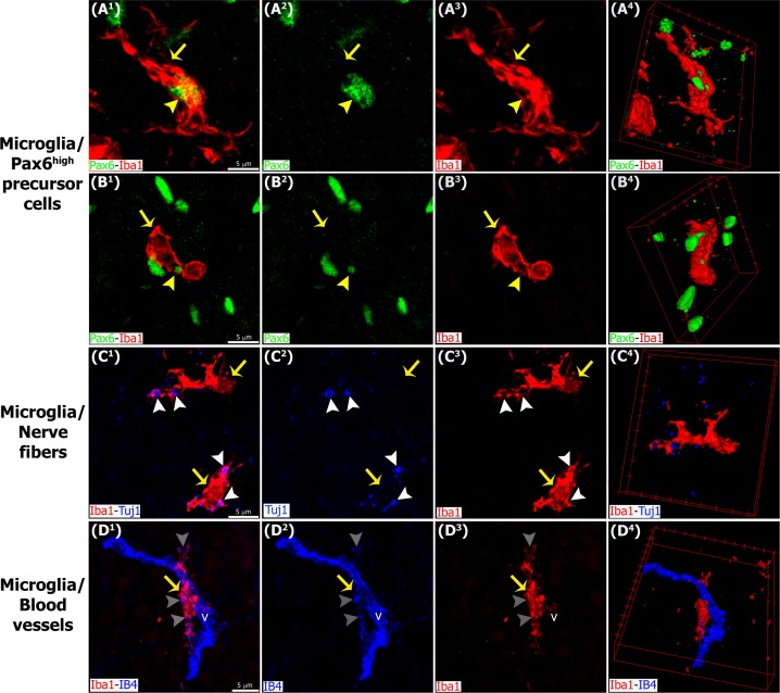
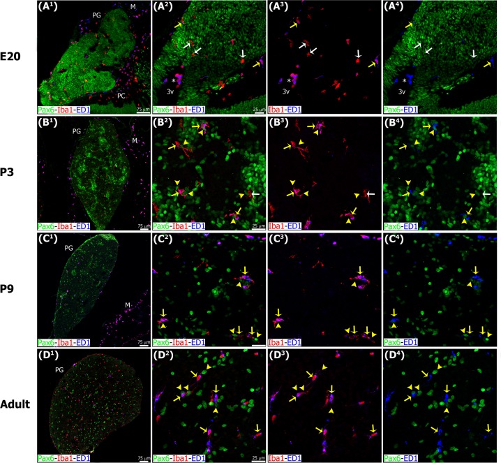
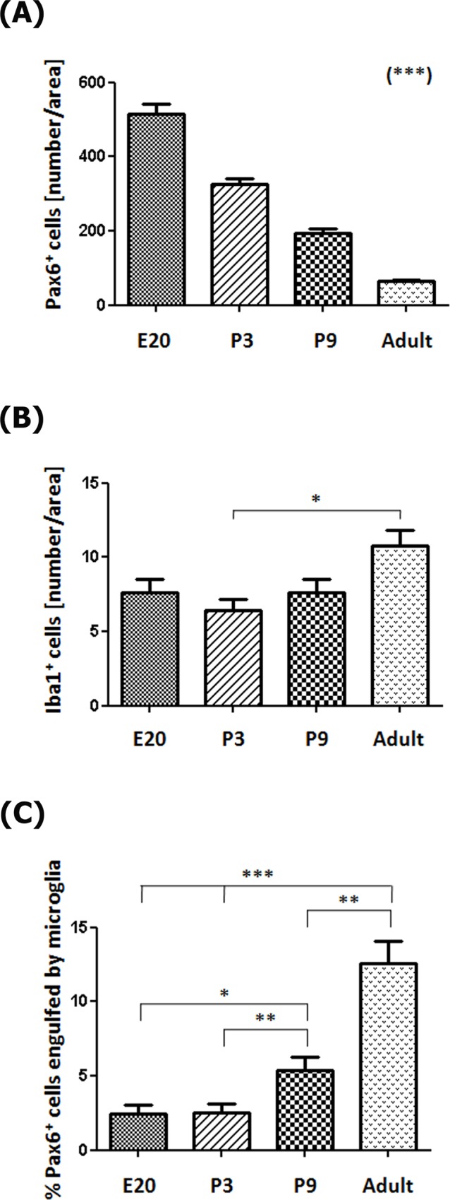
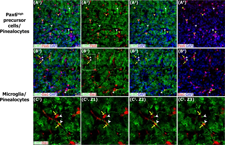
The adult pineal gland is composed of pinealocytes, astrocytes, microglia, and other interstitial cells that have been described in detail. However, factors that contribute to pineal development have not been fully elucidated, nor have pineal cell lineages been well characterized. We applied systematic double, triple and quadruple labeling of cell-specific markers on prenatal, postnatal and mature rat pineal gland tissue combined with confocal microscopy to provide a comprehensive view of the cellular dynamics and cell lineages that contribute to pineal gland development. The pineal gland begins as an evagination of neuroepithelium in the roof of the third ventricle. The pineal primordium initially consists of radially aligned Pax6+ precursor cells that express vimentin and divide at the ventricular lumen. After the tubular neuroepithelium fuses, the distribution of Pax6+ cells transitions to include rosette-like structures and later, dispersed cells. In the developing gland all dividing cells express Pax6, indicating that Pax6+ precursor cells generate pinealocytes and some interstitial cells. The density of Pax6+ cells decreases across pineal development as a result of cellular differentiation and microglial phagocytosis, but Pax6+ cells remain in the adult gland as a distinct population. Microglial colonization begins after pineal recess formation. Microglial phagocytosis of Pax6+ cells is not common at early stages but increases as microglia colonize the gland. In the postnatal gland microglia affiliate with Tuj1+ nerve fibers, IB4+ blood vessels, and Pax6+ cells. We demonstrate that microglia engulf Pax6+ cells, nerve fibers, and blood vessel-related elements, but not pinealocytes. We conclude that microglia play a role in pineal gland formation and homeostasis by regulating the precursor cell population, remodeling blood vessels and pruning sympathetic nerve fibers.


Conflict of interest statement

The authors have declared that no competing interests exist.

Figures

Fig 1
Fig 1. The pineal gland develops from neuroepithelial cells that express the transcription factor Pax6 and the intermediate filament vimentin.
Panels display confocal microscopy of immunolabeled sagittal sections of rat pineal gland (PG) from embryonic day (E) 15 to E21. Both male and female rat embryos were used. Panels A1-G1 show expression of Pax6 (green) and vimentin (VIM, red) in pineal precursor cells at low magnification. Panels A2-G2 display the same structures at higher magnification. Panels A3-G3 and A4-G4 show Pax6 and vimentin expression for each stage of development, respectively. Pineal organogenesis begins around E15 as an evagination of the neuroepithelium in the dorsal diencephalon that is densely populated by Pax6-expressing cells (green). The developing PG becomes a tubular extension at E16. The orientation of Pax6/VIM+ cells is radial at these stages. At E17 the pineal neuroepithelium begins to fold and fuses at the midline. After fusion of the neuroepithelium, double immunolabeled rosette-like structures are visible in the E18-E21 developing PG. At E21 the PG has developed into a recognizable globular structure. Inset boxes are shown at higher magnification in Fig 3. (A1-G1) 20x; scale bar: 75 μm. (A2-G4) 60x; scale bar: 25 μm. PC, posterior commissure. SCO, subcommissural organ. 3v, third ventricle.
Fig 2
Fig 2. Pax6 and vimentin expression persists in the postnatal and adult pineal gland.
Immunoreactivity for Pax6 (green) and vimentin (VIM, red) in pineal glands (PG) from 3-, 7- and 90-day-old male rats (P3, P7 and adult, respectively). The levels of expression of both markers appear to decrease throughout PG development. Pax6+ and/or VIM+ cells are still organized in discrete rosette-like patches at P3, while dispersed immunoreactive cells are more abundant from P7 onwards. Inset boxes are shown at higher magnification in Fig 3. (A1-B1) 20x; (C1) 1.2x digital zoom from a 10x image; scale bar: 75 μm. (A2-B4) 60x; (C2-C4) 40x; scale bar: 25 μm.
Fig 3
Fig 3. Pax6/Vimentin double immunostaining reveals dynamic changes in the organization of precursor cells throughout pineal ontogeny.
Panels include higher magnification images of the insets shown at 60x and 40x in Figs 1 and 2 immunostained for Pax6 (green) and vimentin (VIM, red). (A1-A3) In the earliest stages Pax6/VIM double-positive precursor cells display a radial distribution. (B1-D3) After fusion of the neuroepithelium, Pax6/VIM+ cells are arranged mainly as rosette-like structures. (E1-E3) In the adult pineal gland (PG) individual cells positive for Pax6 and/or vimentin are dispersed throughout the parenchyma. White arrowheads: Pax6high/VIMhigh cells. Yellow arrowheads: Pax6high/VIMlow cells. White arrows: Pax6low/VIMhigh cells. (A1-E3) 3x, 4x, 3x, 2.5x and 2.6x digital zooms of the insets shown in Figs 1B2–1B4, 1F2–1F4 and 1G2–1G4 and 2A2–2A4 and 2C2–2C4, respectively; scale bar: 10 μm. E, embryonic day. P, postnatal day.
Fig 4
Fig 4. Actively dividing precursor cells express Pax6, and they are located at the surface of the ventricular lumen of the developing pineal gland at early stages of development, and dispersed throughout the gland after fusion of the neuroepithelium.
Confocal microscopy of sagittal sections through the rat pineal gland (PG) immunolabeled for Pax6 (green) and the mitotic M-phase marker phospho-histone H3 (PH3, red). All PH3+ mitotic cells expressed Pax6; Pax6 levels, however, varied among dividing pinealocyte precursor cells. The number of mitotic precursor cells increased progressively during embryogenesis, and dropped quickly after birth. White arrowheads: Pax6high mitoses. White arrows: Pax6low mitoses. (A1-C1) 20x; scale bar: 75 μm. (A2-C2) 60x; scale bar: 25 μm. (A3-C5) 3x digital zooms of the insets shown at 60x; scale bar: 10 μm. E, embryonic day. PC, posterior commissure. P, postnatal day. SCO, subcommissural organ. 3v, third ventricle.
Fig 5
Fig 5. Microglia colonize the pineal gland neuroepithelium very early in development and persist across the lifespan, showing affiliation with Pax6+ cells.
Microglia in the developing and adult pineal gland (PG) exhibit an ‘activated’ morphology. Panels show images of confocal microscopy of PG sections immunolabeled for Pax6 (green) and the microglial marker Iba1 (red), and stained with the isolectin IB4 (IB4, blue) to reveal blood vessels (v) and a subpopulation of microglial cells. Iba1+ microglia were present in the meninges (M) and choroid plexus, and appeared to enter the PG from both sources early in development. In a few cases microglial cells were seen phagocytosing Pax6+ precursor cells (yellow arrowheads) in early postnatal development (A1-A5), but this was most common in the postnatal and adult PG. Yellow arrows show Iba1/IB4 double-positive cells. The tight spatial relationship between macrophages and blood vessels suggests that microglia might also colonize the PG through the vasculature (asterisk). White arrows: microglial cells positive for Iba1 and negative for IB4 (red). (A1, B1) 20x; (C1, E1) 1.6x and 1.2x digital zooms from 10x images, respectively; (D1 and F1) 10x; scale bar: 75 μm. (A2-A5, B2-B5, C2-C5, D2-D5, E2-E5) 3x, 1.4x, 1.7x, 1.5x and 1.4x digital zooms from 60x images, respectively; (F2-F5) 1.5x enlargements from a 40x image; scale bar: 10μm. E, embryonic day. M, meninges. P, postnatal day. PC, posterior commissure. SCO, subcommissural organ. 3v, third ventricle.
Fig 6
Fig 6. Microglia express the mitotic cell marker PCNA throughout the entire pineal gland ontogeny, while Pax6+ cells have detectable PCNA levels mainly during early development.
Panels show images of confocal microscopy of pineal gland (PG) sections immunostained for PCNA (blue), Pax6 (green) and Iba1 (red). The combination of these three markers revealed cell heterogeneity in the pineal gland in the different stages analyzed. Yellow arrows: Iba1/PCNA double-positive microglial cells. White arrows: microglia positive for Iba1 and negative for PCNA. Yellow arrowheads: Pax6/PCNA double-immunoreactive cells. White arrowheads: Pax6+ precursor cells negative for PCNA. Grey arrowheads: cells positive only for PCNA. (A1, C1, D1) 1.5x, 1.3x and 1.1x digital zooms from 10x images, respectively; (B1) 20x; scale bar: 75 μm. (A2-C4) 60x; (D2-D4) 1.4x digital zooms from a 40x image; scale bar: 25 μm. E, embryonic day. M, meninges. P, postnatal day.
Fig 7
Fig 7. Microglia interact with Tuj1+ nerve fibers in the postnatal and adult pineal gland.
Sections of postnatal (P) and adult pineal glands (PG) immunolabeled for Pax6 (green), Iba1 (red) and β-tubulin III (Tuj1, blue). Sympathetic nerve fibers and microglia form an intricate network where Pax6+ cells are located. Macrophages (yellow arrows) phagocytosing Pax6+ cells (yellow arrowheads) and/or pruning nerve fibers (white arrowheads) are indicated. (A1-B1) 20x; (C1) 10x; scale bar: 75 μm. (A2-A5, B2-B5, C2-C5) 1.3x, 1.4x and 1.2x digital zooms from 60x images, respectively; scale bar: 25 μm.
Fig 8
Fig 8. Higher magnification images demonstrating interactions between microglia, Pax6+ cells, nerve fibers and blood vessels in the pineal gland.
Panels show confocal images of pineal gland (PG) immunolabeled for Pax6 (green), Iba1 (red) and β-tubulin III (Tuj1, blue), or IB4-stained blood vessels (v, blue). Microglia (yellow arrows) closely affiliate with and in some cases engulf Pax6+ precursor cells (yellow arrowheads) (A1-B4), Tuj1+ nerve fibers (white arrowheads) (C1-C4) and blood vessels (grey arrowheads) (D1-D4) within the PG. (A1-B3, C1-C3, D1-D3) 4x, 3.4x and 3.5x digital zooms from 60x images, respectively; scale bar: 5 μm. (A4, B4, C4, D4) 3D reconstruction of the main interactions.
Fig 9
Fig 9. Microglia phagocytic potential increases throughout pineal ontogeny.
Panels show confocal microscopy of sections immunostained for the lysosomal marker ED1 (CD68, blue), Pax6 (green) and Iba1 (red). (A1-B4) Late embryonic (E20) and early postnatal (P3) pineal gland (PG) sections showing two subpopulations of microglial cells with (yellow arrows) or without (white arrows) ED1 expression. (C1-D4) Later neonatal (P9) and adult PG exhibiting the majority of Iba1/ED1 double-immunoreactive cells in close proximity to or phagocytosing Pax6high cells (yellow arrowheads). Asterisk, cluster of round Iba1/ED1 double-positive cells in the lumen of the third ventricle (3v) and in close proximity to the proximal pineal gland. (A1) 20x; (B1) (C1, D1) 1.2 x and 1.3x digital zooms from 10x images, respectively; scale bar: 75 μm. (A2-A4) 40x; (B2-D4) 60x; scale bar: 25 μm. M, meninges. PC, posterior commissure.
Fig 10
Fig 10. Activated microglia contact and engulf Pax6+ precursor cells.
Higher magnification of pineal gland sections immunolabeled for Iba1 (red, yellow arrows), ED1 (blue, yellow arrows) and Pax6 (green, yellow arrowheads). (A1-A6) Microglial cells with an activated morphology are enriched in cytoplasmic ED1-positive bodies and engulf Pax6high nuclei and elements. (B1-B6) Microglia with few thick projections contacting and surrounding Pax6high precursor cells. (A1-A5, B1-B5) 4x and 3.6x digital zooms from 60x images, respectively; scale bar: 5 μm. (A6, B6) 3D reconstruction of phagocytic microglia/Pax6high precursor cells or elements interactions.
Fig 11
Fig 11. Morphometric analysis shows that Pax6+ cell numbers decrease, while Iba1+ cell numbers, and Pax6+ cells engulfed by microglia increase in the pineal gland through ontogeny.
(A) The number of Pax6+ precursor cells per area (0.05 mm2) decreases throughout pineal development (Post-test for linear trend: P < 0.0001). (B) Microglia density increases slightly between postnatal day 3 (P3) and adulthood (Post-test for linear trend: P < 0.05). (C) The percentage of Pax6+ precursor cells in contact or engulfed by microglial cells is higher in late neonatal stages onwards (Post-test for linear trend: P < 0.0001). Data were expressed as mean ± S.E.M. Statistics: one-way ANOVA followed by the Tukey post-test: *** P < 0.001, ** P < 0.01, * P < 0.05. E, embryonic day.
Fig 12
Fig 12. Pinealocytes appear to be protected from microglia-mediated phagocytosis.
Sections of adult pineal gland immunolabeled for serotonin or 5-hydroxytryptamine (5-HT, green), Pax6 (red) and Iba1 (red). DAPI (blue) was used as general nuclear dye. (A1-A4) Pinealocytes positive for 5–HT (white arrowheads) and Pax6+ cells (yellow arrowheads) represent two distinct cell populations. Few cells positive for both Pax6 and 5-HT were identified (orange arrowheads with white borders). (B1-B4) Microglia (yellow arrows) in close proximity to 5-HT-immunolabeled projections (white arrowheads) and phagocytosing a serotonin-negative cell (grey arrowheads). (C1) Higher magnification of the inset shown in B2. (C1.Z1-C1.Z3) Three consecutive confocal planes (Z) that show the points of contact between microglia and serotonergic processes. (A1-B4) 1.4x digital zooms from 40x images; scale bar: 25 μm. (C1-C1.Z3) 3.2x digital zooms from the inset at 40x shown in B2; scale bar: 10 μm.

References

    1. Ganguly S, Coon SL, Klein DC. Control of melatonin synthesis in the mammalian pineal gland: the critical role of serotonin acetylation. Cell Tissue Res. 2002;309: 127–137. 10.1007/s00441-002-0579-y - DOI - PubMed
    1. Maronde E, Stehle JH. The mammalian pineal gland: known facts, unknown facets. Trends Endocrinol Metab. 2007;18: 142–149. 10.1016/j.tem.2007.03.001 - DOI - PubMed
    1. Yamazaki S, Yoshikawa T, Biscoe EW, Numano R, Gallaspy LM, Soulsby S, et al. Ontogeny of circadian organization in the rat. J Biol Rhythms. 2009;24: 55–63. 10.1177/0748730408328438 - DOI - PMC - PubMed
    1. Furukawa T, Morrow EM, Li T, Davis FC, Cepko CL. Retinopathy and attenuated circadian entrainment in Crx-deficient mice. Nat Genet. 1999;23: 466–470. 10.1038/70591 - DOI - PubMed
    1. Muñoz EM, Bailey MJ, Rath MF, Shi Q, Morin F, Coon SL, et al. NeuroD1: developmental expression and regulated genes in the rodent pineal gland. J Neurochem. 2007;102: 887–899. 10.1111/j.1471-4159.2007.04605.x - DOI - PubMed

MeSH terms