. 2016 Nov 18;11(11):e0166885.
doi: 10.1371/journal.pone.0166885. eCollection 2016.

The Development Of Drosophila Melanogaster under Different Duration Space Flight and Subsequent Adaptation to Earth Gravity

Affiliations

The Development Of Drosophila Melanogaster under Different Duration Space Flight and Subsequent Adaptation to Earth Gravity

Irina V Ogneva et al. PLoS One. .

Abstract

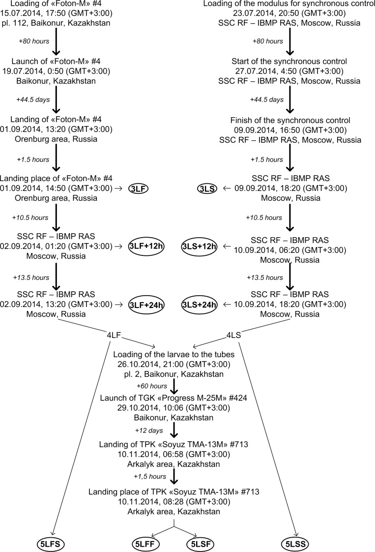
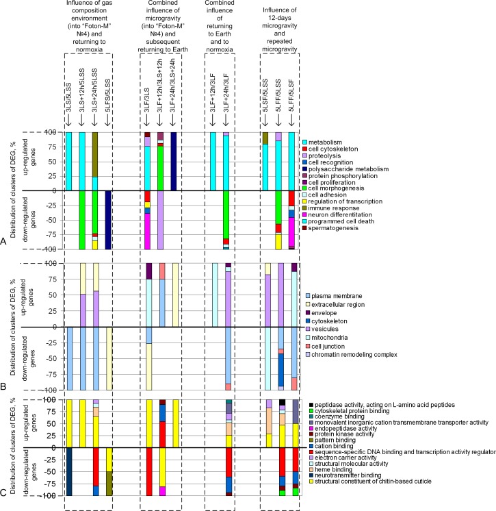
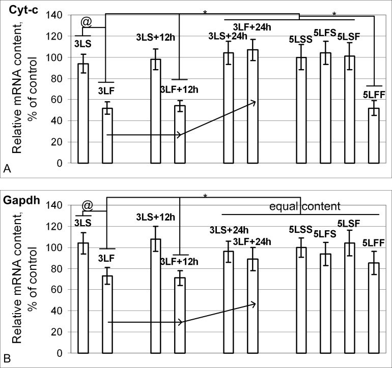
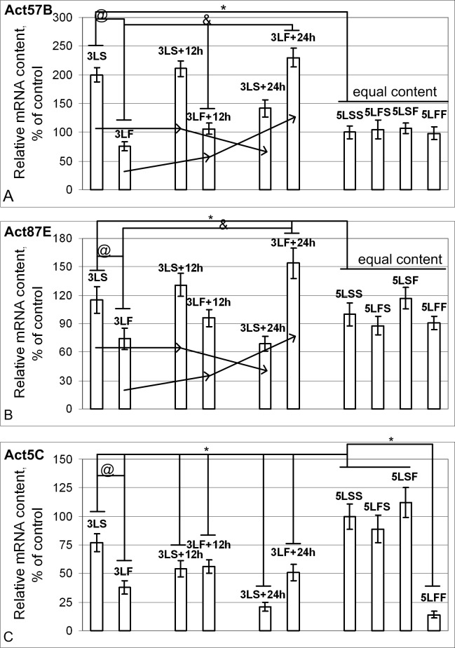
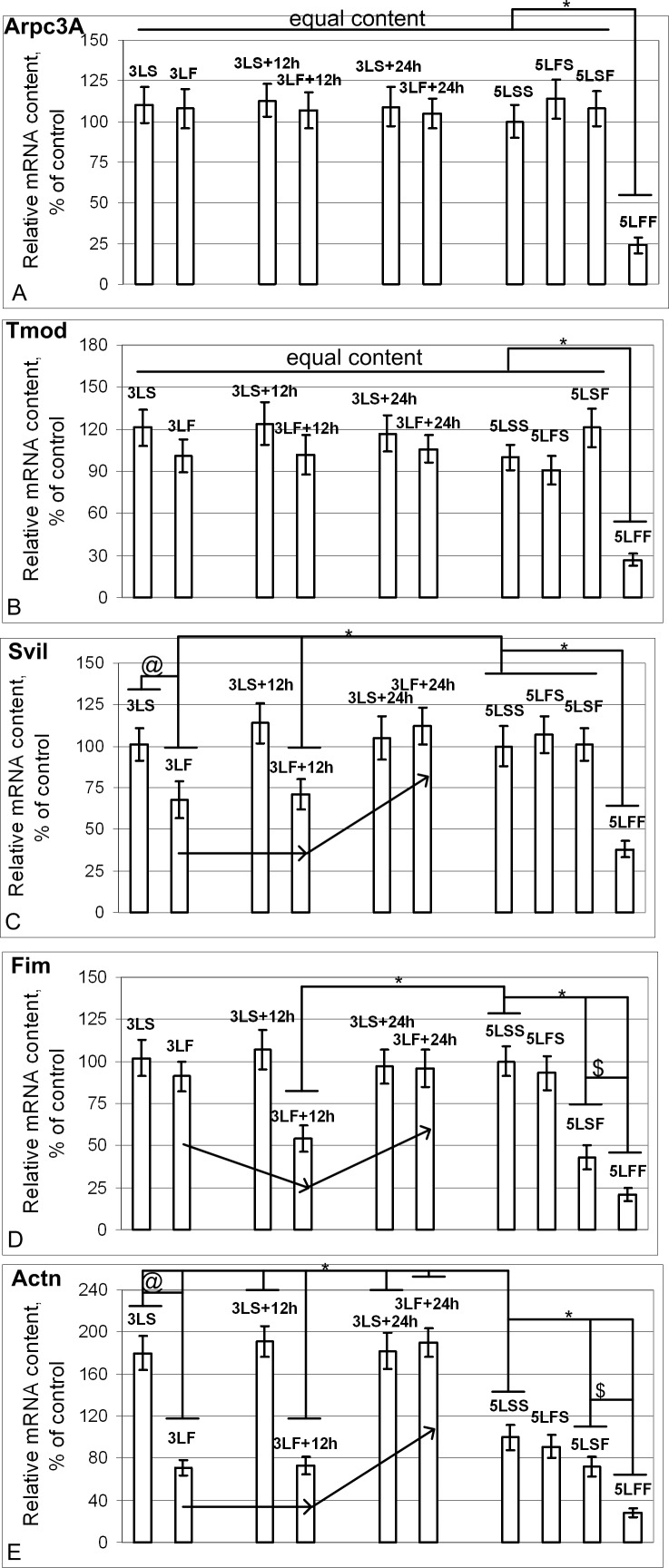
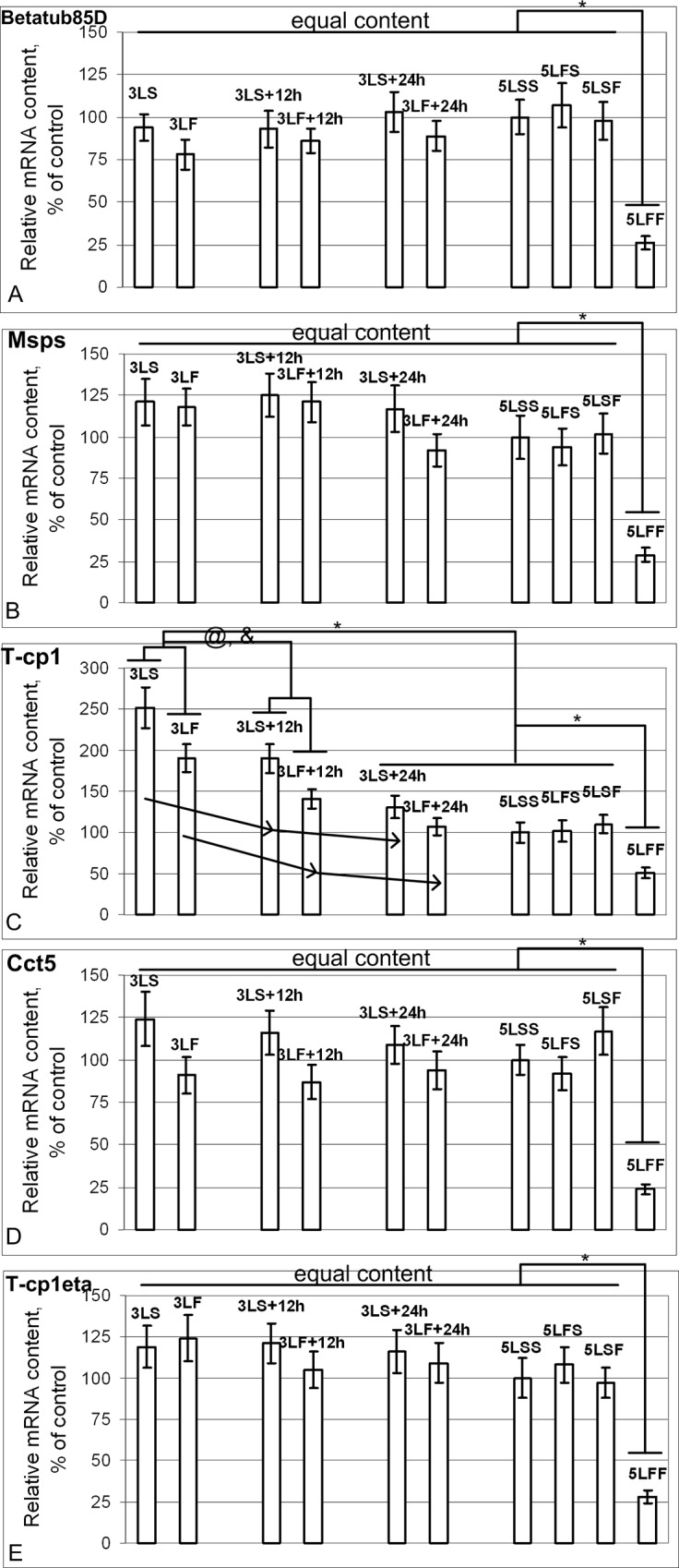
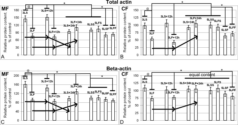
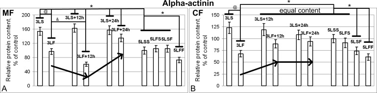
In prospective human exploration of outer space, the need to preserve a species over several generations under changed gravity conditions may arise. This paper demonstrates our results in the creation of the third generation of fruit fly Drosophila melanogaster (third-stage larvae) during the 44.5-day space flight (Foton-M4 satellite (2014, Russia)), then the fourth generation on Earth and the fifth generation again in conditions of the 12-day space flight (2014, in the Russian Segment of the ISS). The species preserves fertility despite a number of changes in the level of expression and content of cytoskeletal proteins, which are the key components of the cleavage spindle and the contractile ring of cells. The results of transcriptome screening and space analysis of cytoskeletal proteins show that the exposure to weightless conditions leads to the increased transcription of metabolic genes, cuticle components and the decreased transcription of genes involved in morphogenesis, cell differentiation, cytoskeletal organization and genes associated with the plasma membrane. "Subsequent" exposure to the microgravity for 12 days resulted in an even more significant increase/decrease in the transcription of the same genes. On the contrary, the transition from the microgravity conditions to the gravity of Earth leads to the increased transcription of genes whose products are involved in the morphogenesis, cytoskeletal organization, motility of cells and transcription regulation, and to the decreased transcription of cuticle genes and proteolytic processes.


Conflict of interest statement

The authors have declared that no competing interests exist.

Figures

Fig 1
Fig 1. Design and cyclogram of the experiment.
The experimental groups are highlighted by the circles.
Fig 2
Fig 2. Distribution of differentially expressed genes among pairwise comparisons of the study groups.
(A) DEG distribution according to the biological processes. (B) DEG distribution according to the cellular compartments. (C) DEG distribution according to the molecular functions.
Fig 3
Fig 3. Relative mRNA content of genes (qPCR data) that encode metabolic proteins.
(A) The Cyt-c mRNA content in 3LS, 3LS+12h, 3LS+24h, 5LSS, 5LFS and 5LSF groups was the same. In the 3LF and 3LF+12h groups, the Cyt-c mRNA was reduced by 48% and 46% compared with the 5LSS group (p<0.05) but was restored to the control level in the 3LF+24h group. In the 5LFF group, the Cyt-c mRNA content was reduced by 48% compared with the 5LSS group (p<0.05). (B) The Gapdh mRNA content in the 3LS, 3LS+12h, 3LS+24h, 3LF+24h, 5LSS, 5LFS, 5LSF and 5LFF groups was the same. In the 3LF and 3LF+12h groups, Gapdh mRNA was reduced by 27% and 29% compared with the 5LSS group (p<0.05).
Fig 4
Fig 4. Relative mRNA content of genes (qPCR data) that encode actin isoforms.
(A) The Act57B mRNA content was increased by 100% in the 3LS group (p<0.05), by 111% in the 3LS+12h group(p<0.05), and by 42% in the 3LS+24h group (p<0.05) compared with the 5LSS group. In 3LF group, Act57B mRNA was reduced by 24% (p<0.05), it was the same in the 3LF+12h group, and it exceeded the level of the 5LSS group by 130% in the 3LF+24h group (p<0.05). In the 5LSS, 5LFS, 5LSF, and 5LFF groups, the mRNA content was the same. (B) The Act87E mRNA content was the same in the 3LS group, was increased by 31% in the 3LS+12h group (p<0.05), and was decreased by 31% in the 3LS+24h group (p<0.05) compared with the 5LSS group. Act87E mRNA was reduced by 26% (p<0.05) in the 3LF group, was the same in the 3LF+12h group, and was increased by 54% in the 3LF+24h group (p<0.05) compared with the 5LSS group. Similar to Act57B, the mRNA content was the same in the 5LSS, 5LF, 5LSF, and 5LFF groups. (C) The Act5C mRNA content in the 3LS, 3LS+12h, 3LS+24h, 3LF, 3LF+12h and 3LF+24h groups, as well as the 5LFF group was reduced by 23%, 46%, 79%, 62%, 44%, 49% and 86% compared with the 5LSS group (p<0.05).
Fig 5
Fig 5. Relative mRNA content of genes (qPCR data) that encode actin-binding proteins.
(A) and (B) The Arpc3A (A) and Tmod (B) mRNA contents were the same in the different groups, with the exception that the contents were reduced by 76% and 73%, respectively, in the 5LFF group compared with the 5LSS group (p<0.05). (C) The Svil mRNA content in the 3LS, 3LS+12h, 3LS+24h, 5LSS, 5LFS, and 5LSF groups was the same. The Svil mRNA content was significantly reduced by 32% and 29% in the 3LF and 3LF+12h groups, respectively, compared with the 5LSS group (p<0.05), whereas the levels were restored to the control level (5LSS group) in the 3LF+24h group. In the 5LFF group, the Svil mRNA content was reduced by 62% compared with the 5LSS group (p<0.05). (D) The Fim mRNA content in the 3LS, 3LS+12h, 3LS+24h, 5LSS, 5LFS groups was the same. The Fim mRNA content was the same in the 3LF group and 5LSS group, it was decreased by 46% in the 3LF+12h group (p<0.05), and was restored to the control level (5LSS group) in the 3LF+24h group. In the 5LSF group, the Fim mRNA content was reduced by 57% compared with the 5LSS group (p<0.05) and by 79% compared with the 5LFF group (p<0.05). (E) The Actn mRNA content in the 3LS, 3LS+12h and 3LS+24h groups was increased by 80%, 91% and 82% (p<0.05), respectively, compared with the 5LSS group. The Actn mRNA content was significantly reduced by 29% and 27% in the 3LF and 3LF+12h groups, respectively, compared with the 5LSS group (p<0.05) and was increased by 90% in the 3LF+24h group (p<0.05). In the 5LSF and 5LFF groups, Actn mRNA content was reduced by 28% and 72% (p<0.05), respectively, compared with the 5LSS group.
Fig 6
Fig 6. Relative mRNA content of genes (qPCR data) that encode proteins of microtubule cytoskeleton.
(A), (B), (D), (E) The Betatub85D (A), Msps (B), Cct5 (D) and T-cp1eta (E) mRNA contents were the same in all study groups, with the exception of the 74%, 71%, 76% and 72% decreases in the 5LFF group compared with the 5LSS group (p<0.05), respectively. (C) The T-cp1 mRNA content in the 3LS and 3LS+12h groups exceeded the level of the 5LSS group by 152% and 90% (p<0.05), respectively, and by 91% and 41% (p<0.05) in the 3LF and 3LF+12h groups, respectively. The T-cp1 mRNA content was the same as the control level in the 3LS+24h, 3LF+24h, and 5LSF groups. The T-cp1 mRNA content in the 5LFF group was reduced by 49% compared with the 5LSS group (p<0.05).
Fig 7
Fig 7. Relative content of actin in the membrane (MF) and cytoplasmic (CF) fractions.
(A) and (B) Total actin. The total actin content in the MF from the 3LS and 3LS+12h groups exceeded the level in the MF from the 5LSS group by 45% and 51% (p<0.05) but was not significantly different in the CF. In the 3LS, 3LS+12h, 5LSF, and 5LFF groups, the total actin contents in the MF and CF were reduced by 24% and 46% (p<0.05), 27% and 58% (p<0.05), 19% and 36% (p<0.05), and 17% and 29% (p<0.05), respectively, compared with the 5LSS group. (C) and (D) Beta-actin. The beta-actin content in the MF from the 3LS and 3LS+12h groups exceeded the level of 5LSS group by 61% and 44% (p<0.05), respectively, but remained unchanged in the CF. The beta-actin content in the CF from the 3LF and 3LF+12h groups was reduced by 27% and 29% (p<0.05) compared with the CF of the 5LSS group but remained unchanged in the MF. In the 3LF+24h group, the beta-actin content in the MF exceeded the level in the MF of the 5LSS group by 28% (p<0.05) but remained unchanged in the CF.
Fig 8
Fig 8. Relative content of alpha-actinin in the membrane (MF) and cytoplasmic (CF) fractions.
(A) membrane fraction (MF). (B) cytoplasmic fraction (CF). The alpha-actinin content in the MF of the 3LS, 3LS+12h and 3LS+24h groups was higher than the MF of the 5LSS group by 54%, 63% and 57% (p<0.05), respectively, but remained unchanged in the CF. The alpha-actinin content in the MF of the 3LF group was the same as the MF of the 5LSS group but was reduced by 32% in the CF (p<0.05). The content in the MF of the 3LF+12h group was reduced by 32% (p<0.05), and in the CF, it was restored to the CF level of the 5LSS group. Then, the alpha-actinin content in the MF of the 3LF+24h group was increased by 35% compared with the MF of the 5LSS group (p<0.05), but it remained at the same level in the CF. The alpha-actinin content in the MF of the 5LSF group was the same as the MF of the 5LSS group, but in the CF, it was decreased by 26% (p<0.05). In the MF and CF of the 5LFF group, the alpha-actinin content was reduced by 27% and 39% compared with the MF and CF of the 5LSS group (p<0.05), respectively.
Fig 9
Fig 9. Relative content of tubulin in the membrane (MF) and cytoplasmic (CF) fractions.
(A) and (B) Beta-tubulin. In all study groups, the beta-tubulin content remained at the same level in both the MF and CF, with the exception of the 46% decrease in the MF of the 5LFF group (p<0.05) compared with the MF of the 5LSS group. (C) and (D) Acetylated tubulin. The acetylated tubulin content remained the same in both fractions from all of the groups.

Similar articles

Cited by

References

    1. Dubinin N, Vaulina E. The evolutionary role of gravity. Life Sci. Space Res. 1976; 14: 47–55. - PubMed
    1. Ross M. The influence of gravity on structure and function of animals. Adv. Space Res. 1984; 4: 305–314. - PubMed
    1. Tairbekov MG, Klimovitskii V, Oganov VS. The role of gravitational force in the evolution of living systems (the biomechanical and energy aspects). Izv. Akad. Nauk. Ser. Biol. 1997; 5: 517–530. - PubMed
    1. Claassen DE, Spooner BS. Impact of Altered Gravity on Aspects of Cell Biology. Int. Rev. Cytol. 1994; 156: 301–373. - PubMed
    1. Buravkova LB, Romanov YaA. The role of cytoskeleton in cell changes under condition of simulated microgravity. Acta Astronaut. 2001; 48: 5–12. - PubMed