. 2017 Mar;150(3):356-363.
doi: 10.1111/imm.12690. Epub 2016 Dec 12.

High Smad7 sustains inflammatory cytokine response in refractory coeliac disease

Affiliations

High Smad7 sustains inflammatory cytokine response in refractory coeliac disease

Silvia Sedda et al. Immunology. 2017 Mar.

Abstract

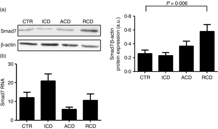
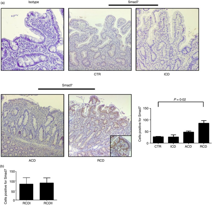
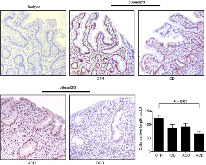
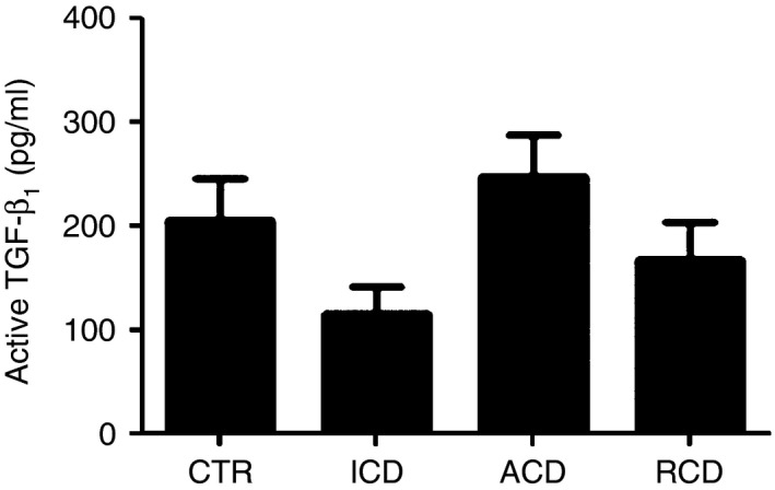
Refractory coeliac disease (RCD) is a form of coeliac disease (CD) resistant to gluten-free diet and associated with elevated risk of complications. Many effector cytokines over-produced in the gut of patients with RCD are supposed to amplify the tissue-destructive immune response, but it remains unclear if the RCD-associated mucosal inflammation is sustained by defects in counter-regulatory mechanisms. The aim of the present study was to determine whether RCD-related inflammation is marked by high Smad7, an intracellular inhibitor of transforming growth factor-β1 (TGF-β1 ) activity. Smad7 was evaluated in duodenal biopsy samples of patients with RCD, patients with active CD, patients with inactive CD and healthy controls by Western blotting, immunohistochemistry and real-time PCR. In the same samples, TGF-β1 and phosphorylated (p)-Smad2/3 were evaluated by ELISA and immunohistochemistry, respectively. Pro-inflammatory cytokine expression was evaluated in RCD samples cultured with Smad7 sense or antisense oligonucleotide. Smad7 protein, but not RNA, expression was increased in RCD compared with active and inactive CD patients and healthy controls and this was associated with defective TGF-β1 signalling, as marked by diminished p-Smad2/3 expression. TGF-β1 protein content did not differ among groups. Knockdown of Smad7 in RCD biopsy samples reduced interleukin-6 and tumour necrosis factor-α expression. In conclusion, in RCD, high Smad7 associates with defective TGF-β1 signalling and sustains inflammatory cytokine production. These results indicate a novel mechanism by which the mucosal cytokine response is amplified in RCD and suggest that targeting Smad7 can be therapeutically useful in RCD.

Keywords: gluten; inflammation; mucosal immune response; transforming growth factor-β.


Figures

Figure 1
Figure 1
Smad7 protein expression is increased in patients with refractory coeliac disease (RCD). (a) Representative Western blots showing Smad7 and β‐actin in total proteins extracted from mucosal samples of one healthy control (CTR), one patient with active coeliac disease (ACD), one patient with inactive coeliac disease (ICD) and one patient with RCD. Right panel shows the quantitative analysis of Smad7/β‐actin ratio in mucosal samples taken from 10 healthy controls (CTR), 10 patients with ACD, 10 patients with ICD and 10 patients with RCD as measured by densitometry scanning of Western blots. Values are expressed in arbitrary units (a.u.) and indicate mean ± SEM of all samples. (b) Smad7 RNA expression is not increased in RCD patients. Smad7 RNA transcripts were evaluated in duodenal biopsies of seven CTR, 10 patients with ACD, eight patients with ICD and six patients with RCD by real‐time PCR, and levels were normalized to β‐actin. Data indicate mean ± SEM of all samples.
Figure 2
Figure 2
Smad7 expression is increased in both epithelial and lamina propria compartments in refractory coeliac disease (RCD). (a) Representative photomicrographs (×100 original magnification) of Smad7‐stained paraffin‐embedded sections of biopsy samples taken from one control (CTR), one patient with active coeliac disease (ACD), one patient with inactive coeliac disease (ICD) and one patient with RCD. In RCD, Smad7‐positive cells are evident in both the epithelial and lamina propria compartments. Higher magnification photomicrograph (× 200) is shown in the insert. Staining with control IgG is also shown. Right panel shows the number of Smad7‐positive cells per high‐power field (hpf) in duodenal sections taken from three CTR, four patients with ACD, four patients with ICD and seven patients with RCD (four RCDI and three RCDII). Data indicate mean ± SE. (b) Number of Smad7‐positive cells in duodenal sections of four patients with RCDI and three patients with RCDII. Data indicate mean ± SE. [Colour figure can be viewed at wileyonlinelibrary.com]
Figure 3
Figure 3
Transforming growth factor‐β 1 (TGFβ 1)/Smad signalling is altered in refractory coeliac disease (RCD). Representative photomicrographs (× 100 original magnification) of p‐Smad2/3‐stained paraffin‐embedded sections of biopsy samples taken from one control (CTR), one patient with active coeliac disease (ACD), one patient with inactive coeliac disease (ICD) and one patient with RCD. In RCD, p‐Smad2/3 staining is reduced in both the epithelial and lamina propria compartments. The right panel shows the number of pSmad2/3‐positive cells per high‐power field (hpf) in duodenal sections taken from five CTR, three patients with ACD, three patients with ICD and six patients with RCD (three RCDI and three RCDII). Data indicate mean ± SE. [Colour figure can be viewed at wileyonlinelibrary.com]
Figure 4
Figure 4
Active transforming growth factor‐β 1 (TGFβ 1) content is not different between patients with refractory coeliac disease (RCD) and controls. Active TGFβ 1 expression was evaluated in total proteins extracted from duodenal biopsy samples of 11 controls (CTR), 12 patients with active coeliac disease (ACD), eight patients with inactive coeliac disease (ICD) and eight patients with RCD by ELISA. Data indicate mean ± SEM.

References

    1. Daum S, Cellier C, Mulder CJ. Refractory coeliac disease. Best Pract Res Clin Gastroenterol 2005; 19:413–24. - PubMed
    1. Abdulkarim AS, Burgart LJ, See J, Murray JA. Etiology of nonresponsive celiac disease: results of a systematic approach. Am J Gastroenterol 2002; 97:2016–21. - PubMed
    1. Al‐Toma A, Verbeek WH, Mulder CJ. Update on the management of refractory coeliac disease. J Gastrointestin Liver Dis 2007; 16:57–63. - PubMed
    1. Ludvigsson JF, Leffler DA, Bai JC, Biagi F, Fasano A, Green PH et al The Oslo definitions for coeliac disease and related terms. Gut 2013; 62:43–52. - PMC - PubMed
    1. Leffler DA, Dennis M, Hyett B, Kelly E, Schuppan D, Kelly CP et al Etiologies and predictors of diagnosis in nonresponsive celiac disease. Clin Gastroenterol Hepatol 2007; 5:445–50. - PubMed