

The molecular basis of breast cancer pathological phenotypes
- PMID: 27861902
- PMCID: PMC5499709
- DOI: 10.1002/path.4847
The molecular basis of breast cancer pathological phenotypes
Abstract
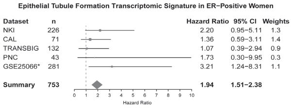
The histopathological evaluation of morphological features in breast tumours provides prognostic information to guide therapy. Adjunct molecular analyses provide further diagnostic, prognostic and predictive information. However, there is limited knowledge of the molecular basis of morphological phenotypes in invasive breast cancer. This study integrated genomic, transcriptomic and protein data to provide a comprehensive molecular profiling of morphological features in breast cancer. Fifteen pathologists assessed 850 invasive breast cancer cases from The Cancer Genome Atlas (TCGA). Morphological features were significantly associated with genomic alteration, DNA methylation subtype, PAM50 and microRNA subtypes, proliferation scores, gene expression and/or reverse-phase protein assay subtype. Marked nuclear pleomorphism, necrosis, inflammation and a high mitotic count were associated with the basal-like subtype, and had a similar molecular basis. Omics-based signatures were constructed to predict morphological features. The association of morphology transcriptome signatures with overall survival in oestrogen receptor (ER)-positive and ER-negative breast cancer was first assessed by use of the Molecular Taxonomy of Breast Cancer International Consortium (METABRIC) dataset; signatures that remained prognostic in the METABRIC multivariate analysis were further evaluated in five additional datasets. The transcriptomic signature of poorly differentiated epithelial tubules was prognostic in ER-positive breast cancer. No signature was prognostic in ER-negative breast cancer. This study provided new insights into the molecular basis of breast cancer morphological phenotypes. The integration of morphological with molecular data has the potential to refine breast cancer classification, predict response to therapy, enhance our understanding of breast cancer biology, and improve clinical management. This work is publicly accessible at www.dx.ai/tcga_breast. Copyright © 2016 Pathological Society of Great Britain and Ireland. Published by John Wiley & Sons, Ltd.
Keywords: PAM50; TCGA; bioinformatics; epithelial tubule formation; genomics; histological grade; mRNA.
Copyright © 2016 Pathological Society of Great Britain and Ireland. Published by John Wiley & Sons, Ltd.
Conflict of interest statement
Conflicts of interest statement CMP is an equity stock holder and Board of Director Member of BioClassifier. CMP is also listed an inventor on patent applications on the Breast PAM50 assay. AHB is an equity stock holder and Board of Director Member of PathAI. The remaining authors declare no competing financial interests.
Figures


References
-
- Elston CW, Elston CW, Ellis IO, et al. Pathological prognostic factors in breast cancer. I. The value of histological grade in breast cancer: experience from a large study with long-term follow-up. Histopathology. 1991;19:403–410. - PubMed
-
- Galea MH, Blamey RW, Elston CE, et al. The Nottingham prognostic index in primary breast cancer. Breast Cancer Res Treat. 1992;22:207–219. - PubMed
-
- Loi S, Sirtaine N, Piette F, et al. Prognostic and predictive value of tumor-infiltrating lymphocytes in a phase III randomized adjuvant breast cancer trial in node-positive breast cancer comparing the addition of docetaxel to doxorubicin with doxorubicin-based chemotherapy: BIG 02-98. J Clin Oncol. 2013;31:860–867. - PubMed
-
- Salgado R, Denkert C, Campbell C, et al. Tumor-infiltrating lymphocytes and associations with pathological complete response and event-free survival in HER2-positive early-stage breast cancer treated with lapatinib and trastuzumab: a secondary analysis of the NeoALTTO Trial. JAMA Oncol. 2015;1:448–454. - PMC - PubMed
MeSH terms
Substances
Grants and funding
LinkOut - more resources
Full Text Sources
Other Literature Sources
Medical
