. 2017 Feb;50(1):e12315.
doi: 10.1111/cpr.12315. Epub 2016 Nov 10.

All-trans retinoic acid preconditioning enhances proliferation, angiogenesis and migration of mesenchymal stem cell in vitro and enhances wound repair in vivo

Affiliations

All-trans retinoic acid preconditioning enhances proliferation, angiogenesis and migration of mesenchymal stem cell in vitro and enhances wound repair in vivo

M Pourjafar et al. Cell Prolif. 2017 Feb.

Abstract

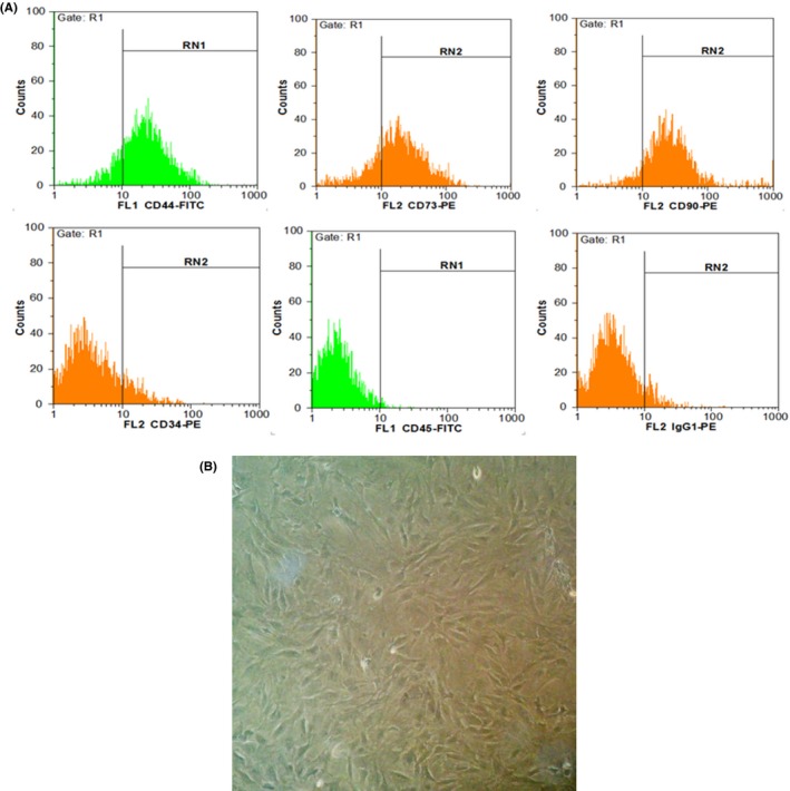
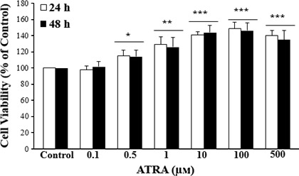
Objectives: Stem cell therapy is considered to be a suitable alternative in treatment of a number of diseases. However, there are challenges in their clinical application in cell therapy, such as to reduce survival and loss of transplanted stem cells. It seems that chemical and pharmacological preconditioning enhances their therapeutic efficacy. In this study, we investigated effects of all-trans retinoic acid (ATRA) on survival, angiogenesis and migration of mesenchymal stem cells (MSCs) in vitro and in a wound-healing model.

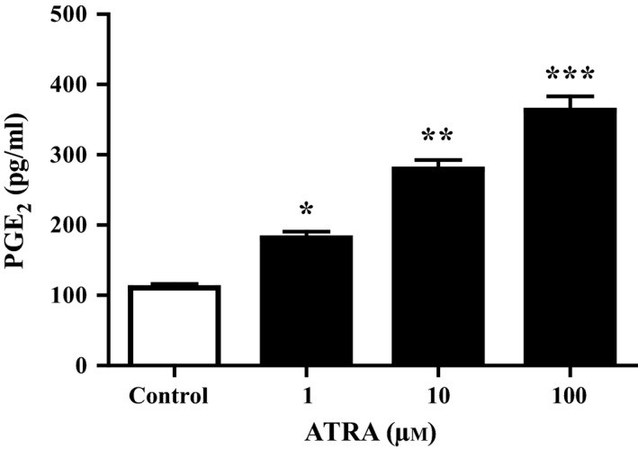
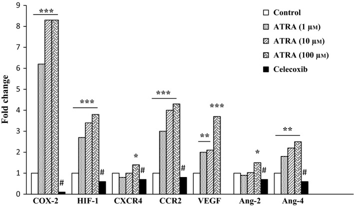
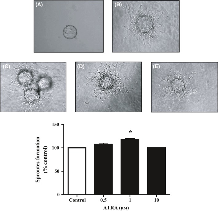
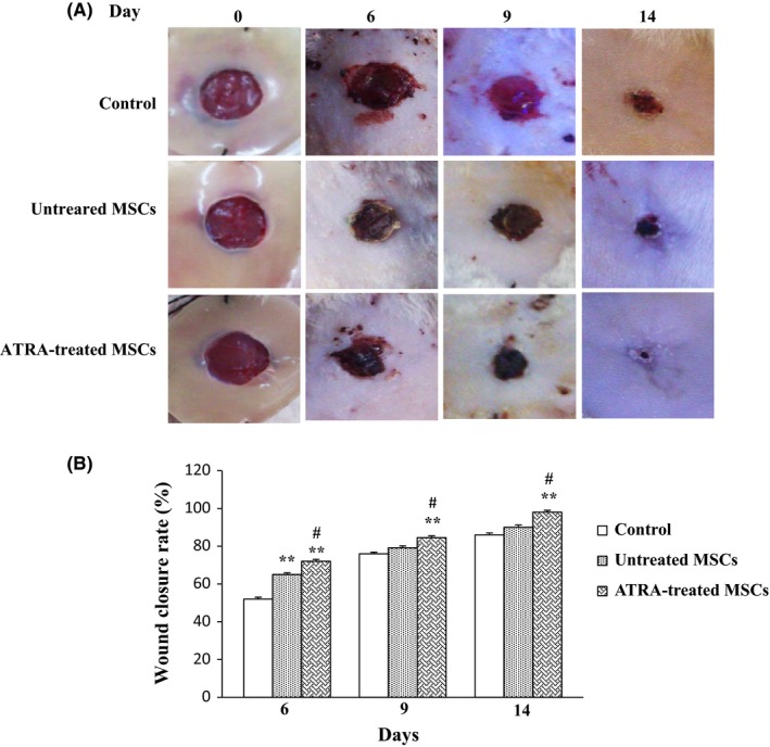
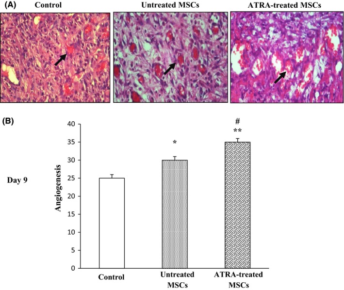
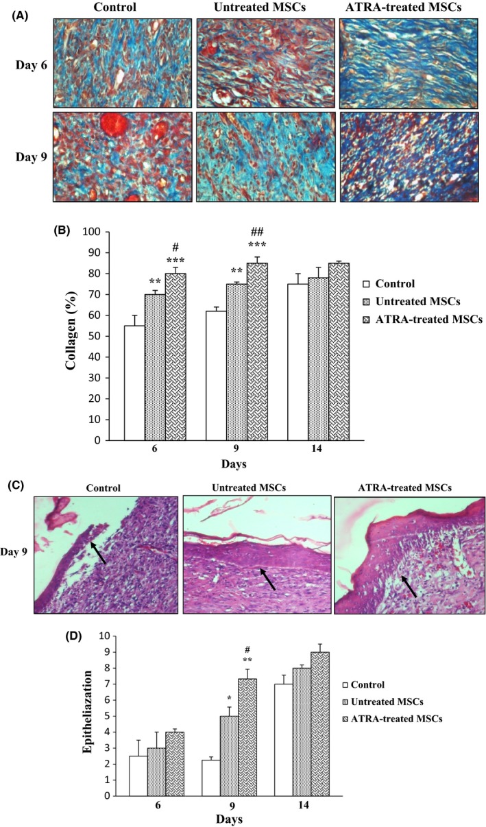
Materials and methods: MSCs were treated with a variety of concentrations of ATRA, and mRNA expression of cyclo-oxygenase-2 (COX-2), hypoxia-inducible factor-1 (HIF-1), C-X-C chemokine receptor type 4 (CXCR4), C-C chemokine receptor type 2 (CCR2), vascular endothelial growth factor (VEGF), angiopoietin-2 (Ang-2) and Ang-4 were examined by qRT-PCR. Prostaglandin E2 (PGE2) levels were measured using an ELISA kit and MSC angiogenic potential was evaluated using three-dimensional tube formation assay. Finally, benefit of ATRA-treated MSCs in wound healing was determined with a rat excisional wound model.

Results: In ATRA-treated MSCs, expressions of COX-2, HIF-1, CXCR4, CCR2, VEGF, Ang-2 and Ang-4 increased compared to control groups. Overexpression of the related genes was reversed by celecoxib, a selective COX-2 inhibitor. Tube formation and in vivo wound healing of ATRA-treated MSCs were also significantly enhanced compared to untreated MSCs.

Conclusion: Pre-conditioning of MSCs with ATRA increased efficacy of cell therapy by activation of survival signalling pathways, trophic factors and release of pro-angiogenic molecules.

Keywords: All-trans retinoic acid; cyclooxygenase-2; mesenchymal stem cells; prostaglandin E2; stem cell therapy.


Conflict of interest statement

The authors declare no conflict of interests related to this study.

Figures

Figure 1
Figure 1
Characterization of MSC using flow cytometry. A, MSCs were positive for CD73, CD44 and CD90, while they were negative for CD34 and CD45 (hematopoietic markers). B, MSCs after fourth passage in 90% confluency. Cells showed fibroblast like morphology (long and thin) under phase contrast microscope
Figure 2
Figure 2
Effect of ATRA on MSCs proliferation. MSCs were treated with various concentrations of ATRA for 24 and 48 hours and their viability were examined by MTT assay. MTT results indicated that MSCs viability and proliferation increased by ATRA compared to the control group. Data are represented as the mean±SEM (n=8). *P<.05, **P<.01, ***P<.001, compared with control
Figure 3
Figure 3
Measurement of PGE2 levels in MSCs. The cells were treated with 0, 1, 10 and 100 μmol/L ATRA for 24 hours. PGE2 levels in the culture medium were measured by ELISA. ATRA significantly increased PGE2 levels. Data are reported as mean±SEM (n=3). *P<.05, **P<.01, ***P<.001 vs control cells
Figure 4
Figure 4
Effect of ATRA on expression of genes involved in MSCs survival, migration and angiogenesis. MSCs were treated with ATRA (0, 1, 10 and 100 μmol/L) for 24 hours, and the expression of genes was measured by qRTPCR. The expression of these genes was significantly increased compared to untreated control. These effects were inhibited by celecoxib. Data were normalized to levels of 18s rRNA. *P<.05, **P<.01 and ***P<.001 vs non‐treated MSCs and # P<.001 vs 100 μmol/L ATRA‐treated MSCs. Cyclooxygenase‐2, COX‐2; hypoxia‐inducible factor‐1, HIF‐1; C‐X‐C chemokine receptor type 4, CXCR4; C‐C chemokine receptor type 2, CCR2; Vascular endothelial growth factor, VEGF; Angiopoietin‐2, Ang‐2; and Angiopoietin‐4, Ang‐4
Figure 5
Figure 5
Effect of ATRA on capillary formation potential of HUVECs. Capillary sprout formation of HUVECs in (A) medium with 2% FBS in the first day, (B) medium with 2% FBS, (C) 0.5 μmol/L (D) 1 μmol/L and (E) 10 μmol/L ATRA‐treated HUVECs were analyzed by inverted microscopy after 48 hours. The alternation in sprout formation determined by NIH image J. Data reported as mean±SEM (n=3); *P<.05 vs control
Figure 6
Figure 6
Effects of MSC on wound closure. A, Images of wounds injected with PBS, untreated MSCs and ATRA‐treated MSCs after 0, 6, 9 and 14 days. B, Representation of average wound closure rate in all groups using NIH ImageJ software. Data are reported as mean±SEM. (n=3; *P<.05, **P<.01 vs control, # P<.05 vs untreated MSCs)
Figure 7
Figure 7
Effect of ATRA on wound vascularity. A, Representative images of histological sections of wounds in rat treated with PBS, untreated MSCs and ATRA‐treated MSCs are shown after 9 days. Arrows signify blood vessels. B, Comparison between angiogenic scores in groups after 9 days. Data are represented as mean±SEM. (n=3); *P<.05, **P<.01 vs control and # P<.05 vs untreated MSCs
Figure 8
Figure 8
Effect of ATRA on collagenization and epithelialization. A, Masson's trichrome staining was used for assessment of collagenization. Fibers of collagen are stained blue and arrows signify collagen fibers. Wounds injected with ATRA‐treated MSCs and untreated MSCs had higher tissue collagenization compared to those injected with PBS at 6 and 9 days. Intensity of staining was compared with normal skin on day 0 was determined by NIH image J. B, Comparison between percent of collagen fibers in groups within 14 days. C, Epithelialization was evaluated using H&E staining of wound sections. Arrows signify epithelium. Wounds injected with ATRA‐treated MSCs had higher tissue epithelialization compared to untreated MSCs and PBS groups at 9 days. D, Comparison between the number of epithelium layers in groups. Data are represented as mean±SEM. (n=30) *P<.05, **P<.01, ***P<.001 vs control group and # P<.05, ## P<.01 vs untreated MSCs

References

    1. Srijaya TC, Ramasamy TS, Kasim NHA. Advancing stem cell therapy from bench to bedside: Lessons from drug therapies. J Transl Med. 2014;12:1. - PMC - PubMed
    1. Herberts CA, Kwa MS, Hermsen HP. Risk factors in the development of stem cell therapy. J Transl Med. 2011;9:1. - PMC - PubMed
    1. Wei X, Yang X, Han Z‐P, Qu F‐F, Shao L, Shi Y‐F. Mesenchymal stem cells: A new trend for cell therapy. Acta Pharmacol Sin 2013;34:747–754. - PMC - PubMed
    1. Kang SK, Shin IS, Ko MS, Jo JY, Ra JC. Journey of mesenchymal stem cells for homing: Strategies to enhance efficacy and safety of stem cell therapy. Stem Cells Int 2012;2012:342968. doi: 10.1155/2012/342968. - DOI - PMC - PubMed
    1. Haider HK, Ashraf M. Strategies to promote donor cell survival: Combining preconditioning approach with stem cell transplantation. J Mol Cell Cardiol. 2008;45:554–566. - PMC - PubMed

MeSH terms

Associated data