Review
. 2016 Dec;172(4):349-366.
doi: 10.1002/ajmg.c.31534. Epub 2016 Nov 8.

Gene and cell-based therapies for inherited retinal disorders: An update

Review

Gene and cell-based therapies for inherited retinal disorders: An update

Jesse D Sengillo et al. Am J Med Genet C Semin Med Genet. 2016 Dec.

Abstract

Retinal degenerations present a unique challenge as disease progression is irreversible and the retina has little regenerative potential. No current treatments for inherited retinal disease have the ability to reverse blindness, and current dietary supplement recommendations only delay disease progression with varied results. However, the retina is anatomically accessible and capable of being monitored at high resolution in vivo. This, in addition to the immune-privileged status of the eye, has put ocular disease at the forefront of advances in gene- and cell-based therapies. This review provides an update on gene therapies and randomized control trials for inherited retinal disease, including Leber congenital amaurosis, choroideremia, retinitis pigmentosa, Usher syndrome, X-linked retinoschisis, Leber hereditary optic neuropathy, and achromatopsia. New gene-modifying and cell-based strategies are also discussed. © 2016 Wiley Periodicals, Inc.

Keywords: CRISPR; Usher syndrome; achromatopsia; choroideremia; gene therapy; iPSCs; leber congenital amaurosis; leber hereditary optic neuropathy; retinal degeneration; retinitis pigmentosa; retinoschisis; stem cells.


Conflict of interest statement

Conflicts of interest: The authors declare no conflicts of interest.

Figures

Figure 1.
Figure 1.. Schematic illustration of retinal cell layers.
RPE cells (brown) maintain close contact with and phagocytose rod (purple) and cone (light blue) photoreceptor outer segments. Bipolar cells (yellow) synapse with and transfer information between photoreceptors and ganglion cells (green). Axons of the ganglion cell layer converge to form the optic nerve. Horizontal (red) and amacrine (dark blue) cells serve multiple functions by integrating and regulating signal transduction throughout the retina.
Figure 2.
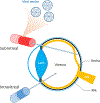
Figure 2.. Vector delivery.
Schematic illustration of subretinal and intravitreal delivery of therapeutic gene-containing vectors. Vector delivery can be achieved by subretinal injection of the viral vector, which involves the formation of a transient retinal detachment that spontaneously resolves. Intravitreal injection is less invasive and produces fewer iatrogenic complications, but may deliver therapeutic genes less efficiently. ON, optic nerve; RPE, retinal pigment epithelium.
Figure 3.
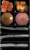
Figure 3.. Retinal imaging of patients with inherited retinal disease.
Color fundoscopy of a normal patient (A) compared to patients with choroideremia (B), asymptomatic RPGR-associated RP female carrier (C) and retinoschisis (D). OCT of a normal patient depicting high resolution image of the retinal layers (E) compared to patients with retinoschisis (F) and achromatopsia (G). In B, note the characteristic pale fundus of choroideremia as a result of the sclera transilluminating through a degenerating RPE and choroid. In C, a typical tapetal-like reflex of the retina in an RPGR-associated RP carrier can be seen sparing the macula. In D, the subtle spoke-wheel appearance of retinoschisis is seen on color fundoscopy, and characteristic foveal and perifoveal cysts are seen on OCT in F. Patients with achromatopsia can show a foveal optical gap with loss of the inner and outer segment junction on OCT as depicted in G.
Figure 4.
Figure 4.. Non-homologous end joining (NHEJ) versus homology-directed repair (HDR).
Illustration depicting differences between NHEJ (left) and HDR (right). A double strand break (DSB) may undergo subsequent error-prone NHEJ via ligation of the ends of the DNA without regard for the sequence accuracy, leading to indels. HDR occurs using a wild type donor template that is homologous to the target site and serves as a template for precise correction.
Figure 5.
Figure 5.. CRISPR/Cas9.
Schematic illustration of CRISPR/Cas9 associated with a target sequence. gRNA complexes with the Cas9 endonuclease and directs it to the site of interest, where it creates a DSB. gRNA, guide RNA; Cas9, CRISPR-associated protein 9; PAM, protospacer adjacent motif.
Figure 6.
Figure 6.. CRISPR/Cas9 delivery.
Illustration depicting how CRISPR/Cas9 can be packaged for gene-modifying therapy. Cas9 and gRNA are delivered via a vector without donor template (left) or with donor template (right). When given without donor template, gene ablation can be achieved at a target sequence specified by gRNA. Cas9 variants can be used to create a shorter sequenced, single construct. Precise homology-directed repair is achieved when the vector includes a donor template. ITR sequences allow identification and encapsulation of the construct by the viral vector. ITR, inverted terminal repeat; gRNA, guide RNA; Cas9, CRISPR-associated protein 9.
Figure 7.
Figure 7.. CRISPRi strategy.
In the absence of CRISPRi, transcription factors and RNA polymerase bind to the transcription start site (TSS), which leads to gene expression. Using CRISPRi, a “dead” Cas9 (dCas9) fused to a Krüppel associated box (KRAB) domain complexes with a gRNA that is complementary to a promoter or exonic sequence. This complex does not create a DSB and instead sterically blocks gene transcription.

References

    1. Acland GM, Aguirre GD, Ray J, Zhang Q, Aleman TS, Cideciyan AV, Pearce-Kelling SE, Anand V, Zeng Y, Maguire AM, Jacobson SG, Hauswirth WW, Bennett J. 2001. Gene therapy restores vision in a canine model of childhood blindness. Nat Genet 28:92–95. - PubMed
    1. Alexander JJ, Umino Y, Everhart D, Chang B, Min SH, Li Q, Timmers AM, Hawes NL, Pang JJ, Barlow RB, Hauswirth WW. 2007. Restoration of cone vision in a mouse model of achromatopsia. Nat Med 13:685–687. - PMC - PubMed
    1. Algvere PV, Berglin L, Gouras P, Sheng Y. 1994. Transplantation of fetal retinal pigment epithelium in age-related macular degeneration with subfoveal neovascularization. Graefes Arch Clin Exp Ophthalmol 232:707–716. - PubMed
    1. Allocca M, Doria M, Petrillo M, Colella P, GarciaHoyos M, Gibbs D, Kim SR, Maguire A, Rex TS, Di Vicino U, Cutillo L, Sparrow JR, Williams DS, Bennett J, Auricchio A. 2008. Serotype-dependent packaging of large genes in adeno-associated viral vectors results in effective gene delivery in mice. J Clin Invest 118:1955–1964. - PMC - PubMed
    1. Anand V, Barral DC, Zeng Y, Brunsmann F, Maguire AM, Seabra MC, Bennett J. 2003. Gene therapy for choroideremia: In vitro rescue mediated by recombinant adenovirus. Vision Res 43:919–926. - PubMed

Publication types

MeSH terms