. 2018 Jan;12(1):59-69.
doi: 10.1002/term.2363. Epub 2017 May 31.

Scaffold-free tissue engineering of functional corneal stromal tissue

Affiliations

Scaffold-free tissue engineering of functional corneal stromal tissue

Fatima N Syed-Picard et al. J Tissue Eng Regen Med. 2018 Jan.

Abstract

Blinding corneal scarring is predominately treated with allogeneic graft tissue; however, there is a worldwide shortage of donor tissue leaving millions in need of therapy. Human corneal stromal stem cells (CSSC) have been shown produce corneal tissue when cultured on nanofibre scaffolding, but this tissue cannot be readily separated from the scaffold. In this study, scaffold-free tissue engineering methods were used to generate biomimetic corneal stromal tissue constructs that can be transplanted in vivo without introducing the additional variables associated with exogenous scaffolding. CSSC were cultured on substrates with aligned microgrooves, which directed parallel cell alignment and matrix organization, similar to the organization of native corneal stromal lamella. CSSC produced sufficient matrix to allow manual separation of a tissue sheet from the grooved substrate. These constructs were cellular and collagenous tissue sheets, approximately 4 μm thick and contained extracellular matrix molecules typical of corneal tissue including collagen types I and V and keratocan. Similar to the native corneal stroma, the engineered corneal tissues contained long parallel collagen fibrils with uniform diameter. After being transplanted into mouse corneal stromal pockets, the engineered corneal stromal tissues became transparent, and the human CSSCs continued to express human corneal stromal matrix molecules. Both in vitro and in vivo, these scaffold-free engineered constructs emulated stromal lamellae of native corneal stromal tissues. Scaffold-free engineered corneal stromal constructs represent a novel, potentially autologous, cell-generated, biomaterial with the potential for treating corneal blindness. Copyright © 2016 John Wiley & Sons, Ltd.

Keywords: cell sheet; cornea; human cells; ocular; scaffold-free; stem cells; transplantation.


Figures

Figure 1
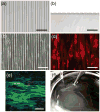
Figure 1. Formation of scaffold-free tissue sheet with parallel cell and matrix organization
Light micrographs of (a) top view and (b) cross sectional view of the PDMS substrate show grooves approximately 10 μm wide, 10 μm apart, and 5 μm deep. (c) Phase contrast image shows CSSC cultured on the grooved substrate. (e) For better visualization, CSSC were labeled with DiI (red) and cultured on grooved substrate. (e) Two-photon micrograph of 10-day cultures of CSSC on grooved substrates in keratocyte differentiation medium (KDM) shows deposition of parallel organized collagenous matrix (green). Nuclei (blue) were stained by SYTOX-green (blue). (f) After 10 days of culture a robust tissue sheet is formed that can be separated from the substrate using forceps. Scale bars: (a) and (b) = 50 μm, (c)–(e) = 100 μm
Figure 2
Figure 2. Transmission electron microscopy of scaffold-free tissue sheets generated in vitro
Scaffold-free tissue sheets harvested after 12 days culture in KDM were fixed and imaged by TEM as described in Methods. (a) Lower and (b) higher magnification images of the top view of scaffold-free tissue sheets shows that the constructs contain long, parallel organized collagen fibrils. (c) Lower and (d) higher magnification images of engineered corneal stroma tissues in cross section show collagen fibrils are approximately uniform in diameter. (e) Size distribution of collagen fibril diameters as measured from TEM images of cross section of engineered tissues show that collagen fibril diameter are similar to the collagen fibril diameter seen in native, human corneal stromal tissue. Scale bars: (a) = 2 μm, (b) = 500 nm, (c) 2 μm, and (d) = 100 nm
Figure 3
Figure 3. Immunostaining of scaffold-free tissue sheets produced in vitro
Paraffin sections of scaffold-free tissue sheets produced after 12 days culture in KDM were immunostained as described in Methods. (a) Presence of for type I collagen (green) was detected in the matrix of scaffold-free tissue sheets, (a′) with corresponding nuclear DAPI stain (blue), (a″) merged image of (a) and (a′), and (a″′) corresponding merged image of negative control. (b) Type V collagen expression was seen in engineered tissue sheets with (b′) corresponding nuclear DAPI stain (blue), (b″) merged image of (b) and (b′), and (b″′) corresponding merged image of negative control. (c) Keratocan expression was detected in scaffold-free tissue sheets, with (c′) corresponding nuclear DAPI stain (blue), (c″) merged image of (c) and (c′), and (c″′) corresponding merged image of negative control. Scale bars: 20 μm
Figure 4
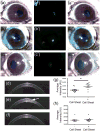
Figure 4. Histological analysis of scaffold-free tissue sheets in mouse stromal pockets in vivo
Mouse eyes were imaged in vivo after lamellar transplantation of scaffold-free tissue sheets as described in Methods. (a) Light micrograph image of mouse eye containing scaffold-free tissue sheet with DiO-labeled cells, (a′) with corresponding fluorescent image showing human cells (green), and (a″) the merged image of (a) and (a′). (b) Light micrograph image of mouse eye containing scaffold-free tissue sheet with DTAF labelled matrix, (b′) with corresponding fluorescent image showing matrix (green) from scaffold-free sheet, and (b″) the merged image of (b) and (b′). (c) A light micrograph image of control mouse eye lacking a scaffold-free tissue sheet is shown, (c′) with corresponding fluorescent image, and (c″) the merged image of (c) and (c′). Optical coherence tomography (OCT) was used to assess light scatter in the corneal stroma. (d) Cross-sectional projection image of untreated control mouse cornea. (e) Cross sectional image of a mouse eye with implanted tissue sheet (arrow) 1 week after implantation. (f) Cross-sectional projection image of mouse eye, 5 weeks after tissue sheet implantation. (g) Quantification of light scatter from OCT scans of the stroma showing light scatter by implanted tissue sheet 1-week post-implantation. (h) Quantification of light scatter from OCT scans of the stroma showing light scatter by implanted tissue sheet 5-weeks post-implantation.
Figure 5
Figure 5. Persistence of human cells and matrix in mouse cornea stroma after implantation
Five weeks after implantation of CSSC generated tissue sheets, mouse corneas were harvested and analyzed as described under Methods. (a) Confocal image of whole mount of cornea shows DiO labelled human cells (green). All cells were stained using nuclear dye DAPI (blue). (b) Higher magnification of boxed region in (a) shows region where human cells are located and in (c) a cross sectional projection of dotted line in (b) shows that the human cells are localized to the central stroma. (d) Immunostaining human keratocan (red) in cryosections from corneas 5 weeks after tissue sheet implantation. (d′) Micrograph of green channel in same region as (d) shows DiO labelled human cells (green), (d″) micrograph of DAPI nuclear staining of all cells in same region as (d), and (d″′) a merged image of (d)–(d″) show keratocan staining localization with respect to the human cells. (e) Immunostaining human keratocan (red) in a section of control mouse cornea lacking scaffold-free tissue sheets. (e′) Micrograph of same region as (e) showing lack of DiO labeled green cells (green), (e″) micrograph in same region as (e) of DAPI nuclear staining of all cells (blue), and (e″′) merged image of (e)–(e″). Scale bars: (a) = 500 μm, (b) = 100 μm, (d)–(e) = 20 μm

Similar articles

Cited by

References

    1. Meek KM, Leonard DW. Ultrastructure of the corneal stroma: a comparative study. Biophys J. 1993;64:273–80. - PMC - PubMed
    1. Levin LA, Adler FH. Adler’s physiology of the eye : clinical application. 11. Edingburg: Saunders/Elsevier; 2011.
    1. Smolin G, Foster CS, Azar DT, Dohlman CH. Smolin and Thoft’s the cornea : scientific foundations and clinical practice. 4. Philadelphia: Lippincott Williams & Wilkins; 2005.
    1. Hassell JR, Birk DE. The molecular basis of corneal transparency. Exp Eye Res. 2010;91:326–35. - PMC - PubMed
    1. Fini ME. Keratocyte and fibroblast phenotypes in the repairing cornea. Progress in retinal and eye research. 1999;18:529–51. - PubMed

Publication types

LinkOut - more resources